 |
| About Bioline | All Journals | Testimonials | Membership | News |
|
||||||
|
||||||
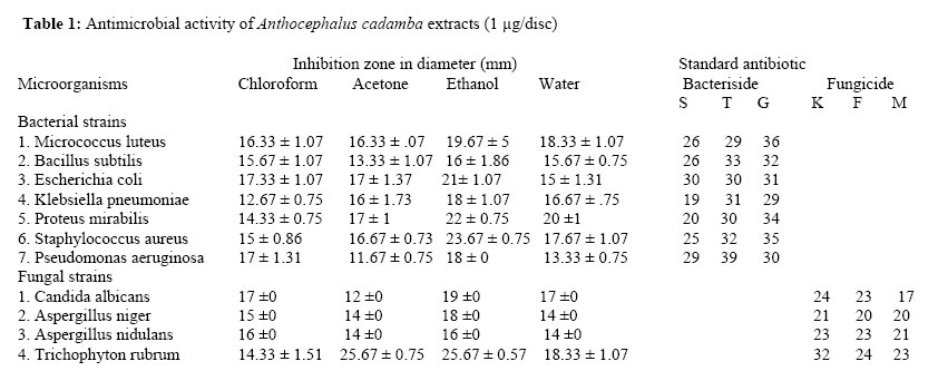
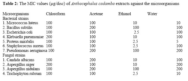
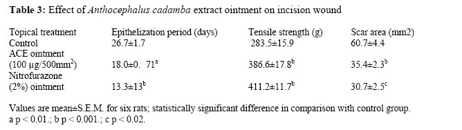
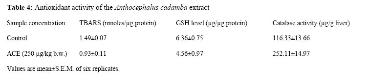
African Journal of Traditional, Complimentary and Alternative Medicines, Vol.4, No. 4, 2007, pg. 481 – 487 Research Paper ANTIMICROBIAL, WOUND HEALING AND ANTIOXIDANT ACTIVITIES OF ANTHOCEPHALUS CADAMBA Sanjay.Prahalad Umachigi*, Kumar. G. S1., Jayaveera K.N3., Kishore kumar D.V1., Ashok kumar C.K2., Dhanapal.R1.*,1Rural College of Pharmacy, D.S.Road, PO Box-10, Devanahalli-562110, Bangalore Rural Dist, Karnataka, India. 2SLN College of Pharmacy, Chitoor, AP, India. 3Department of Chemistry, Jawaharlal Nehru Technological University College of Engineering, Ananthpur- 01, Andhra Pradesh, India. *E-mail: sanjayumachigi@gmail.com Code Number: tc07064 AbstractAnthocephalus cadamba (Roxb.) Miq. Syn A. chinensis (Lamk) A. Rich (Rubiaceae) is ethnomedicinally widely used in the form of paste by tribe in western Ghats for treating skin diseases. In this context, antimicrobial potential of A. cadamba against a wide range of microorganisms was studied. To validate the ethnotherapeutic claims of the plant in skin diseases, wound healing activity was studied, besides antioxidant activity to understand the mechanism of wound healing. The alchoholic and aqueous extract of this plant showed significant antibacterial and antifungal activity against almost all the organisms: Micrococcus luteus, Bacillus subtilis, Staphylococcus aureus, Escherichia coli, Klebsiella pneumoniae, Proteus mirabilis, Pseudomonas aeruginosa, and four fungi Candida albicans, Trichophyton rubrum—dermatophyte fungi, Aspergillus niger, Aspergillus flavus and Aspergillus nidulans—systemic fungi, with especially good activity against the dermatophyte (Trichophyton rubrum) and some infectious bacteria (Escherichia coli, Proteus mirabilis and Staphylococcus aureus) with an MIC of 2.5 µg/disc. The results show that A. cadamba extract has potent wound healing capacity as shown from the wound contraction and increased tensile strength. The results also indicated that A. cadamba extract possesses potent antioxidant activity by inhibiting lipid peroxidation and increase in the superoxide dismutase (SOD) and catalase activity. Key words: Anthocephalus cadamba; Antimicrobial activity; Antioxidant; Wound healing; IntroductionAnthocephalus cadamba (Roxb.) Miq. Syn A. chinensis (Lamk) A. Rich (Rubiaceae) is widely distributed throughout the greater part of India and is used as a folk medicine in the treatment of fever, anaemia, uterine complaints, blood diseases, skin diseases, leprosy, dysentery, and for improvement of semen quality. The leaves are recommended as a gargle in cases of stomatitis (Slkar et al., 1996). Some scientific studies have been carried out to reveal its antimalarial (Sianne and Fanie 2002) and antihepatotoxic activities (Kapi, et al., 1995). The major constituents of bark are triterpenes, tripernoid glycosides, saponins, indole alkaloids cadambine,3a-dihydrocadambine, cadamine, isocadamine and isodihydrocadambine (Niranjan et al., 2000; Kitagawa et al., 1996; Mahato and Garai 1998; Malinow, 1984; Brown and Chapple, 1976). In recent years, many possible sources of natural antibiotics are used for several infectious diseases, mostly bacterial and fungal infections. In this respect, the most investigated taxa are from angiosperms whereas very little data is currently available about other groups of plants, especially bryophytes (Madsen and Pates, 1952; McCleary et al., 1960; Banerjee and Sen, 1978; Latiff et al., 1989; Basile et al., 1999). Wounds are physical injuries that result in an opening or breaking of the skin. Proper healing of wounds is essential for the restoration of disrupted anatomical continuity and disturbed functional status of the skin. It is a product of the integrated response of several cell types to injury. Wound healing is a complex multifactorial process that results in the contraction and closure of the wound and restoration of a functional barrier (Chattopadhyay et al., 2002). Repair of injured tissues occurs as a sequence of events, which includes inflammation, proliferation and migration of different cell types (Sidhu et al., 1999). It is consented that reactive oxygen species (ROS) are deleterious to wound healing process due to the harmful effects on cells and tissues. Absorbable synthetic biomaterials are considered to be degraded via ROS (Aliyeva et al., 2004). Free-radical- scavenging enzymes (FRSE) are a cytoprotective enzymal group that has an essential role in the reduction, deactivation and removal of ROS as well as in the regulation of the wound healing process. These cells, through their characteristic “respiratory burst” activity, produce free radicals (Baboir, 1979). Wound related non-phagocytic cells also generate free radicals by involving non-phagocytic NADPH oxidase mechanism (Griendling, 2000). Topical applications of compounds with free-radical-scavenging properties in patients have shown to improve significantly wound healing and protect tissues from oxidative damage (Thiem and Grosslinka, 2003). In this context, antimicrobical potential of A. cadamba against a wide range of microorganisms was studied. The study includes some organic and aqueous extracts of A. Cadamba against bacterial and fungal strains including a dermatophyte. Preliminary phytochemical screening was also conducted in order to observe the chemical nature of active extractives. Besides the minimum inhibitory concentration (MIC), dosage against each bacteria and fungi was also evaluated and calculated. To validate the ethnotherapeutic claim of the plant in skin diseases, wound healing activity was studied. The antioxidant activity was also investigated to reveal the mechanism behind the wound healing activity. Materials and methods Anthocephalus cadamba whole plant (barks, leaves, flowers and fruits) was collected from Western Ghats of Karnataka, India in October 2005. The plants were identified and voucher specimens (RCP/PCOG/02/2005-06) have been deposited in Pharmacognosy Department Herbarium, Know for future reference. Extraction procedure and phytochemical screeningAir-dried and powdered drug of A. cadamba were successively percolated with petroleum ether (6.8%, w/w), acetone (10.4%, w/w), chloroform (13.2%, w/w), ethanol (19.7%, w/w), water (21.4%, w/w) and whole plant was separately percolated with 50% hydro alcohol (20.2%, w/w) (ACE). The extracts were decanted, filtered with Whatman No. 1 filter paper and concentrated under reduced pressure below 40oC to obtain dry extract ACE. Qualitative analysis of phytochemical constituents was carried out, viz. alkaloids (Dragendorff's test), anthraquinones, saponins (foam formation), flavonoids (using magnesium and dil. HCl), sesquiterpenes and terpenes (Liebermann-Burchard's test) according to standard methods (Sofowora, 1982; Trease and Evans, 1987). MicroorganismsThe test microorganisms used for the antimicrobial activity screening were 7 bacteria and 4 fungi: (Gram positive and Gram negative)-Micrococcus luteus MTCC (106), Bacillus subtilis MTCC (121), Staphylococcus aureus MTCC (96), Escherichia coli MTCC (443), Klebsiella pneumoniae MTCC (109), Proteus mirabilis MTCC (1429), Pseudomonas aeruginosa MTCC (429), and 4 fungi Candida albicans MTCC (183), Trichophyton rubrum MTCC (296)-dermatophyte fungi, Aspergillus niger MTCC (16404), and Aspergillus nidulans MTCC (11267)- systemic fungi. These organisms were identified and procured from Institute of Microbial Technology (IMTECH-CSIR), Chandigarh, India. Antimicrobial activityThe agar diffusion method (Murray et al., 1995) was used to evaluate the antimicrobial activity. Bacteria were cultured overnight at 37 oC in Mueller Hinton Broth (MHB, Oxoid) and fungi at 28 oC for 72 h in Potato Dextrose Broth (PDB, Oxoid) and used as inoculum. A final inoculum, using 100 _l of suspension containing 108 CFV/ml of bacteria and 104 spore/ml of fungi spread on Mueller Hinton Agar (MHA) and Potato Dextrose Agar (PDA) medium, respectively. The disc (6mm in diameter) was impregnated with 10 µl of 100 mg/ml (1 mg/disc) extracts placed on seeded agar. Gentamicin (10µg/disc), streptomycin (10 µg/disc) and tetracycline (10µg/disc) were used as positive controls for bacteria and fluconazole (10 µg/disc), ketoconzole (10 µg/disc) and metronidazole (5 µg/disc) for fungi. The test plates were incubated at 37 oC for 24 h for bacteria and at 28 oC for 72 h for fungi depending on the incubation time required for a visible growth. MIC values were also studied for microorganisms, which were determined as sensitive to the extract in disc diffusion assay. Sterile filter paper discs (6mm in diameter) containing 2.5-1000 µg/disc of plant extracts were placed on the surface of a medium. MIC was defined as the lowest concentration of extract that inhibited visible growth on agar Pharmacological activity AnimalsInbred house Wistar rats of either sex were used in the study. The range of the weight of the animals was between 200-250 g. they were housed individually in standardized environmental condition. All the animals were provided with water food ad libitum. Gold Mohur Lipton India Ltd supplied the standard rat pellet food. Ethical clearance for the animal study was obtained from the institutional animal ethics committee. In the experiment, the rats were divided into three groups (n = 6): group 1 was the control group that received simple ointment base, group 2 was treated with reference standard (0.2%, w/w nitrofurazone ointment) and group 3 received ACE ointment (100 mg/500mm2) topically on wound created on the dorsal back of rats daily till the wounds completely healed (Chatterjee and Chakravorty, 1993). For antioxidant activity, rats were divided into two groups of control and experimental rats. Experimental rats received 1.25 ml (250 mg/kg b.w.) of ACE while control rats received normal saline. Excision wound modelAn impression was made on the dorsal thoracic region 1 cm away from vertebral column and 5 cm away from ear using a round seal of 2.5 cm diameter on the anaesthetized rat. The skin of impressed area was excised to the full thickness to obtain a wound area of about 500 mm2 diameter. Haemostasis was achieved by blotting the wound with cotton swab soaked in normal saline. Contractions, which contributed to wound closure in the first 2 weeks, were studied by tracing the raw wound. Wound area was measured by retracing the wound on a millimeter scale graph paper. The degree of wound healing was calculated (Werner et al., 1994) and hydroxyproline was measured using the method of Neuman and Logan (1950). Incision wound modelRats were anaesthetized and two paravertebral-long incisions were made through the skin and cutaneous muscles at a distance of about 1.5 cm from the midline on each side of the depilated back of the rat. Full aseptic measures were not taken and no local or systemic antimicrobial was used throughout the experiment (Udupa et al., 1995). All the groups were treated in the same manner as mentioned in the case of the excision wound model. No ligature was used for stitching. After the incision was made, the parted skin was kept together and stitched with black silk at 0.5 cm intervals. Surgical thread (No. 000) and a curved needle (No. 11) were used for stitching. The continuous thread on both wound edges were tightened for good closure of the wounds. The wound was left undressed, ACE ointment, along with water soluble base ointment (control) and nitrofurazone ointment were applied topically twice a day for 9 days. When wounds were thoroughly cured, the sutures were removed on the 9th day and tensile strength was measured with a tensiometer. Tensile strengthThe tensile strength of a wound represents the degree of wound healing. Usually wound healing agents promote a gain in tensile strength. The sutures were removed on the 9th day after wounding and the tensile strength was measured on the 10th day. The herbal ointment along with standard and control were applied throughout the period, twice daily for 9 days. The mean tensile strength on the two paravertebral incisions on both sides of the animals were taken as the measures of the tensile strength of the wound for an individual animal. The tensile strength of ACE ointment treated wounds was compared with control and nitrofurazone ointment as standard. The tensile strength increment indicates better wound healing stimulated by the applied herbal formulation. Further epithelization period and scar area were measured daily for 25 days after determination of tensile strength (Werner et al., 1994). Antioxidant activity Lipid peroxidation was assayed by the measurement of malondialdehyde (MDA) levels in liver on the basis of reaction with thiobarbituric acid (Okhawa et al., 1979). The activity of SOD was determined in liver by monitoring the inhibition of the autoxidation of pyrogallol (Marklund and Marklund, 1974). CAT activity in liver was determined according to the standard method (Aebi, 1974). Proteins were determined according to Lowry method (1951) using bovine serum albumin as a standard. Values were represented as mean±S.E.M and data were analyzed by paired t-test using SPSS software package. Statistical analysisPharmacological data were subjected to statistical analysis using SPSS 11.0 for Windows. The values are represented as mean ± S.E.M. for six rats. Paired t-test was used for reporting the p-value and significance with respect to the control group. Results and DiscussionThe disc diffusion method was used to determine the inhibition zones of A. cadamba extracts (organic and aqueous). The plant showed significant antibacterial and antifungal activity against almost all the organisms (Table 1) and especially good activity was found against the dermatophytes. However, the petroleum ether extracts of this plant showed little antimicrobial activity. Significant antimicrobial activity was observed in ethanolic and aqueous extracts. Amongst the test organisms used, a dermatophyte Trichophyton rubrum was found to be most sensitive, Staphylococcus aureus came next, followed by Proteus mirabilis, Escherichia coli, Micrococous luteus, Aspergillus niger, Klebsiella pneumoniae, Pseudomonas aeruginosa, Candida albicans, Aspergillus nidulans, Aspergillus flavus and Bacillus subtilis. Increased inhibition was found at higher levels of extract concentration. MICs of these extracts are summarized in Table 2. Some of the extracts like the ethanolic extract of A. cadamba gave very low MIC values, and inhibited the growth of Escherichia coli, Proteus mirabilis, Staphylococcus aureus and Trichophyton rubrum with a concentration of 2.5 µg/disc. Preliminary phytochemical screening of A. cadamba showed the presence of saponins, terpenes, sesquiterpenes glycosides, alkaloids and absence of anthraquinones and flavonoids. The antimicrobial activity could be due to the presence of terpenes (Toyota and Asakawa, 1999). Since MIC of A. cadamba against Trichophyton rubrum (dermatophyte) was substantially low (2.5 µg/disc), the wound healing activity was done on it. The wound healing activity results showed that upon application of ACE ointment there was a decrease in the epithelization period, along with a visibly decreased scar area (Table 3). There was also a significant increase in the tensile strength and hydroxyproline content (Table 3). The crude extract of ACE significantly stimulated wound contraction. Thus, the plant extract might be useful as a wound healing agent. The potent wound healing capacity of ACE as shown from the wound contraction and increased tensile strength has thus validated the ethnotherapeutic claim. This also validates the potent inhibition of the dermatophyte (Trichophyton rubrum). Some of the bryophytes like Lunularia cruciata have also been reported for antioxidant activity (Ielpo et al., 1998). The results indicate that ACE possesses potent antioxidant activity by inhibiting lipid peroxidation (Table 4). ConclusionThe efficacy of this plant in wound healing may be due to its action against dermatophyte and the effect on antioxidant enzymes. References:
© Copyright 2007 - African Journal of Traditional, Complementary and Alternative Medicines The following images related to this document are available:Photo images[tc07064t4.jpg] [tc07064t3.jpg] [tc07064t1.jpg] [tc07064t2.jpg] |
| |||||||||
