 |
| About Bioline | All Journals | Testimonials | Membership | News |
|
||||||
|
||||||
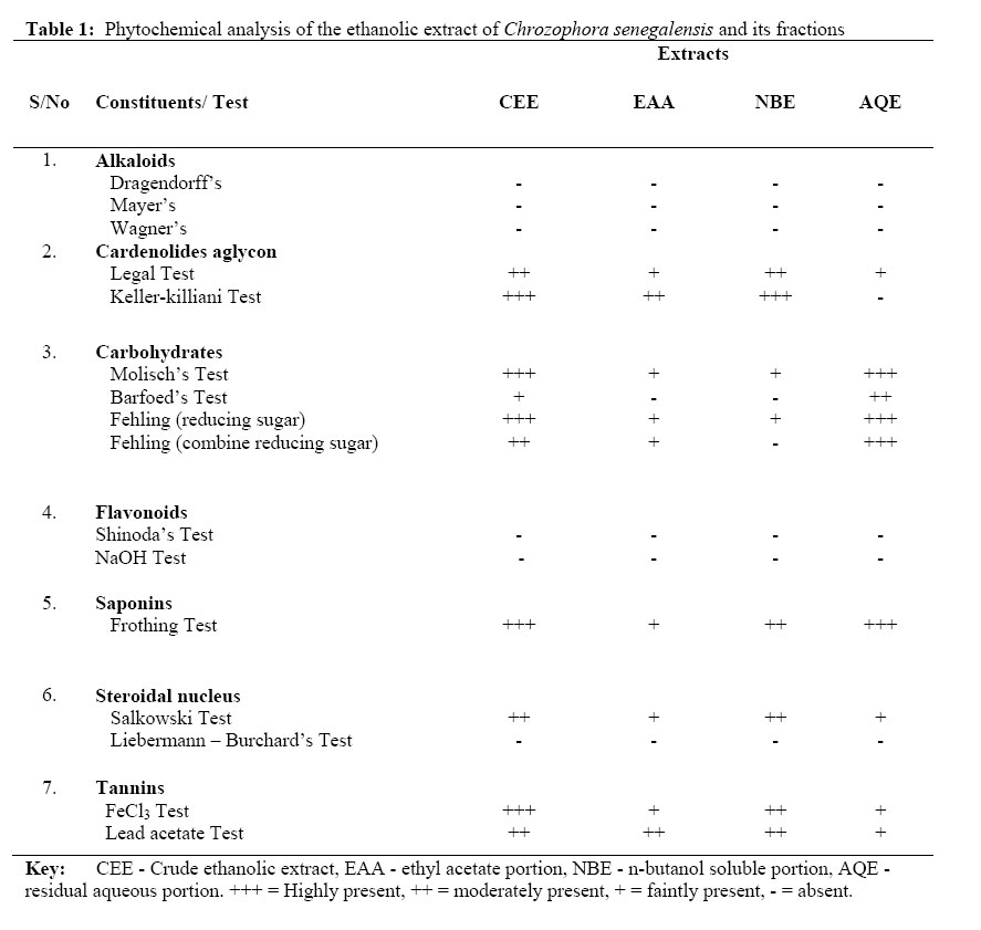
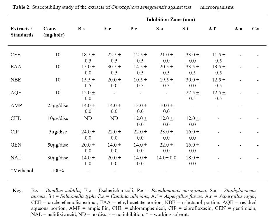
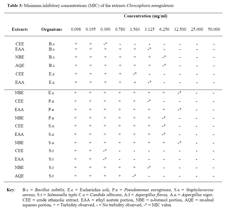
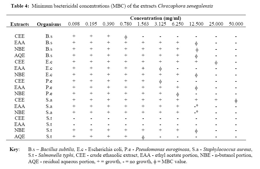
African Journal of Traditional, Complimentary and Alternative Medicines, Vol.4, No. 4, 2007, pg. 488 – 494 Research Paper PHYTOCHEMICAL AND ANTIMICROBIAL EFFECTS OF CHROZOPHORA SENEGALENSIS *Usman, H1., Musa, Y. M2., Ahmadu, A. A3, Tijjani, M. A1. 1Department of Chemistry, University of Maiduguri, Maiduguri, Nigeria. Code Number: tc07065 AbstractThe in vitro antimicrobial activities of the whole plant extract (ethanolic-CEE) of Chrozophora senegalensis and its fractions (ethyl acetate-EAA, n-butanol-NBE, aqueous-AQE) were assayed using the agar plate diffusion and nutrient broth dilution methods. Test microorganisms were Bacillus subtilis (NCTC 8326 B76), Escherichia coli (ATCC 11775), Pseudomonas aeruginosa (ATCC 10145), Staphylococcus aureus (ATCC 021001). Aspergillus flavus, Aspergillus niger, Candida albicans and Salmonella typhi - laboratory isolates. CEE, EAA and NBE inhibited all the test bacterial organisms and a fungus-Aspergillus flavus. AQE inhibited only Salmonella typhi and Bacillus subtilis. None of the extracts had activity on other 3 fungal organisms tested. CEE and EAA showed minimum inhibition concentration (MIC) of 0.390 and 3.125 mg/ml against S. typhi and E. coli, while NBE and AQE had MIC of 3.125 and 1.563 mg/ml against S. typhi respectively. NBE had an MIC of 12.500 mg/ml against E. coli. The minimum bactericidal concentration (MBC) of CEE and EAA was found to be <0.098 against S. typhi. The MBC of AQE was 12.5 mg/ml against E. coli and S. aureus, and 6.25 mg/ml towards P. aeruginosa. CEE and EAA exhibited similar antibacterial activities, followed by AQE. The extracts revealed the presence of carbohydrates, tannins, saponins, sterols determined by utilizing standard methods of analysis. This study has justified the traditional use of the plant for treating diarrhea, boils and syphilis. Key words: Antimicrobial activity, Chrozophora senegalensis, Extracts, Phytochemical Screening, Euphorbiaceae. IntroductionPlant is man's friend in survival, giving him food and fuel, shelter and medicine from the days beyond the dawn of civilization (Bose and Choudhary, 1991). Although, the use of herbs as remedy for treatment of disease ailments has declined in the West, it continues to exist throughout the developing World (Rahman and Choudhary, 1999). Many African plants are used in traditional medicine as antimicrobial agents but only few are documented (Bellomaria and Kacou, 1995; Lewis and Elvin-Lewis, 1997; Ahmad, et al., 1998). However, over 80% of the world's population use plant as their primary source of medication (Cordell, 2000) and in view of the fact that antibiotics are sometimes associated with adverse side effects to the host including hypersensitivity, immunosuppressive and allergic reactions, it is of interest to develop alternative antimicrobial drugs such as medicinal plants for the treatment of infectious diseases (Clark, 1996). The plant Chrozophora senegalensis A. Juss (family: Euphorbiaceae) is herb with a deep-red flowers and violet tingled capsules; found common in dried-up inundated flats or sandy river beds (Dalziel, 1955). In Northern Nigeria, the plant is an astringent for diarrhea taken boiled with cereal foods and also to treat boils. A decoction is used as a body wash by pregnant women. It is chiefly used as a remedy for syphilis; also as an ingredient in a mixture called 'Rigakafi'- some other ingredients are roots and leaves of Crotalaria spp. Portulaca Oleracea, Feretia Canthiothes, etc. is taken as a reputed preventive and cure for the treatment of syphilis (Dalziel, 1955).In some districts, it is used as black dye. The plants of the genus Chrozophora have extra-floral nectarines; as one of the Hausa names suggests (Dalziel, 1955). The plant has been reported to serve as a remedy for intestinal pain, for conjunctivitis as well as cicatrizant (Tignokpa et al., 1986; Etkin, 1997). The plant is known locally as: Damagi, Damangi or Bauren kiyashi, also as Walkin machiji in 'Hausa'language (Dalziel, 1955). There was no report on the extensive phytochemical studies of this plant species. Meanwhile, n this investigation, the antimicrobial effects of the crude ethanolic extract of the whole plant and its partitioned fractions against organisms commonly implicated in diseases reported earlier including S. typhi-the major causative agent for typhoid fever were reported. To the best of our knowledge, this is the first time the whole plant extracts was screened for antimicrobial activities. Experimental Plant MaterialThe whole plants of Chrozophora senegalensis A. Juss (voucher No. 579) were collected in April 2004, from Kudingi village, Samaru-Zaria, Kaduna State, Nigeria. The plant specimen was identified by Mr. U.A. Gallah and authenticated at the Herbarium in Biological Science Department, Ahmadu Bello University, Zaria - Nigeria, where a herbarium specimen was deposited. Extraction and preparation of plant extractThe plant material (whole) was air-dried at room temperature for five days and pulverized with mortar and pestle. Four hundred grams of the powered plant material was defatted with petroleum ether (60-80oC) using continuous soxhlet extraction method to exhaustion. The extract was then concentrated in vacuo and greenishyellow viscous oil 15.0 g (3.75% w/w) coded 'PEE' was obtained. The resulting marc was exhaustively extracted by same method using 95% ethanol in water and a greenish-brown gummy mass 66.0 g (16.5% w/w) coded 'CEE'was obtained after concentration in vacuo. About 35 g of the crude ethanolic extract (CEE) was completely suspended in 250 ml distilled water and then filtered. The resulting filtrate was then partitioned with ethyl acetate until the resulting organic layer was visibly clear, the ethyl acetate soluble portion was then concentrated in vacuo to generate a yellowish-brown mass 2.25 g (6.43% w/w) coded 'EAA'; then the resulting aqueous layer partitioned with n-butanol until the resulting organic layer was visibly clear; the n-butanol soluble portion was also concentrated in vacuo to generate a dark-brown mass 10.01 g (28.6% w/w) coded 'NBE'. The residual aqueous layer was finally concentrated in vacuo to yield a reddish-brown mass 14.18 g (40.5% w/w) coded 'AQE'. The extracts were stored aseptically in a desiccator at room temperature until demanded. Phytochemical screeningThe crude ethanolic extract and its partitioned extracts were screened phytochemically for the presence of its constituents utilizing standard methods of analyses (Sofowora, 1993; Harborne, 1993; Trease and Evans, 2002). Bacterial and Fungal strainsThe Gram-positive organisms used in this study are Bacillus subtilis (NCTC 8326 B76) and Staphylococcus aureus (ATCC 021001). Gram-negative organisms are Escherichia coli (ATCC 11775), Pseudomonas euroginosa (ATCC 10145) and Salmonella typhi. The fungal strains, Candida albicans, Aspergillus flavus and Aspergillus niger, are clinical isolates. All the organisms were obtained from the Department of Pharmaceutics and Pharmaceutical Microbiology, Ahmadu Bello University, Zaria - Nigeria. Antimicrobial testsAntimicrobial activities of the 4 extracts (CEE, EAA, NBE and AQE) were determined using disc diffusion technique as described by Odama, et al.,(1986); Chung, et al.,(1990), using a stock concentration of 100 mg/ml, prepared by dissolving l g of each extract into 10 ml of sterilized distilled water. The microorganisms were maintained on agar slants. The inocula were prepared by inoculating the test organisms in nutrient broth and incubating them for 24 hours at 37 oC while for A. flavus and A. niger, sabouraud dextrose broth was used which was incubated for 48hours. After incubation, the broth cultures were diluted to 1:1000 for Gram-positive bacteria and 1:5000 for the Gram-negative bacteria. One milliliter of the diluted cultures was inoculated into 19 ml sterile molten nutrient agar (48 oC) and poured into sterile petri dishes. Similarly, 1ml of the diluted fungal suspensions was poured unto sterile sabourand dextrose agar plates and the excess sucked up with sterile Pasteur pipette. These were gently swirled and allowed to solidify. Afterwards, wells were bored into the solidified and inoculated nutrient agar plates using number 4 cork borer. All the wells were filled with equal volumes of 0.1 ml (10 mg/hole) of each extract. Ampicillin (25 µg), Chloramphenicol (10 µg), Ciprofloxacin (5 µg), Gentimicin (50 µg), Nalidixic acid (30 µg) standard discs were placed on the agar plate. About an hour was allowed for the extract to diffuse into the agar. Plates were then incubated overnight at 25 oC and 37 oC for fungi and bacterial strains respectively. At the end of the incubation period, inhibition zones were recorded in millimeters as the diameter of growth free zones around the bored holes using a transparent metre rule. Each extract and standard antibiotics were independently tested in duplicate. Diameters of zones of inhibition > 10 mm were considered active (Zwadyk, 1972). Minimum inhibitory concentration (MIC)MIC was determined using the broth dilution technique as described by Sidney et al., (1978); Vollekovà et al., (2001). The minimal inhibitory concentration value was determined for the microorganisms that were sensitive to the extracts under study (CEE, EAA, NBE and AQE). The microorganisms were prepared as described earlier. Each extract was first diluted to the highest concentration (100 mg/ml) in sterile distilled water, and then two-fold serial dilution of each extracts were made to a concentration ranging from 0.098 - 50 mg/ml using nutrient broth. The extracts were inoculated 0.2 ml suspension of the organisms. MIC is defined as the lowest concentration where no visible turbidity was observed in the test tubes. Minimum bactericidal concentration (MBC)MBC were determined by using the broth dilution technique previously described by Vollekovà et al., (2001) by assaying the test tubes resulting from MIC determinations. A 1oopful of the content of each test tube was independently inoculated by streaking on a solidified nutrient agar plate incubated at 37oC for 24 hours and then observed for bacterial growth. The lowest concentration of the subculture with no growth was considered the minimum bactericidal concentration. Results and DiscussionAs shown in Table 1, the phytochemical screening of the crude ethanolic-extract (CEE) and its fractions [EAA, NBE and AQE] revealed the presence of carbohydrates, saponins, tannins and steroids. Alkaloids and flavonoids were not detected in the extracts of Chrozophora senegalensis. These classes of compounds are known to show curative activity against several pathogens (Hassan et al., 2004) and therefore could explain its use traditionally for the treatment of wide array of illnesses. The antimicrobial screenings are recorded in Table 2 expressing the zones of inhibition of bacterial and fungal growth. The extracts showed considerable amount of inhibition against > Bacillus subtilis, Staphylococcus aureus, Escherichia coli, and Pseudomonas euroginosa; with much activity on Salmonella typhi. All the extracts had appreciable activities against Aspergillus flavus but none on other fungal organisms, Candida albicans and Aspergillus niger. The standard antibiotics disc used in this study inhibited the growth of the test bacteria but inactive against fungal organisms. The zones of inhibition produced by most antibiotic discs against E. coli and S. typhi were found to be smaller than those produced by some extracts especially CEE and EAA. From the results of the MIC and MBC presented in Tabls 3 and 4 respectively, it can be surmised that the active constituents responsible for the widest activities in this plant was residing in the mid-polar (EAA) fraction of the ethanolic extracts compared with other fractions (NBE and AQE); this was basically because the CEE and EAA fraction notably exhibited minimal inhibitory concentration of 0.390 and 3.125 mg/ml against the Gram-negative organisms (>S. typhi and E. coli). NBE had an MIC of 12.500 mg/ml against E. coli. The minimum bactericidal concentration of ethanolic and ethyl acetate extracts was found to be <0.098 against S. typhi. For n-butanol extract, the MBC was 12.5 mg/ml against E. coli and S. aureus, and 6.25 mg/ml towards P. aeruginosa. The ethanolic and ethyl acetate extract exhibited similar antibacterial activities, followed by the n-butanol extract. The AQE had a good activity on S. typhi, giving 1.563 mg/ml as MIC and MBC. All the extracts exhibited appreciable activity against S. aureus, a pyogenic bacterium known to play a significant role in invasive skin diseases including superficial and deep follicular lesion (Srinivasan, et al., 2001). The broadspectrum antibacterial activity exhibited by the CEE and EAA could not be unrelated with the high concentrations of tannins, saponins and sterols in these extracts. In line with these findings, Trease and Evans, (1978) reported that tannins had been widely used as an application to sprains, bruises and superficial wounds. The inability of AQE to inhibit the growth of some Gram-negative organisms could be probably due to little or no much left from the exhaustive partitioning using the less polar solvents. In conclusion, the fact that the extracts produced inhibitory activities against almost all the test bacteria and a fungus and marked higher activities than most of the reference drugs on the microbes provides some scientific basis for some of the uses in traditional medicine against some of the claim mentioned earlier. It is also of interest to report for the first time that extract from this plant could serve as a remedy for typhoid fever. This finding lends credence to the traditional use of the plant as a remedy for boils, diarrhea, syphilis and intestinal pain. The plant could be a potent anti-typhoid preparation. We therefore, suggest the isolation and possible characterization of the active constituent(s) from the extracts of this plant species as possible antibacterial agents. Acknowledgements The authors are grateful to Messrs Fine Akawo, Department of Chemistry, University of Maiduguri, and Ezekiel Dangana, Department of Pharmaceutics and Pharmaceutical Microbiology, Ahmadu Bello University, Zaria for their technical assistance. References
© Copyright 2007 - African Journal of Traditional, Complementary and Alternative Medicines The following images related to this document are available:Photo images[tc07065t1.jpg] [tc07065t4.jpg] [tc07065t2.jpg] [tc07065t3.jpg] |
| |||||||||
