 |
| About Bioline | All Journals | Testimonials | Membership | News |
|
||||||
|
||||||
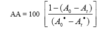
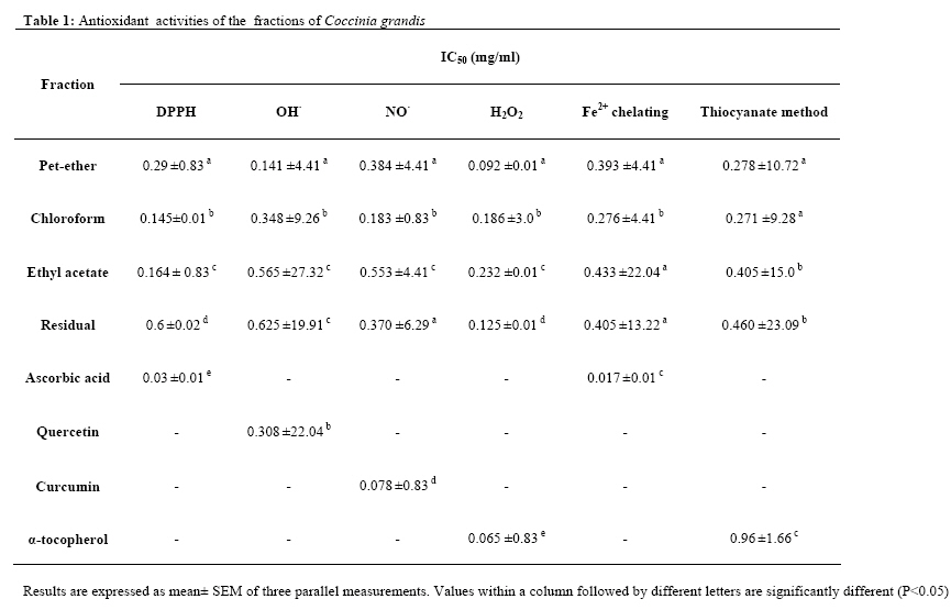
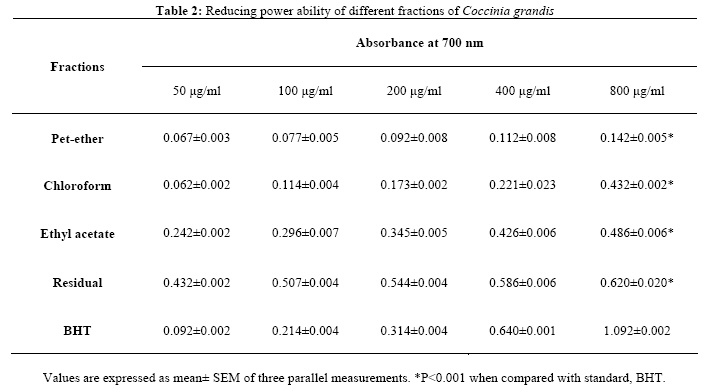
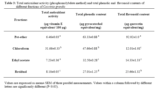
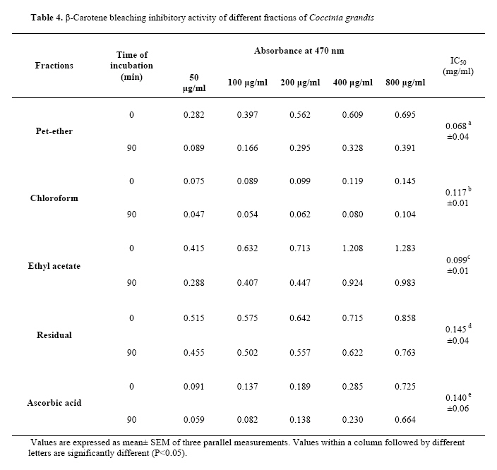
African Journal of Traditional, Complimentary and Alternative Medicines, Vol.5, No. 1, 2008, pg. 61-73 Research Paper IN VITRO ANTIOXIDANT ACTIVITIES OF THE FRACTIONS OF COCCINIA GRANDIS L. LEAF EXTRACT M. Umamaheswari and T. K. Chatterjee* Division of Pharmacology, Department of Pharmaceutical Technology,Jadavpur University, Kolkata, India. *E-mail: tkchatterjee_81@rediffmail.com Code Number: tc08011 AbstractThe present study was aimed at investigating the antioxidant activities of the various fractions of the hydromethanolic extract of the leaves of Coccinia grandis L. Voigt. (Cucurbitaceae). The antioxidant activities of the fractions have been evaluated by using nine in vitro assays and were compared to standard antioxidants such as ascorbic acid, α-tocopherol, curcumin and butylated hydroxyl toluene (BHT). All the fractions showed effective Hdonor activity, reducing power, free radical scavenging activity, metal chelating ability and inhibition of β-carotene bleaching. None of the fractions exerted an obvious pro-oxidant activity. The antioxidant property depends upon concentration and increased with increasing amount of the fractions. The free radical scavenging and antioxidant activities may be attributed to the presence of phenolic and flavonoid compounds present in the fractions. The results obtained in the present study indicate that the leaves of C. grandis are a potential source of natural antioxidant. Key words: Coccinia grandis (Cucurbitaceae), free radicals, antioxidant, pro-oxidant. IntroductionThere is an increased evidence for the participation of free radicals in the etiology of various diseases like cancer, diabetes, cardiovascular diseases, autoimmune disorders, neurodegenerative diseases, aging, etc. (Bandopadhyay et al., 1999). A free radical is defined as any atom or molecule possessing unpaired electrons. The primary oxygen derived free radicals are superoxide anion (O2·), hydroxyl (OH·), hydroperoxyl ( OOH·), peroxyl (ROO·) and alkoxyl (RO·) radicals and non free radicals are hydrogen peroxide (H2O2), hypochlorous acid (HOCl), ozone (O3) and singlet oxygen (1O2). These reactive intermediates are collectively termed as reactive oxygen species (ROS). Similarly, reactive nitrogen species (RNS) are mainly nitric oxide (NO·), peroxynitrite (ONOO·) and nitrogen dioxide (NO2). Free radicals can cause a wide range of toxic oxidative reactions like initiation of the peroxidation of the membrane lipids leading to the accumulation of lipid peroxides, direct inhibition of mitochondrial respiratory chain enzymes, fragmentation or random cross linking of molecules like DNA, enzymes and proteins which ultimately leads to cell death (Halliwell and Gutteridge, 1999). ROS can be formed in living organisms by both endogenous and exogenous sources. Endogenous sources of free radicals include normal aerobic respiration, peroxisomes and stimulation of polymorphonuclear leucocytes and macrophages. The exogenous sources include ionizing radiation, tobacco smoke, pollutants, pesticides and organic solvents (Irshad and Chaudhuri, 2002). Antioxidants are agents which scavenge the free radicals and prevent the damage caused by them. They can greatly reduce the damage due to oxidants by neutralizing the free radicals before they can attack the cells and prevent damage to lipids, proteins, enzymes, carbohydrates and DNA (Fang et al., 2002). Antioxidants can be classified into two major classes i.e., enzymatic and non-enzymatic. The enzymatic antioxidants are produced endogenously and include superoxide dismutase, catalase, and glutathione peroxidase. The non-enzymatic antioxidants include tocopherols, carotenoids, ascorbic acid, flavonoids and tannins which are obtained from natural plant sources (Lee et al., 2004). A wide range of antioxidants from both natural and synthetic origin has been proposed for use in the treatment of various human diseases (Cuzzocrea et al., 2001). There are some synthetic antioxidant compounds such as butylated hydroxytoluene, butylated hydroxyanisole and tertiary butylhydroquinone which are commonly used in processed foods. However, it has been suggested that these compounds have shown toxic effects like liver damage and mutagenesis (Grice, 1986; Wichi, 1988). Flavonoids and other phenolic compounds of plant origin have been reported as scavengers of free radicals (Formica and Regelson, 1995; Rice-Evans et al., 1997). Hence, nowadays search for natural antioxidant source is gaining much importance. C. grandis L. Voigt. commonly known as “Ivy gourd” is a tropical plant belonging to the family Cucurbitaceae. It has been found in many countries in Asia and Africa. The roots, stems, leaves and whole plant of C. grandis are used in the treatment of jaundice, bronchitis, skin eruptions, burns, insect bites, fever, indigestion, nausea, eye infections, allergy, syphilis, gonorrhoea, etc. (Kirthikar and Basu, 1987; Wasantwisut and Viriyapanich, 2003). The leaves of this species are widely used in Indian folk medicine for reducing the amount of sugar in urine of patients suffering from diabetes mellitus. Literature suggests the use of this plant in the treatment of diabetes (Venkateswaran and Pari, 2003). The crude hydromethanolic extract of the leaves of C. grandis has been reported for its xanthine oxidase inhibitory and hypouricaemic activities (Umamaheswari et al., 2007). The objective of the present study was to investigate the antioxidant activity of the different fractions of the hydromethanolic extract of the leaves of C. grandis using nine in vitro models. Total phenolic and flavonoid content of the fractions were also determined in order to evaluate a relationship between the antioxidant activity and the phytochemical constituents. Materials and methods Plant material The plant material consists of dried powdered leaves of C. grandis L. Voigt. Cucurbitaceae. The leaves were collected in and around Coimbatore district, Tamil Nadu, India during the month of October 2006 and was authenticated by Dr. G.V.S Murthy, Joint Director, Botanical Survey of India, Tamil Nadu Agricultural University Campus, Coimbatore (Ref No BSI/SC/5/23/06-07/Tech.1951). Preparation of the extract and fractionationThe air-dried powdered leaves of C. grandis (100 g) was extracted with methanol-water (7:3) mixture using a mechanical shaker for 4 h. The resultant extract was concentrated under reduced pressure to yield a residue. The hydromethanolic extract was then extracted successively with equal volumes of petroleum-ether, chloroform and ethyl acetate. Each fraction was then concentrated under reduced pressure to obtain the pet-ether fraction (PEF1.6%w/w), chloroform fraction (CF-1.8%w/w), ethyl acetate fraction (EAF-0.8%w/w) and residual fraction (RF5.3%w/w). Drugs and chemicalsDPPH was obtained from Hi Media Laboratories Pvt. Ltd., Mumbai. Quercetin and pyrocatechol were purchased from Sisco Research Laboratories Pvt. Ltd., Mumbai. Folin-Ciocalteu reagent was obtained from SD Fine Chemicals Pvt. Ltd, Mumbai. Calf thymus DNA was purchased from Genei Chemicals, Bangalore. Ferrozine was obtained from Sigma Aldrich, USA. All other drugs and chemicals used in the study were obtained commercially and were of analytical grade. Phytochemical screeningPreliminary phytochemical screening of the crude hydromethanolic extract of the leaves was carried out. In vitro antioxidant activity DPPH radical scavenging assay The free radical scavenging activity of the fractions was measured in vitro by 1,1-diphenyl-2-picrylhydrazyl (DPPH) assay (Mensor et al., 2001). About 0.3 mM solution of DPPH in 100% ethanol was prepared and 1 ml of this solution was added to 3 ml of the fraction dissolved in ethanol at different concentrations (25-400 µg/ml). The mixture was shaken and allowed to stand at room temperature for 30 min and the absorbance was measured at 517 nm using a spectrophotometer. The % scavenging activity at different concentrations was determined and the IC50 value of the fractions was compared with that of ascorbic acid, which was used as the standard. Reducing power abilityThe reducing power was investigated by the Fe3+-Fe2+ transformation in the presence of the fractions as described by Fejes et al., (2000). The Fe2+ can be monitored by measuring the formation of Perl’s Prussian blue at 700 nm (Meir et al., 1995). One ml of the fraction (50-800 µg/ml), 2.5 ml of phosphate buffer (pH 6.6) and 2.5 ml of 1% potassium ferricyanide were incubated at 50°C for 30 min and 2.5 ml of 10% trichloroacetic acid was added to the mixture and centrifuged for 10 min at 3000g. About 2.5 ml of the supernatant was diluted with 2.5 ml of water and shaken with 0.5 ml of freshly prepared 0.1% ferric chloride. The absorbance was measured at 700 nm. Butylated hydroxy toluene (50-800 µg/ml) was used as the standard. All tests were performed in triplicate and the graph was plotted with the average of the three determinations. Hydroxyl radical scavenging assayHydroxyl radical scavenging activity was measured by the ability of the different fractions of C. grandis to scavenge the hydroxyl radicals generated by the Fe3+-ascorbate-EDTA-H2O2 system (Fenton reaction) (Halliwell et al., 1987; Ilavarasan et al., 2005). The reaction mixture in a final volume of 1.0 ml contained 100 µl of 2-deoxy2ribose (28 mM in 20 mM KH2PO4 buffer, pH 7.4), 500 µl of the fractions at various concentrations (50-800 µg/ml) in buffer, 200 µl of 1.04 mM EDTA and 200 µM FeCl3 (1:1v/v), 100 µl of 1.0 mM hydrogen peroxide (H2O2) and 100 µl of 1.0 mM ascorbic acid. Test samples were kept at 37oC for 1 h. The free radical damage imposed on the substrate, deoxyribose was measured using the thiobarbituric acid test. One ml of 1% thiobarbituric acid (TBA) and 1.0 ml 2.8% trichloroacetic acid (TCA) were added to the test tubes and were incubated at 1000C for 20 min. After cooling, the absorbance was measured at 532 nm against a blank containing deoxyribose and buffer. Quercetin (50800 µg/ml) was used as a positive control. Hydrogen peroxide scavenging assayHydrogen peroxide solution (2 mM/L) was prepared with standard phosphate buffer (pH 7.4). Different concentration of the fractions (25-400 µg/ml) in distilled water was added to 0.6 ml of hydrogen peroxide solution. Absorbance was determined at 230 nm after 10 min against a blank solution containing phosphate buffer without hydrogen peroxide. The percentage scavenging activity at different concentrations of the fractions was determined and the IC50 values were compared with the standard, α-tocopherol (Oktay et al., 2003). Nitric oxide radical scavenging assayThis assay was performed according to the method described by Sreejayan et al., (1997). Nitric oxide generated from sodium nitroprusside in aqueous solution at physiological pH interacts with oxygen to produce nitrite ions, which was measured by Griess reagent. The reaction mixture (3 ml) containing 10 mM sodium nitroprusside in phosphate buffered saline, and the fractions or the reference compound (curcumin) at different concentrations (50-800 µg/ml) were incubated at 25°C for 150 min. About 0.5 ml aliquot of the incubated sample was removed at 30 min intervals and 0.5 ml Griess reagent was added. The absorbance of the chromophore formed was measured at 546 nm. Inhibition of the nitric oxide generated was measured by comparing the absorbance values of control, fractions and curcumin (50-800 µg/ml). Thiocyanate methodThe peroxy radical scavenging activity was determined by thiocyanate method using α- tocopherol (50-800 µg/ml) as standard (Yildirim et al., 1999). Increasing concentration of the fractions (50-800 µg/ml) in 0.5 ml of distilled water was mixed with 2.5 ml of 0.02 M linoleic acid emulsion (in 0.04 M phosphate buffer pH 7.0) and 2 ml phosphate buffer (0.04M, pH 7) in a test tube and incubated in darkness at 37oC. At intervals during incubation, the amount of peroxide formed was determined by reading the absorbance of the red colour developed at 500 nm by the addition of 0.1 ml of 30% ammonium thiocyanate solution and 0.1 ml of 20 mM ferrous chloride in 3.5% hydrochloric acid to the reaction mixture. The percentage scavenging activity was calculated and the IC50 values of the fractions were compared with the standard, α-tocopherol. Phosphomolybdate methodThe total antioxidant capacity of the fractions was determined by phosphomolybdate method using αtocopherol as the standard (Jayaprakasha et al., 2002). An aliquot of 0.1ml of the fractions (100 µg) solution was combined with 1.0 ml of reagent (0.6 M sulfuric acid, 28 mM sodium phosphate and 4 mM ammonium molybdate). The tubes were capped and incubated in a boiling water bath at 95oC for 90 min. After the samples had cooled to room temperature, the absorbance was measured at 695 nm against the blank using an UV spectrophotometer. The blank solution contained 1.0 ml of reagent solution and the appropriate volume of the same solvent used for the sample and it was incubated under same conditions as rest of the sample. The total antioxidant capacity was expressed as µg equivalents of α-tocopherol by using the standard tocopherol graph. Ferrous chelating abilityThe ferrous chelating ability of the fractions was monitored by measuring the formation of the ferrous ionferrozine complex. The reaction mixture containing 1.0 ml of different concentrations of the fractions (50-800 µg/ml) was mixed with 3.7 ml of methanol, 0.1 ml of 2 mM ferrous chloride and 0.2 ml of 5 mM ferrozine to initiate the reaction and the mixture was shaken vigorously and left to stand at room temperature for 10 min. The absorbance of the solution was measured at 562 nm. The percentage chelating effect on ferrozine-Fe2+ complex was calculated. The IC50 values were compared with ascorbic acid (Huang and Kuo, 2000). β-carotene bleaching assayA solution of β-carotene was prepared by dissolving 2 mg of β-carotene in 10 ml chloroform and 1.0 ml of this solution was then pipetted out into a flask containing 20 mg of linoleic acid and 200 mg of Tween 40 emulsifier. Chloroform was completely evaporated using a vacuum evaporator. Aliquots of 5.0 ml of this emulsion were transferred into a series of tubes containing various concentrations of the fractions (25-400 µg/ml) or αtocopherol. The absorbance of the fractions and the standard was measured immediately (t=0) and after 90 min at 470 nm. The tubes were incubated at 500C in a water bath during the test. The antioxidant activities (AA) of the samples were evaluated in terms of bleaching of β-carotene using the following formula:
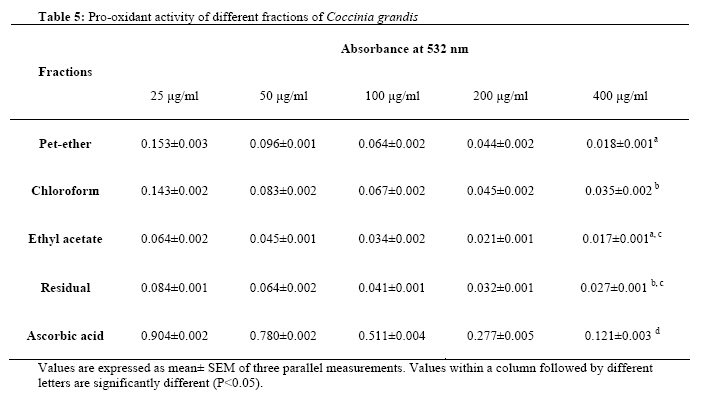
where A0 and A0• are the absorbance values measured at zero time of incubation for test sample and control respectively and At and At• are the absorbance values of the test sample and control respectively, after incubation for 90 min. (Jayaprakasha et al., 2002). Bleomycin-dependent DNA damageThe pro-oxidant activity of the fractions was determined by bleomycin-dependent DNA damage. The reaction mixture (4.5 ml) contained calf thymus DNA (10 µg/ml), 50 µg of 1.0 ml bleomycin sulfate, 1.0 ml of 5 mM magnesium chloride, 1.0 ml of 50 mΜ ferric chloride and 1.0 ml of different concentrations of the fractions. Ascorbic acid was used as the positive control. The mixture was incubated at 37oC for 1 h. The reaction was terminated by the addition of 0.05 ml EDTA (0.1 M). The colour was developed by adding 0.5 ml of 1% w/v thiobarbituric acid (TBA) and 0.5 ml of 25% v/v HCl followed by heating at 37oC for 15 min. After centrifugation, the extent of DNA damage was measured at 532 nm using an UV-spectrophotometer. All the determinations were carried out in triplicate (Ng et al., 2003). Estimation of total phenolic contentTotal soluble phenolics of the fraction were determined with Folin-Ciocalteu reagent using pyrocatechol as the standard (Gulcin et al., 2004). An aliquot of 0.1 ml suspension of 1 mg of the fractions in water was totally transferred to a 100 ml Erlenmeyer flask and the final volume was adjusted to 46 ml by the addition of distilled water. Folin-Ciocalteu reagent (1 ml) was added to this mixture, followed by 3 ml of 2% sodium carbonate 3 min later. Subsequently, the mixture was shaken for 2 h at room temperature and the absorbance was measured at 760 nm. The concentration of total phenolic compounds in the fractions was determined as µg pyrocatechol equivalent by using the standard pyrocatechol graph. Estimation of total flavonoid contentTotal soluble flavonoid content of the fractions was determined with aluminium nitrate using quercetin as the standard (Hsu, 2006). One mg of the fraction was added to 1ml of 80 % ethanol. An aliquot of 0.5 ml was added to test tubes containing 0.1 ml of 10 % aluminium nitrate, 0.1 ml of 1 M potassium acetate and 4.3 ml of 80 % ethanol. The absorbance of the supernatant was measured at 415 nm after incubation at room temperature for 40 min. The total flavonoid content in the fractions was determined as µg quercetin equivalent by using the standard quercetin graph. Calculation of 50% Inhibitory Concentration (IC50)The concentration (mg/ml) of the fractions that was required to scavenge 50% of the radicals was calculated by using the percentage scavenging activities at five different concentrations of the fractions. Percentage inhibition (I%) was calculated using the formula,
where Ac is the absorbance of the control and As is the absorbance of the sample. Statistical analysisAll experiments were performed in triplicate (n=3) and results were expressed as mean ± SEM. Statistical analysis was carried out with (SPSS package version 10.0) using ANOVA followed by Turkey’s test (P<0.05). Results Phytochemical screening Phytochemical screening of the crude hydromethanolic extract of the leaves of Coccinia grandis revealed the presence of flavonoids, saponins, phenols, tannins and terpenoids. DPPH assayAll the fractions of C. grandis demonstrated H-donor activity. The highest DPPH radical scavenging activity was detected in chloroform fraction (IC50 0.145 mg/ml), followed by ethyl acetate, pet-ether and residual fractions (IC50 0.164, 0.29 and 0.6 mg/ml respectively) (Table 1). These activities are less than that of ascorbic acid. The scavenging ability increased towards the ethyl acetate fraction with increasing polarity of the solvent. Reducing power abilityTable 2 shows the reductive capabilities of different fractions of C. grandis when compared to the standard, BHT. Like the antioxidant activity, the reducing power increased with increasing amount of the fractions. The residual fraction of C. grandis showed the highest reducing ability (absorbance 0.620) than all the other fractions tested. However, the activity was less than the standard, BHT (absorbance 1.092). The pet-ether, chloroform and ethyl acetate fractions also showed significant activity indicating its reductive ability. Hydroxyl radical scavenging assayHydroxyl radical scavenging activity was quantified by measuring the inhibition of the degradation of deoxyribose by the free radicals generated by the Fenton reaction. All the fractions of C. grandis and the standard (quercetin) inhibited the production of hydroxyl radicals. The scavenging activity of the pet-ether fraction (IC50 0.141mg/ml) was higher than that of quercetin (0.308 mg/ml). The IC50s values of the chloroform, ethyl acetate and residual fractions were 0.348, 0.565 and 0.625 mg/ml respectively (Table 1). Hydrogen peroxide scavenging assayAll the fractions of C. grandis scavenged hydrogen peroxide in a concentration-dependent manner. The pet-ether fraction (PEF) of C. grandis showed strong H2O2 scavenging activity (IC50 0.092 mg/ml) whereas that of the standard, α-tocopherol was 0.065 mg/ml. The residual, chloroform and ethyl acetate fractions also showed significant scavenging activities (IC50 of RF, CF and EAF were 0.125, 0.186 and 0.232 mg/ml respectively) when compared to the standard (Table 1). Nitric oxide radical scavenging assayThe fractions of C. grandis effectively reduced the generation of nitric oxide from sodium nitroprusside. The chloroform fraction showed strong nitric oxide scavenging activity (IC50 0.183 mg/ml) and that of standard curcumin was 0.078 mg/ml. The residual fraction (0.37 mg/ml), pet-ether fraction (0.384 mg/ml) and ethyl acetate fraction (0.553 mg/ml) also showed good scavenging activities (Table 1). Thiocyanate methodThe total antioxidant activity of the fractions of C. grandis was determined by the thiocyanate method and compared with the standard, α-tocopherol. The absorbance decreased with the increasing concentrations of the fractions, which indicate that the fractions could effectively decrease the amount of formed peroxides. The total antioxidant activity of the pet-ether and chloroform fractions were almost similar (IC50 0.278 and 0.271 mg/ml respectively) and that of the standard, α-tocopherol was 0.096 mg/ml. The ethyl acetate and residual fractions also showed good antioxidant activity but at higher concentrations (IC50 0.405 and 0.46 mg/ml respectively) (Table 1). Phosphomolybdate methodThe phosphomolybdate method is quantitative, since the total antioxidant capacity is expressed as αtocopherol equivalents. Among the fractions tested, the chloroform fraction contains 31.66µg vitamin E equivalent/ 100 µg. The antioxidant activity increased in the order of chloroform fraction > residual fraction > ethyl acetate fraction > pet-ether fraction (Table 3). Ferrous chelating abilityAddition of the fractions of C.grandis interferes with the ferrous-ferrozine complex and the red colour of the complex decreased with the increasing concentrations of the fractions. All the fractions captured ferrous ions before ferrozine and thus have ferrous chelating ability. Among the fractions tested, the chloroform fraction showed the highest ferrous ion chelating ability (IC50 0.276 mg/ml). The abilities shown by pet-ether, ethyl acetate and residual fractions were almost similar (IC50 0.393, 0.433 and 0.405 mg/ml respectively). Ascorbic acid (IC50 0.017 mg/ml) showed the highest ferrous ion chelating ability (Table 1). β-carotene bleaching assayAddition of the fractions of C.grandis reduced the discolouration of β-carotene thereby preventing its bleaching. The pet-ether, ethyl acetate and chloroform fractions showed strong inhibition on β-carotene bleaching and their IC50 values are 0.068, 0.099 and 0.117 mg/ml respectively. These activities were significantly higher than that of α-tocopherol (IC50 0.14 mg/ml). The IC50 of the residual fraction of C. grandis was 0.145 mg/ml, which was similar to that of standard (Table 4). Bleomycin-dependent DNA damageThe pro-oxidant activity of the fractions of C. grandis was assessed by their effects on damage to DNA in the presence of a bleomycin-Fe3+ complex. The absorbance of all the fractions decreased with increasing concentrations, which proves that none of the fractions exhibited pro-oxidant activity (Table 5). Total phenolic and flavonoid contentTotal phenolic content was estimated by using Folin-Ciocalteu reagent. Total phenolic content of the different fractions of C. grandis were solvent dependent and expressed as µg pyrocatechol equivalent.. The content of the total phenolics in the fractions decreased in the order of pet-ether > chloroform > ethyl acetate > residual fractions. The total flavonoid content in the fractions was expressed as µg quercetin equivalent. The pet-ether fraction of C. grandis showed highest amount of flavonoids among the fractions tested. The content of total flavonoids in the fractions decreased in the order of pet-ether fraction > residual fraction > ethyl acetate fraction > chloroform fraction (Table 3). DiscussionFree radicals are known to play a definite role in a wide variety of pathological manifestations. Antioxidants fight free radicals and protect us from various diseases. They exert their action either by scavenging the reactive oxygen species or protecting the antioxidant defence mechanisms. DPPH assay is one of the most widely used methods for screening antioxidant activity of plant extracts (Nanjo et al., 1996). DPPH is a stable, nitrogen-centered free radical which produces violet colour in ethanol solution. It was reduced to a yellow coloured product, diphenylpicryl hydrazine, with the addition of the fractions in a concentration-dependent manner. The reduction in the number of DPPH molecules can be correlated with the number of available hydroxyl groups. All the fractions showed significantly higher inhibition percentage (stronger hydrogen –donating ability) and positively correlated with total phenolic content. The transformation of Fe3+ into Fe2+ in the presence of various fractions was measured to determine the reducing power ability. The reducing ability of a compound generally depends on the presence of reductones (antioxidants), which exert the antioxidant activity by breaking the free radical chain by donating a hydrogen atom (Meir et al., 1995) The antioxidant principles present in the fractions of C. grandis caused the reduction of Fe3+/ ferricyanide complex to the ferrous form, and thus proved the reducing power ability. Hydroxyl radical is the most deleterious and reactive among the ROS and it bears the shortest half-life compared with other free radicals. The oxygen derived hydroxyl radicals along with the added transition metal ion (Fe2+) causes the degradation of deoxyribose into malondialdehyde which produces a pink chromogen with thiobarbituric acid (Halliwell et al., 1987). All the fractions of C. grandis when added to the reaction mixture, scavenged the hydroxyl radicals and prevented the degradation of deoxyribose. Hydrogen peroxide itself is not particularly reactive with most biologically important molecules, but is an intracellular precursor of hydroxyl radicals which is very toxic to the cell (Halliwell, 1991). Thus, scavenging of H2O2 is a measure of the antioxidant activity of the fractions. All the fractions of C. grandis scavenged hydrogen peroxide which may be attributed to the presence of phenolic groups that could donate electrons to hydrogen peroxide, thereby neutralising it into water. In vitro inhibition of nitric oxide radical is a measure of antioxidant activity of plant drugs. Nitric oxide is a free radical which plays an important role in the pathogenesis of pain, inflammation, etc. Scavenging of nitric oxide radical is based on the generation of nitric oxide from sodium nitroprusside in buffered saline, which reacts with oxygen to produce nitrite ions that can be measured by using Griess reagent (Marcocci et al., 1994). The absorbance of the chromophore is measured at 546 nm in the presence of the fractions. All the fractions of C. grandis decreased the amount of nitrite generated from the decomposition of sodium nitroprusside in vitro. This may be due to the antioxidant principles in the fractions which compete with oxygen to react with NO· thereby inhibiting the generation of nitrite. The amount of formed peroxides was measured by the thiocyanate method. The fractions were incubated with linoleic emulsion in dark at 37°C and the amount of peroxides was determined spectrophotometrically by measuring the absorbance at 500 nm (Yen and Chen, 1995). A decrease in absorbance indicated the antioxidant activity of the fractions which might be due to the inactivation of the free radicals and the presence of flavonoid like phytochemicals. The phosphomolybdate method has been routinely used to evaluate the total antioxidant capacity of the extracts (Prieto et al., 1997). In the presence of the fractions, the Mo(VI) is reduced to Mo(V) and forms a green coloured phosphomolybdenum V complex which shows maximum absorbance at 695 nm. All the fractions possessed antioxidant activity. The metal chelating ability of the fractions of C. grandis was measured by the formation of ferrous ionferrozine complex. Ferrozine combines with ferrous ions forming a red coloured complex which absorbs at 562 nm (Yamaguchi et al., 2000). It was reported that the chelating agents which form σ bond with a metal, are effective as secondary antioxidants, because they reduce the redox potential thereby stabilising the oxidised form of the metal ion (Duh et al., 1999). The results of our study demonstrate that the fractions have an effective capacity for iron binding, suggesting its antioxidant potential. In addition, the metal chelating ability of the fractions demonstrated that they reduce the concentration of the catalysing transition metal involved in the peroxidation of lipids. The β-carotene bleaching assay is a commonly used model to analyze the antioxidant activity of the plant extracts because β-carotene is extremely sensitive to free radical mediated oxidation of linoleic acid. In this assay, oxidation of linoleic acid, an unsaturated fatty acid occurs due to the production of reactive oxygen species formed from halogenated water. The reactive oxygen species will initiate β-carotene oxidation leading to discolouration (Gutierrez et al., 2006). All the fractions of C. grandis inhibited β-carotene oxidation, suggesting that the antioxidant activity could be related to high level of phenolic compounds. Bleomycin-dependent DNA damage has been adopted as a sensitive and specific method to examine the potential pro-oxidant drugs. Degradation of DNA occur if the samples to be tested reduce the bleomycin-Fe3+ to bleomycin-Fe2+ resulting in the formation of a product similar to MDA which reacts with TBA to give a pink colour (Liu and Ng, 2000). All the fractions decreased the absorbance and bleomycin-Fe3+ is not converted into bleomycin-Fe2+, thereby preventing the DNA degradation. These results confirm that the fractions of C. grandis are devoid of pro-oxidant activity. Phenolics are ubiquitous secondary metabolites in plants and possess a wide range of therapeutic uses such as antioxidant, antimutagenic, anticarcinogenic, free radical scavenging activities and also decrease cardiovascular complications (Yen et al., 1993). The scavenging ability of the phenolics is mainly due to the presence of hydroxyl groups. Total phenolic assay by using Folin-Ciocalteu reagent is a simple, convenient and reproducible method. It is employed routinely in studying phenolic antioxidants (Huang et al., 2005). Flavonoids are a group of polyphenolic compounds, which exhibit several biological effects such as antiinflammatory, antihepatotoxic, antiulcer, antiallergic, antiviral, anticancer activities. They also inhibit enzymes such as aldose reducatse and xanthine oxidase. They are capable of effectively scavenging the reactive oxygen species because of their phenolic hydroxyl groups and are potent antioxidants (Cao et al., 1997). In view of their wide pharmacological and biological actions, they have a greater therapeutic potential. The presence of high phenolic and flavonoid content in the fractions has contributed directly to the antioxidant activity by neutralising the free radicals. Based on the results obtained, it may be concluded that all the fractions of the hydromethanolic extract of the leaves of C. grandis showed strong antioxidant activity, reducing power ability, free radical scavenging activity, metal chelating ability and inhibition of β-carotene bleaching when compared to standards such as ascorbic acid, αtocopherol, curcumin, and butylated hydroxytoluene. As the various fractions of C. grandis exhibited different reactive oxygen species scavenging activities, there may be different percentages of phytochemical constituents present in the fractions. Further studies to evaluate the in vivo potential of the fractions in various animal models and the isolation and identification of the antioxidant principles in the leaves of Coccinia grandis are being carried out. References
© Copyright 2008 - African. Journal. Traditional, Complementary and Alternative Medicines The following images related to this document are available:Photo images[tc08011t4.jpg] [tc08011t2.jpg] [tc08011t5.jpg] [tc08011t3.jpg] [tc08011t1.jpg] |
| |||||||||
