 |
| About Bioline | All Journals | Testimonials | Membership | News |
|
||||||
|
||||||
African Journal of Traditional, Complimentary and Alternative Medicines, Vol. 5, No. 2, 2008, pg. 141-146 Research PaperAnti-Nociceptive and Anti-Inflammatory Activities of Aqueous Leaves Extract of Ocimum Gratissimum ( Labiate ) in Rodents. * Tanko, Y1. Magaji, G. M2. Yerima, M2. Magaji, R. A3.Mohammed, A1 1 Department
of Human Physiology, Ahmadu Bello University, Zaria 2Department of
Pharmacology and Clinical Pharmacy, Ahmadu Bello University, Zaria Nigeria 3Departments
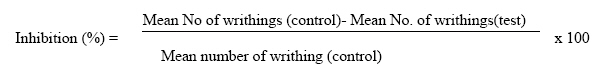
of Human Physiology, Bayero University, Kano, Nigeria Code Number: tc08019 Abstract The aqueous leaves extract of Ocimum gratissimum was investigated for anti-nociceptive and anti-inflammatory effects in mice and rats. The models used to study the effect on nociception are the acetic acid-induced abdominal constriction test, hot- plate method in mice. The anti-inflammatory effect was investigated employing the formalin-induced hind–paw oedema in rats. The extract caused a significant (p<0.05), dose-dependent inhibition of acetic acid-induced writhing and hot-plate method .The extract also exhibited anti-inflammatory effect which was significant (P<0.001) at all the three doses. .The intraperitoneal LD50 value of the extract was 1264.9mg/kg body weight in mice. Preliminary phytochemical screening revealed the presence of alkaloids, saponins, tannins and flavonoids. The results suggest the extract contained pharmacologically active principles, and supports the local application of the plant in painful and inflammatory conditions. Key words: Ocimum grassitimum, anti-nociceptive, anti-inflammatory. Introduction Ocimum gratissimum is commonly known as fever leaf in general but is has different native names in different part of the country .In Yoruba language, it is known as Ewfirin ajase, Nchu-nwu in Ibo, Bunsuru daji in Hausa, Ireru in Ebira, Ebaubokho in Benin, ufuo-yibo in Urhobo and ntion in Efik ( Iwu, 1993).. There are about 60 or more species of Ocimum and numerous varieties, belonging to the Family Labiatae. This different types of species are represented by the five most important representatives of the more that 60 Ocimum species and these include (i) Ocimum gratissimum, (ii) Ocimum basilicum, (iii) Ocimum americanum, (iv) Ocimum sanctum and (v) Ocimum americanum.(Martin and Salguerio, 1999;Mandal and Pattnaik, 2000). Ocimum gratissimum Linn. (Labiaceae) is a herbaceous plant commonly found in tropical Asia especially India. It is used in the treatment of epilepsy in the coastal area of Nigeria (Osito, 1992), High fever (Oliver, 1980), and Diarrhoea (Oliver 1980; Sofowora, 1993). The plant is also used to treat typhoid fever and diabetes (Adjanahoun et al 1991; Igoli et al., 2002; Tor-Anyin et al., 2003). Today, basil is used mainly as a culinary herb. It medicinal value is not as widely appreciated in Western World. In France it is used in perfumes and cosmetics (Ross, 2003). This research was aimed at investigating the possible anti-nociceptive and anti-inflammatory activities of aqueous leaves extract of the plant in order to support or refute the claims by traditional herbalists. Materials and Methods Fresh leaves of Ocimum gratissimum were collected from Ahmadu Bello University Main campus Zaria in January 2007. The plant was identified by Mal. M. Musa at the herbarium unit of Biological Science Department A.B.U., Zaria where a voucher specimen (No. 2637) has been deposited.The fresh leaves collected were dried under the shade and ground into powder. The powder (100 g) was boiled in 500ml of distilled water for 1 hour. The extract was cooled, filtered using a filter paper and evaporated to dryness in water bath of 600 C. A brownish residue weighing 15.7 % (w/w) was obtained and kept in air tight bottles in a refrigerator until use. The aqueous extract obtained was subjected to preliminary phytochemical screening, to identify the chemical constituents. The methods of analysis employed were those described by (Trease and Evans, 1989). Experimental animals Adult Wister rats of both sexes weighing between 150-200g and adult swiss albino mice of both sexes weighing between 25-30g were used for the experiments. The animals were obtained from animal house of Department of Veterinary Medicine, Ahmadu Bello University, Zaria. They were kept in well ventilated room and fed with standard grower mash. Excel feeds Plc, Kaduna and allowed water ad libitum. The “ Principle of laboratory animal care “ ( NIH publication No 85- 23 ) guideline are procedures were following in this study ( NIH publication reserved 1985 ). Acute toxicity study This was conducted by using the method described by Lorke (1983). In the initial phase, mice were divided into 3 groups of three and treated with the aqueous leaves extract of the plant at doses of 10, 100 and 1000mg extract/ kg body weight intraperitoneally (i.p. ) and were then observed for 24 hrs for signs of toxicity including death .In the final phase, mice were divided into 4 groups of one mouse each and treated with the ethanol extract at doses of 600, 1000, 1600 and 2900 mg / kg body weight i.p. The median lethal dose (LD50) was calculated from the second phase. Tests for anti-nociceptive studies (a) Acetic acid-induced abdominal contraction The Acetic acid induced writhing test in mice as described by Koster et al. (1959) was employed. Twenty five (25) Swiss albino mice were divided into 5 groups of 5 mice each. The first group was given 10 ml/kg of Normal saline i.p. and served as control, groups 2 received Piroxicam 20mg/kg as a positive control, 3, 4 and 5 received 50, 100 and 200 mg of extract per kg of body weight i.p.respectively. Thirty mins later, mice in all the groups were treated with Acetic acid (0.6%v/v, 1ml per 100g body weight i.p.). Five minutes after Acetic acid injection, mice were placed in individual cages and the number of abdominal contractions was counted for each mouse for a period of 10 mins. Percentage inhibition of writhing was calculated using the formula:
(b) Hot-plate test in mice Analgesic activity was tested in mice using the hot plate method described by Janssen and Jagneau (1957). Twenty five (25) Swiss albino mice were divided into 5 groups of 5 mice in each group. Group 1 served as control which was given 10 ml/kg of Normal saline i.p., group 2 received Piroxicam 20mg/kg as a reference drug, group 3, 4 and 5 received 50, 100 and 200 mg/kg of extract per kg of body weight i.p.respectively. A 500 ml of glass beaker was placed on a Heidolph® MR 2002 hot-plate (with adjustable temperature). The temperature of the hot-plate was then regulated to 50 ± 20 C (on the hot-plate) in order to obtain the animal’s response to electric heat-induced nociceptive pain stimulus (licking of the forepaws and eventually jumping out of the glass beaker). Jumping out of the beaker was taken as an indicator of the animal’s response to heat-induced nociceptive pain stimulus. The time taken for each mouse to jump out of the beaker (i.e. reaction time) recorded in seconds. Readings were taken at intervals of 30, 60 and 90 mins after extract administration. Cut off time in the absence of response was 60 s to avoid tissue damage to the mice paws (Sharma et al., 1982). Test for anti-inflammatory study Increases in the rat hind linear paw diameter induced by subplanter injection of a phlogistic agent was used as the measure of acute inflammation( Winter et al.,1963) The phlogistic agent employed in this study was formalin (50µl of 2.5% v/v ).. The rats were randomly divided into five groups (n=5). Thirty mins before the injection of formalin, the groups were treated i.p. as follows: group 1, normal saline (10ml/kg as a negative control); group 2, 20mg/kg of Diclofenac; groups 3, 4 and 5 received extract of O. gratissimum at the doses of (50, 100 and 200mg/kg) respectively. Inflammation of the hind paw was induced by injecting formalin into the subplanter surface of the left hind paw. Paw linear diameter (cm) was measured using vernier caliper at 1, 2, 3 4 and 5 hrs after formalin injection.
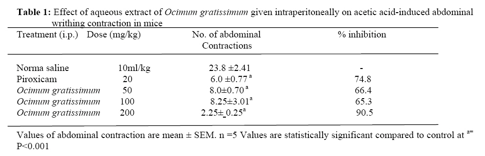
Statistical analysis Results were expressed as mean ± Standard Error of Mean (SEM). The data was statistically analyzed using the one-way ANOVA to determine whether results in a particular group were significantly different from those in the corresponding control groups. Results were statistically significant when P values are less than 0.05 (P < 0.05) as described by Duncan et al. (1977). Results The freshly prepared extracts were subjected to preliminary phytochemical screening test for various constituents. This revealed the presence of alkaloids, tannins, saponins, flavonoids, terpenoids and steroids. The sign of toxicity was first noticed after 4-6 hrs of extract administration. There was decreased locomotor activity and decreased sensitivity to touch and jerking. Also there was decreased feed intake, and prostration after 10 hrs of extract administration. The median lethal dose (LD50) in mice was calculated to be 1264.9 mg/kg body weight The extract demonstrated a significant (P< 0.05) anti-nociceptive activity at all the doses (50,100 and 200 mg/kg body weight i.p.) tested compared to control normal saline. The activity resides more at the highest dose 200mg/kg body weight i.p.that was found to have the highest percentage (90.5 %) of inhibition of the abdominal contriction induced by acetic acid in mice (Table 1). Also the extract was found to have a significant (p <0.05) inhibitory effect on hot-plate method at all the three doses as shown in Table 2. The extract was also found to have a significant (p <0.001) inhibitory effect on the formalin-induced oedema in rats at all the doses (50, 100, and 200mg/kg body weight i.p.) tested in rats when compared to the normal saline control (Table 3). Table 2: Effectof the extract on latency of pain in mice using hot-plate
Each value is mean ±SEM of 5 rats ap<0.05; compared to control. NS: statistically not significant Table 3: Effect of aqueous leaves extract of Ocimum gratissimum on formalin -induced paw Oedema in rats
Each value is mean ±SEM of 5 rats. ap<0.05; bp<0.01; cp<0.001 compared to control. NS: statistically not significant Discussion The aqueous leaves extract of O. gratissimum was found to have significant (P<0.05) antinociceptive effect at all the doses tested. Although, the inhibitory effect on the acetic-induced writhings in mice was dose-dependent, the percentage inhibition at a dose of 200mg/kg body weight of extract was found to be highest with 90.5% inhibition while that of the Diclofenac was 74.8%. The abdominal constriction method is a very sensitive one and can detect anti-nociceptive effect of substances at a dose that cannot be detected by other methods, such as the tail-flick test (Collier et al., 1968). Abdominal constriction responses were found to partly involve local peritoneal receptors (Bentley et al., 1981). The method is associated with prostanoids in general, e.g. increased levels of PGE2 and PGEF2α in peritoneal fluids (Derardt et al., 1980) as well as lipoxygenase products by some researchers (Levini et al., 1984; Dhara et al., 2000). The anti-nociceptive potency of the extract is comparable to that of piroxicam, a standard non-steroidal anti-inflammatory drug (NSAID). Although acetic acid-induced pain (also called the abdominal constriction response or writhing movement) is a non-specific model, it is widely used for the evaluation of peripheral anti-nociceptive activity (Gene et al., 1998). It is very sensitive and able to detect anti-nociceptive effects of compounds at dose level that may appear inactive in the other methods like the tail flick test (Bentley, 1983). Therefore, the results of the acetic acid-induced writhing strongly suggest that the mechanism of action of this extract may be linked partly to lipoxygenases and/or cyclo-oxygenases. Suppression of both phases of pain as observed with the extract in this study lends strong credence to the presence of both central and peripheral effects. This speculation of dual activity is further buttressed by the significant activity observed on both the acetic acid induced abdominal constriction and hot-plate tests .The activity demonstrated by the extract might be due to the presence of flavonoids and tannins that were present in the extract. This was supported by other workers who found that flavonoids and tannins were found to have antinociceptive and /or anti-inflammatory activities (Ahmadiani,et al., 1998). The significant (P< 0.001) anti-inflammatory activity exhibited by the extract at all the doses used (50, 100, and 200mg/kg body weight extract i.p.) against oedema induced by formalin in rats compared to the control group was an indication that, the plant might serve as a useful source of anti-inflammatory agent. This anti-inflammatory effect of the extract observed might be due to the presence of flavonoids in the plant. This was supported by other workers, who found that flavonoids inhibited phosphodiesterases which are involved in cell activation, and their effect depend upon the biosynthesis of protein cytokines that mediate adhesion of circulating leucocytes to the sites of injuries (Duke, 1992). Finally the aqueous leaves extract does possess significant antinociceptive and anti-inflammatory effects in laboratory animals at the doses investigated. The results support the traditional use of this plant in some painful and inflammatory conditions and suggest the presence of biologically active components which may worth further investigation and elucidation. Further studies may reveal the exact mechanisms of action responsible for the analgesic and anti-inflammatory activities of O. gratissimum leaves extract. Acknowledgement The authors wish to thank Malam Bala Mohammed of the Department of Human Physiology of A.B .U, Zaria for the care of the experimental animals throughout the period of this research work. References
© Copyright 2008 - African. Journal. Traditional, Complementary and Alternative Medicines The following images related to this document are available:Photo images[tc08019t1.jpg] | ||||||||||||||||||||||||||||||||||||||||||||||||||||||||||||||||||||||||||||||||||||
| |||||||||

