 |
| About Bioline | All Journals | Testimonials | Membership | News |
|
||||||
|
||||||
African Journal of Traditional, Complimentary and Alternative Medicines, Vol. 5, No. 2, 2008, pg. 158-164 Research PaperHepatoprotective and Antioxidant Effects of Argyreia Speciosa in Rats P. V. Habbu*, R. A. Shastry, K. M. Mahadevan1, Hanumanthachar Joshi, S.K. Das *Department of Pharmacognosy and Phytochemistry S.E.T’s
College of Pharmacy, S.R.Nagar,Dharwad, Karnataka, India, Abstract The present study has been designed to evaluate the liver protective and in-vivo antioxidant role of Ethanolic extract (EtAS) and Ethyl acetate extract (EAAS) of roots of Argyreia speciosa, an important ‘rasayana’ herb in Indian System of medicine, in CCl4-induced hepatotoxicity and oxidative stress in rats.Animals were treated with EtAS and EAAS at doses of 200 mg and 400 mg / kg body weight p.o. along with CCl4 (0.7 ml / kg in olive oil, 1:1 v/v i.p. on every alternate days) for seven days. Serum biochemical parameters such as SGOT, SGPT, ALP, cholesterol, total and direct bilirubin were determined. Antoixidant status in liver was determined by measuring the activities of Super oxide dismutase (SOD), Catalase and Peroxidase. Histopathological study of isolated liver specimens was also carried out to know the protection offered by the extracts. There was a significant rise in the levels of serum GOT, GPT, and ALP and other biochemical parameters, decrease in the levels of SOD, Catalase and Peroxidase after administration of CCl4. Suspensions of EtAS and EAAS (200 and 400 mg/ kg) successfully prevented the alterations of these effects in rats (p< 0.001). Histopathological examination demonstrated that CCl4 treated group induces ballooning degeneration and centrilobular necrosis. Groups treated with EtAS and EAAS showed recovery on ballooning degeneration and centrlobular bridging necrosis was occasionally present. Data also showed that these extracts possessed strong antioxidant activity, and were comparable to Silymarin, a well known liver protecting herbal formulation. Key words: Argyreia speciosa, antioxidant, CCl4, Hepatoprotective, Vruddhadaruka. Introduction Oxidative stress resulting from the toxic effects of free radicals on the tissue plays an important role in the pathogenesis of various diseases such as Alzheimer’s disease, Parkinson’s disease, and those involving anti-inflammatory processes (Watanbe et al., 2004). Free radical induced lipid peroxidation is believed to be one of the major causes of cell membrane damage leading to a number of pathological situations (Haliwell, 1993, Oberley, 1988, Slater, 1984). A number of recent reports clearly demonstrated that in addition to hepatic problems, CCl4 also causes disorders in kidney, lungs, testis and brain as well as in blood generating free radicals (Charbonneau et al., 1986, Ahamad et al., 1987, Ohta et al., 1997, Ozturk et al., 2003). Lipid peroxides produced from unsaturated fatty acids via radicals, cause histotoxicity and promote the formation of additional free radicals in a chain reaction type. It is thought that, if the in vivo activity of enzymes or scavengers is not high enough to inhibit the radicals, various diseases such as liver disease, diabetes and accelerated aging may result (Niki, 1995). In the modern medicine, plants occupy significant birth as raw materials for some important drug preparations (de Mejia et al., 2002, Iwu et al., 1994, Chopra et al., 1986). The traditional Indian medicinal plants act as antiradicals and DNA cleavage protectors (Russo et al., 2001).Moringa oleifera(Pari et al., 2002), Eclipt alba (Singh et al., 2001), Phyllanthus niruri(Syamasundar et al., 1985, Venkateswaran et al., 1987, Unander et al., 1995), Picrorhiza kurroa(Chauhan et al., 1992)possess hepatoprotective property against toxins and drugs induced hepatotoxicity. Argyreia speciosa Burm. F. (Convolvulaceae), commonly known as Vruddhadaruka is a rasayana herb used in many ayurvedic preparations in Indian system of medicine. Roots of A. speciosa are used in Ayurveda as aphrodisiac, rejuvenating, intellect promoting, brain tonic, nervine tonic, in hepatomegaly, and tonic for general debility (Warrier et al., 1994, Kirtikar et al., 1984, Sharma et al., 2004). Previous investigations on A. speciosa roots revealed the presence of Aryl esters (Srivastava et al., 1998), coumarin glucoside (Shukla et al., 2000), p-hydroxycinnamate and scopoletin (Shukla et al., 1999) as important phytoconstituents. The plant has been reported to possess anti-inflammatory (Gokhale et al., 2002), wound healing (Kartik et al., 2003), antimicrobial, (Shukla et al., 1999), immunomodulatory (Gokhale et al., 2003) and nootropic (Joshi et al., 2007) activity. The present study was designed to assess the in-vivo antioxidant and hepatoprotective and effects of A. speciosa roots in rats. Materials and Methods Chemicals All the solvents and chemicals used were of analytical grade/HPLC grade and obtained from Qualligens, Mumbai. Standard kits for SGOT, SGPT and Bilirubin were obtained from Teco Diagnostics, USA, and Cholesterol from Span Diagnostics, India. Standard drug Silymarin was obtained from Micro Laboratory India. UV-Visible Spectrophotometer (Genway 6505, UK) was used to measure the absorbance. Animals All the experiments were carried out using male, Swiss albino mice (25-30 g) and Wistar rats of either sex (180-200 g) procured from animal house of BLDEA Medical College, Bijapur, Karnataka. The animals had free access to food and water, and they were housed in a natural (12 h each) light-dark cycle. Food given to animals consisted of wheat flour kneaded with water and mixed with a small amount of refined vegetable oil. The animals were acclimatized for at least 5 days to the laboratory conditions before conducting experiments. The experimental protocol was approved by the Institutional Animal Ethics Committee (IAEC) and the care of the laboratory animals was taken as per the CPCSEA regulations. Extraction of plant material Roots of A. speciosa were collected from hilly areas surrounding Dharwad, Karnataka, and authenticated by qualified taxonomist, Department of Botany, Karnataka University, Dharwad. A herbarium specimen was kept in Department of Pharmacognosy (SETCPD/Ph.cog/herb/33/2006). The roots were dried under shade and powdered. Dried powder was exhaustively extracted successively using Ethyl acetate and Ethanol (95%) respectively. Both extracts were concentrated under vacuum at 40°C to yield a semisolid mass and stored in a descicator. The percentage yield of extracts was found to be 1.76% and 0.82% for Ethanolic extract (EtAS) and Ethyl acetate extract (EAAS) respectively. Suspensions of the extracts were prepared in Tween-80 and distilled water (2:8) and used to assess hepatoprotective and antioxidant activity. Acute toxicity studies Acute toxicity study was carried out using Swiss albino mice (25-30 g) by up and down / staircase method as per CPCSEA guidelines. Both the extracts were orally administered to different groups of mice at doses of 50 mg, 300 mg, 1000 mg and 2000 mg / kg body weight respectively. Animals were observed for 48 h to study the general behavior of animals, signs of discomfort and nervous manifestations (Reed et al. 1992). Hepatoprotective and antioxidant activity Hepatoprotective and antioxidant activity was carried out using Wistar strain albino rats of either sex (180-200 g). Animals were divided into 7 groups of six animals in each group. Group I served as control (normal saline 5 ml/kg, p.o.). Group II: Served as negative control (CCl4/olive oil (1:1), 0.7 ml/kg, i.p.) on alternate days for period of seven days. Group III: Treated with Silymarin 100 mg/kg, p.o for successive seven days. All the animals except control group received CCl4 (0.7 ml/kg, i.p.) on every alternate days. Group IV and VI: EtAS (200 and 400 mg/kg) was orally administered for successive seven days. Group V and VII: EAAS (200 and 400 mg/kg) orally for seven days. On the seventh day 2 hr after the administration of the last dose, the animals were sacrificed by cervical dislocation, blood was withdrawn by intracardiac puncture. It was allowed to coagulate for 30 mins, serum was separated by centrifugation and used to estimate serum glutamate pyruvate transaminsase (SGPT), serum glutamate oxaloacetate transaminase (SGOT), alkaline phosphatase (ALP), serum cholesterol, total bilirubin and direct bilirubin. Livers were isolated to measure the levels of antioxidant enzymes and for histopathological studies (Singh et al 1999, Mujumdar et al., 1998, Rege et al., 1984). Table.1. Effect of A. speciosa root extracts on serum biochemical parameters in CCl4 induced hepatotoxicity in rats.
Values are mean ± SEM, n = 6, one way ANOVA followed by Dunnet’s multiple comparison test. a p<0.001when compared with control, b p<0.001 when compared with control and CCl4 group. Measurement of antioxidant enzymes Five percent liver homogenate was prepared with 0.15 M KCl and centrifuged at 1000 rpm for 10 min. The cell free supernatant was used for the estimation of Super oxide dismutase (SOD) (Beauchamp, 1971), Catalase (Aobi, 1984) and Peroxidase (Nicholas, 1962). SOD assay: Liver homogenate (0.5 ml) was taken, and 1 ml of 50 mM sodium carbonate, 0.4 ml of 24µm NBT, and 0.2 ml of 0.1mM EDTA were added. The reaction was initiated by adding 0.4 ml of 1mM hydroxylamine hydrochloride. Zero time absorbance was taken at 560 nm followed by recording the absorbance after 5 min at 25º C. The control was simultaneously run without liver homogenate. Units of SOD activity were expressed as the amount of enzyme required to inhibit the reduction of NBT by 50%. The specific activity was expressed in terms of units per mg of proteins. Catalase assay: 1 ml of liver homogenate was taken with 1.9 ml of phosphate buffer in test tubes (50 mM, PH 7.4). The reaction was initiated by the addition of 1ml of H2O2 (30 mM). Blank without liver homogenate was prepared with 2.9 ml of phosphate buffer and 1 ml of H2O2. The decrease in optical density due to decomposition of H2O2 was measured at the end of 1 min against the blank at 240 nm. Units of Catalase were expressed as the amount of enzyme that decomposes 1µM H2O2 per min at 25º C. The specific activity expressed in terms of units per mg of proteins. Peroxidase assay: Liver homogenate (0.5ml) was taken, and to this were added 1ml of 10mM KI solution and 1ml of 40mM sodium acetate. The absorbance of potassium per iodide was read at 353 nm, which indicates the amount of peroxidase. Then 20µl of H2O2 (15 mM) was added, and the change in the absorbance in 5 min was recorded. Units of Peroxidase activity were expressed as the amount of enzyme required to change the optical density by 1 unit per min. The specific activity expressed in terms of units per mg of proteins. Table. 2: Effect of A. speciosa root extracts on antioxidant enzymes in CCl4 induced hepatotoxicity in rats.
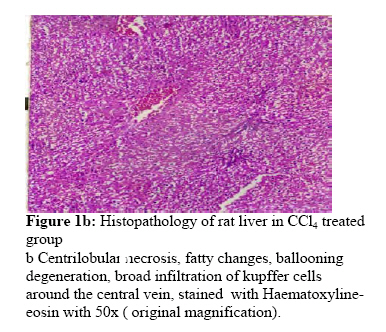
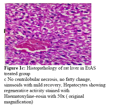
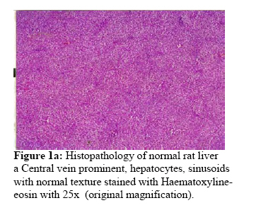
Values are mean ± SEM, n = 6, one way ANOVA followed by Dunnet’s multiple comparison test. a p<0.001when compared with control, b p<0.001 when compared with control and CCl4 group. Histopathological studies For histopathopathological observation, sections were taken from each lobe of liver immediately. The tissues were fixed in 10% neutral formalin, dehydrated in graded alcohol and embedded in paraffin, cut into 4-5 µm thick sections and stained with Haematoxylin-Eosin for photomicroscopic assessment (Galigher, 1971). Statistical analysis The data were expressed as the mean ± SEM, (n = 6). Data were analyzed using One way ANOVA followed by Dunnet’s multiple comparison test. Values of p< 0.05 were considered statistically significant. Results Argyreia speciosa extracts did not show any toxicity and behavioral changes in mice and hence doses of 200 and 400 mg/kg were selected for hepatoprotective and in vivo antioxidant activity. Preliminary phytochemical analysis of EAAS and EtAS revealed the presence of ergoline alkaloids, steroids, tannins and flavanoids as major active constituents. The activities of SGOT, SGPT, ALP and other biochemical parameters after administration of CCl4 and test extracts are summarized in Table 1. There was a significant rise in the levels of all biochemical parameters after administration of CCl4. In contrast, treatment with suspensions of EtAS (200 mg/kg and 400 mg/kg) and EAAS (200 mg/kg and 400 mg/kg ) of A. speciosa roots exhibited an ability to counteract the CCl4 induced hepatotoxicity by decreasing serum enzyme levels compared to control and CCl4 group (p<0.001). Pretreatment of rats with suspensions of EtAS and EAAS at a dose of 200 mg/kg and 400 mg/kg preserved Catalase, SOD, and Peroxidase activity compared to control and CCl4 group (p <0.001), thus providing protection against CCl4 toxicity (Table 2). Histopathological examination demonstrated that CCl4 treated group induces ballooning degeneration, centrilobular necrosis and apoptosis in hepatocytes (Fig 1. b). Groups treated with EtAS and EAAS showed recovery on ballooning degeneration and centrlobular bridging necrosis was occasionally present (Fig 1. c and Fig 1. d). Discussion The hepatic damage induced by CCl4 is well known to be mediated by its free radical metabolites such as CCl·3 and CCl3COO-, which interact with unsaturated lipid membrane to produce lipid peroxidation and other cellular macromolecules leading to cell damage (Gulcin, et al., 2004, Snyder et al., 1996). The free radicals in the presence of oxygen, leads to auto oxidation of the fatty acids present in the cytoplasmic membrane phospholipids (Recknagel, 1989) and causes functional and morphological changes in the cell membrane. Further more, influx of extracellular Ca+ ions into cell is claimed to be an important step leading to cell death. Hepatocellular necrosis leads to very high level of GOT and GPT released from liver in the blood. Among the two, GPT is a better index of liver injury, as liver GPT represents 90% of total enzyme present in the body (Achliya, et al., 2003). ALP activities on the other hand are related to functioning of hepatocytes, increase in its activity is due to increased synthesis in presence of increased biliary pressure (Moss, 1974). Reduction in the levels of SGOT, SGPT towards the respective normal value is an indication of stabilization of plasma membrane as well as repair of hepatic tissue damages caused by CCl4. Suppression of increased ALP activity with concurrent depletion of raised bilirubin level suggests the stability of biliary dysfunction in rat liver during chronic hepatic injury with CCl4 (Mukherjee, 2002). The effect of free radical metabolites on the mean liver detoxificant enzymes like Catalase, Super oxide dismutase (SOD) and Peroxidase, reduced enzyme activity, due to enzyme inactivation during catalytic cycle. The results of the present work indicate that Ethanolic extract and Ethyl acetate extract (200 mg/kg, 400 mg/kg) of A. speciosa roots decreased the elevated enzyme levels induced by CCl4, thus protecting the structural integrity of hepatocyte cell membrane or regeneration of damaged liver cells. These two extracts are found to be capable of enhancing or maintaining the activity of hepatic enzymes which are involved in combating Reactive Oxygen Species. The hepatoprotective effect of A. speciosa roots was evidenced by the amelioration of biochemical indicators of liver damage and pathological disturbances caused by CCl4. From the study we can conclude that root extracts of A. speciosa protects liver from oxidative damage and could be used as an effective protector in CCl4 induced damage. Future plans are in progress to isolate phytoconstituent(s) responsible and possible mode of action of these extracts. Acknowledgements Authors are thankful to President, Soniya Education Trust, Dharwad and Principal S. E. T’s college of Pharmacy, Dharwad for kind support and encouragement to carry out this research work. References
© Copyright 2008 - African. Journal. Traditional, Complementary and Alternative Medicines The following images related to this document are available:Photo images[tc08022f1d.jpg] [tc08022f1c.jpg] [tc08022f1a.jpg] [tc08022f1b.jpg] |
| |||||||||
