 |
| About Bioline | All Journals | Testimonials | Membership | News |
|
||||||
|
||||||
African Journal of Traditional, Complimentary and Alternative Medicines, Vol. 5, No. 2, 2008, pg. 213-222 Research PaperClerodendron inerme Protects Cellular Integrity During 7,12-dimethylbenz[a]-anthracene induced Hamster Buccal Pouch Carcinogenesis Shanmugam Manoharan*, Kannan Kavitha,Subramanian Balakrishnan, and Kasinathan Rajalingam Department of Biochemistry & Biotechnology,
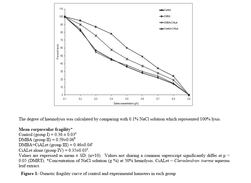
Faculty of Science, Annamalai University, Annamalainagar-608002, Tamil Nadu, India. Code Number: tc08030 Abstract Aim of the present study was to investigate the protective effect of Clerodendron inerme on cellular integrity by measuring the status of glycoconjugates, lipids, osmotic fragility, and membrane bound enzyme activity in 7, 12-dimethylbenz (a) anthracene (DMBA)-induced oral carcinogenesis. Oral squamous cell carcinoma was induced in the buccal pouch of Syrian golden hamsters by painting with 0.5% DMBA in liquid paraffin thrice a week for 14 weeks. The levels of glycoconjugates, lipids, osmotic fragility and membrane bound enzyme activity were analyzed by using specific colorimetric methods. We observed 100% tumor formation in DMBA painted hamsters. Altered glycoconjugates and lipid pattern were observed in DMBA painted hamsters as compared to control hamsters. Erythrocytes from DMBA painted hamsters were more fragile than those from control hamsters. The activity of membrane bound enzyme (Na+ K+ ATPase) decreased in DMBA painted hamsters as compared to control hamsters. Oral administration of aqueous leaf extract of Clerodendron inerme (CiALet) at a dose of 500mg/kg body weight significantly prevented the tumor formation and histopathological abnormalities as well as normalized the above said biochemical variables in DMBA painted hamsters. Our results thus demonstrate the protective effect of Clerodendron inerme on cellular integrity during DMBA induced oral carcinogenesis. Key words: Oral cancer, DMBA, hamster, Clerodendron inerme, osmotic fragility, lipids, glycoconjugates. Introduction The term oral cancer refers to a diverse group of tumors arising from lip, cheek, floor of the mouth, hard and soft palate, tongue, pharynx and oral cavity. Oral cancer is the fifth most frequent cancer worldwide and over 300,000 new cases of oral cancer are diagnosed every year. India has recorded the highest incidence of oral cancer where it accounts for 40-50% of all cancers. Cancer of the oral cavity is frequently associated with chewing of betel quid containing tobacco in addition to smoking and alcohol consumption. Oral cavity cancers can be prevented by avoiding known risk factors (Moore et al.,2000; Johnson, 2001). The carcinogen, 7, 12-dimethylbenz (a) anthracene (DMBA) can initiate and promote the development of oral carcinoma of the buccal mucosa through distinct premalignant lesions. DMBA induced oral carcinogenesis is the most widely accepted experimental model for oral cancer since it has many morphological and histological similarities with human oral carcinoma. This experimental model also expresses many biochemical and molecular markers that are expressed in human oral carcinoma (Miyata et al., 2001). Lipids serve as an integral component of the cell membrane and have a profound role in the regulation of cellular processes. They play an important role in maintaining the structural integrity and function of cell membranes. Lipids have also influence on the activity of membrane bound enzymes. Abnormalities in the levels of lipids and altered cholesterol phospholipids molar ratio in the cell membranes have been implicated as an important aspect of malignant transformation. Alterations in lipids and lipoproteins composition have also been well documented in oral carcinogenesis (Kolanjiappan et al., 2003; Manoharan et al.,1995). Measurement of osmotic fragility can help to assess the integrity of red blood cells. Changes in red blood cell osmotic fragility have been documented in several pathological conditions including carcinogenesis. The membrane bound enzyme, Na+/K+ ATPase (Na+/K+ pump), maintains the electro-chemical gradients of Na+ and K+ and the ion gradient produced by Na+K+ ATPase influences cell volume and osmotic pressure. Altered Na+/K+ ATPase activity has been documented in carcinogenesis (Kolanjiappan et al.,2002; Selvendiran and Sakthisekaran, 2004). Cellular membranes contain 2-10% carbohydrates as glycolipids and glycoproteins. Glycoconjugates, the vital components of the cell surface, play a significant role in contributing to the surface properties of the cells and also play a crucial role in tumorigenesis and as mediators of immunological specificity. Altered glycoconjugates pattern in cell membranes alter the structural integrity, rigidity and function of cells (Manoharan et al., 2004). Sialic acids are terminal sugar component of the oligosaccharide chains of glycoproteins and glycolipids. Abnormalities of glycoproteins and different forms of sialic acids have been well documented in several pathological conditions including various malignancies (Manoharan et al., 1995; Aranganathan et al.,2005). Altered levels of lipid bound sialic acid and fucose are considered as markers of malignant transformation (Suresh et al.., 2007; Manoharan et al., 2004). Clerodendron inerme (L.) Gaertn is a perennial herb, widely distributed throughout India, South and South East Asia, Australia and Pacific islands. It is popularly known as “Seaside Clerodendron” in English. It is used in the indigenous system of medicine for the treatment of various diseases including cancer. The juice of the leaves and roots are used for the treatment of beriberi, veneral infections and skin diseases (Kiritikar and Basu, 1975). The phytochemical examination of C. inerme revealed the presence of flavanoids, sterols, flavones, triterpenes, diterpenes, quinone and neolignans (Rehman et al., 1997). Previous studies from our laboratory have shown the chemopreventive and antilipidperoxidative potential of aqueous extract of C. inerme (L.)Gaertn leaves in 7,12-dimethylbenz(a)anthracene-induced hamster buccal pouch carcinogenesis (Manoharan et al.,2006). The present study demonstrates the protective effects of C. inerme on cellular integrity by examining the status of glycoconjugates, lipids, osmotic fragility and membrane bound enzyme activity during 7,12-dimethylbenz(a)anthracene induced hamster buccal pouch carcinogenesis. Materials and Methods Animals Male golden Syrian hamsters 8-10 weeks old weighing 80-120g were purchased from National Institute of Nutrition, Hyderabad, India, and maintained in Central Animal House, Rajah Muthaiah Medical College and Hospital, Annamalai University. The hamsters were housed four or five in a polypropylene cage and provided standard pellet diet (Agro Corporation Private Limited, Bangalore, India) and water ad libitum. The standard pellet diet is composed of 21% protein, 5% lipids, 4% crude fiber, 8% ash, 1% calcium, 0.6% phosphorus, 3.4% glucose, 2% vitamin, and 55% nitrogen-free extract (carbohydrates). The hamsters were maintained under controlled conditions of temperature and humidity with a 12h light/dark cycle as per the principles and guidelines of the ethical committee for animal care of Annamalai University in accordance with Indian National Law on animal care and use. Chemicals 7, 12-dimethylbenz (a) anthracene (DMBA), γ-glutamylparanitroanilide, galactose, galactosamine and N-acetyl neuraminic acid were purchased from Sigma Aldrich Chemical Pvt Ltd., Bangalore, India. All other chemicals used were of analytical grade, purchased from Hi-media Laboratories, Mumbai, India. Plant Material C. inerme (voucher No. AU05105)leaves were collected in and around Cuddalore, Tamil Nadu, India. A voucher specimen was also deposited in the Department of Botany, Annamalai University. Hundred gram of fine powder of C. inerme leaves was suspended in 250 ml of water for two hrs and then heated at 60-65°C for 30 min. The extract was preserved and the process was repeated for three times with the residual powder, each time collecting the extract. The collected extract was pooled and passed through fine cotton cloth. The filtrate upon evaporation at 40°C yielded 14% semisolid extract. This was stored at 0-4°C until used. A known volume of the residual extract was suspended in distilled water and orally administered to the hamsters by gastric intubation using a force-feeding needle during the experimental period. Experimental protocol The experimental design was approved by the Annamalai University animal ethical committee [Reg.No:160/1999/CPCSEA], Annamalai University, Annamalainagar. A total number of 40 hamsters were randomized into four groups of ten hamsters in each group. Group I hamsters served as control and were painted with liquid paraffin thrice a week for 14 weeks on their left buccal pouches. Groups II and III hamsters were painted with 0.5% DMBA in liquid paraffin thrice a week for 14 weeks on their left buccal pouches. Group II hamsters received no other treatment. Group III hamsters were orally administered with C. inerme aqueous leaf extract (CiALet) [500mg/kg body weight], starting one week before the exposure to the carcinogen and continued on days alternate to DMBA painting, throughout the experimental period. Group IV hamsters received oral administration of CiALet alone [500mg/kg body weight] throughout the experimental period. The experiment was terminated at the end of 14 weeks and all hamsters were sacrificed by cervical dislocation. Biochemical studies were conducted on plasma, erythrocyte membrane and buccal mucosa of control and experimental hamsters in each group. For histopathological examination, buccal mucosal tissues were fixed in 10% formalin and routinely processed and embedded with paraffin, 2-3mm sections were cut in a rotary microtome and stained with haematoxylin and eosin. Biochemical analysis After plasma separation, the erythrocyte membrane was prepared by the method of Dodge et al., (1968) modified by Quist (1980). The precipitate obtained after treating the plasma with 95% ethanol was used for the estimation of protein bound hexose and hexosamine. Similarly, the precipitate obtained after treating the erythrocyte membranes with 1% phosphotungstic acid followed by 5% trichloro acetic acid (TCA) was used for the estimation of protein found hexose and hexosamine. The defatted tissues obtained after treating buccal mucosa tissues with methanol and chloroform was used for the estimation of glycoproteins. The protein bound hexose, hexosamine, total sialic acid and fucose in plasma and erythrocyte membrane were estimated by the methods of Niebes et al, (1972), Wagner (1979), Warren (1959) and Dische and Shettles (1948) respectively. Plasma lipid bound sialic acid level was determined by the method of Katopodis and Stock (1980). Lipid extraction from the tissues was done by the method of Folch et al. (1957). The level of total cholesterol and high-density lipoproteins (HDL) cholesterol was estimated by the methods of Parekh and Jung (1970) and Gidez and Webb, (1950) respectively. The estimation of phospholipids was done by the method of Zilversmit and Davies (1950). Free fatty acids and triglycerides were measured by the methods of Falholf et al. (1973) and Foster and Dunn (1973) respectively. Osmotic fragility was determined by the method of Parpart et al. (1946) and mean corpuscular fragility (MCF) was calculated by recording the saline concentration, which would have resulted in 50% hemolysis. Statistical analysis The data are expressed as mean ± SD. Statistical comparisons were performed by One way analysis of variance (ANOVA) followed by Duncan’s Multiple Range Test (DMRT). The results were considered statistically significant if the p values were less than 0.05. Results Table 1 shows the levels of lipids in plasma, erythrocyte membrane and buccal mucosa tissues and lipoproteins in plasma of control and experimental hamsters in each group. The levels of total cholesterol, low-density lipoproteins (LDL) cholesterol and free fatty acids were high whereas high-density lipoproteins (HDL) cholesterol, very low-density lipoproteins (VLDL) cholesterol, phospholipids and triglycerides levels were low in plasma of DMBA painted hamsters as compared to control hamsters. The total cholesterol level was increased whereas phospholipid level was decreased in red blood cell (RBC) membrane of DMBA painted hamsters as compared to control hamsters. The level of total cholesterol and free fatty acids were increased whereas phospholipid was decreased in buccal mucosal tissues of DMBA painted hamsters as compared to control hamsters. Increase in cholesterol and phospholipid ratio (C/P ratio) was observed in plasma, RBC membrane and in the tumor tissues of DMBA alone painted hamsters as compared to control hamsters. Oral administration of CiALet restored the above mentioned lipid abnormalities in DMBA painted hamsters. No significant difference in lipid and lipoproteins levels was observed in hamsters treated with CiALet alone as compared to control hamsters. The level of total cholesterol and free fatty acids were increased whereas phospholipid was decreased in buccal mucosal tissues of DMBA painted hamsters as compared to control hamsters. Increase in cholesterol and phospholipid ratio (C/P ratio) was observed in plasma, RBC membrane and in the tumor tissues of DMBA alone painted hamsters as compared to control hamsters. Oral administration of CiALet restored the above mentioned lipid abnormalities in DMBA painted hamsters. No significant difference in lipid and lipoproteins levels was observed in hamsters treated with CiALet alone as compared to control hamsters. Osmotic fragility curves for control and experimental hamsters in each group are shown in Figure 1. The fragility curve of DMBA painted hamsters was shifted to the right for control hamsters. The mean corpuscular fragility was also significantly higher in cancer hamsters as compared to controls. Treatment of DMBA painted hamsters with CiALet shifted the curve to the left of cancer hamsters. Mean corpuscular fragility values did not differ significantly in hamsters treated with CiALetalone as compared to control hamsters. Table 2 shows the activity of membrane bound Na+K+ ATPase in control and experimental hamsters in each group. The activity of membrane bound Na+K+ ATPase was significantly decreased in cancer hamsters as compared to control hamsters. The activity of Na+K+ ATPase was restored in DMBA treated hamsters after treatment with CiALet. No significant difference was observed in hamsters treated with CiALet alone as compared to control hamsters Table 1: Plasma, erythrocyte membrane and buccal mucosa tissue lipid and lipoprotein profile in control and experimental hamsters in each group.
Values are expressed as mean ± SD, (n=10). Values not sharing a common superscript significantly differ at p < 0.05 (DMRT). CiALet – Clerodendron inerme aqueous leaf extract. *LDL-cholesterol = Total cholesterol – HDL Table 2: The activity of erythrocyte membrane Na+K+ATPase in control and experimental hamsters in each group.
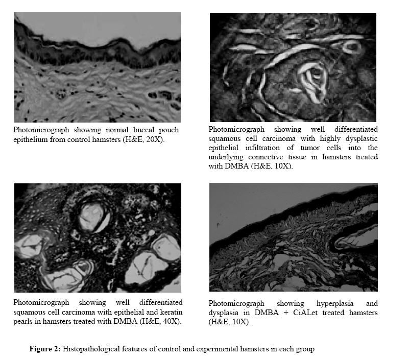
Values are expressed as mean ± SD, (n=10). Values not sharing a common superscript significantly differ at p < 0.05 (DMRT). A= μmoles of inorganic phosphorus formed/ hour/ mg protein. CiALet – Clerodendron inerme aqueous leaf extract. Table 3 shows the levels of glycoconjugates in plasma, erythrocyte membrane and buccal mucosa tissues of control and experimental hamsters in each group. The levels of glycoconjugates were significantly higher in plasma and buccal mucosa tumor tissues whereas low in erythrocyte membrane of DMBA painted hamsters as compared to control hamsters. Oral administration of CiALet to DMBA painted hamsters reversed the levels of glycoconjugates. No significant difference was noticed in the levels of plasma, erythrocyte membrane and buccal mucosa tissue glycoconjugates in CiALet alone treated hamsters as compared to control hamsters. Discussion In the present study, we have observed hyperplasia, dysplasia and severe keratosis at eighth to tenth week and well and moderately differentiated squamous cell carcinoma at 14th week in DMBA painted hamsters. Oral administration of CiALet to DMBA painted hamsters on days alternate to DMBA painting for 14 weeks significantly prevented the premalignant lesions and tumor formation (Figure 2). This indicates that C. inerme aqueous extract has suppressing effects on cell proliferation in DMBA induced hamster buccal pouch carcinogenesis. The chemopreventive effect of C. inerme is probably due to the presence of several bioactive chemopreventive principles and their synergistic effects. Lipids, an integral part of biomembrane, are essential for several biological functions including normal cell growth and division, as well as for cellular proliferation (Allampallam et al.,2000). Although abnormalities of lipids have been implicated in the pathogenesis of cardiovascular diseases, altered levels of lipids and lipoproteins in circulation have also been found to be associated with the etiopathogenesis of several cancers (Manoharan et al., 1995; Kolanjiappan et al.,2003). Elevated plasma cholesterol in DMBA painted hamsters can be related to decrease in HDL cholesterol and increase in VLDL, LDL cholesterol and free fatty acids observed in the present study. Increase in cholesterol-phospholipids molar ratio in plasma, erythrocyte membranes and tumor tissues indicate that the structural integrity and function of the cell is severely destructed. Elevated erythrocyte membrane cholesterol is probably due to ineffective exchange mechanism with plasma cholesterol. Increase in plasma free fatty acids in DMBA painted hamsters indicates that tumor cells utilize free fatty acids as a metabolic substrate for their growth. Tumor cells sequester lipids from circulation for their demand. Increase in total cholesterol has been reported in several tumor tissues as compared to their adjacent tissues (Siemianowicz et al., 2000). Several studies have reported that tumor tissues utilize lipids including total cholesterol, triglycerides and lipoproteins for the biogenesis of newer membranes (Kolanjiappan et al., 2003). Our results corroborate these observations. Lowered fatty acids in tumor tissues are partly responsible for decreased phospholipids levels. Oral administration of CiALet to DMBA painted hamstersnormalized the levels of lipids and lipoprotein which indicates their membrane stabilizing effect during oral carcinogenesis. Table 3: Plasma, erythrocyte membrane and buccal mucosa tissues glycoconjugates in control and experimental hamsters in each group.
Values are expressed as mean ± SD, (n=10). Values not sharing a common superscript significantly differ at p < 0.05 (DMRT). CiALet – Clerodendron inerme aqueous leaf extract. Red blood cells obtained from DMBA painted hamsters were found to be more fragile than those obtained from control hamsters. An imbalance in oxidant and antioxidant status can cause an increase in red blood cell fragility and reduction in red blood cell fluidity (Kolanjiappan et al.,2002). Previous studies from our laboratory have reported an elevated lipidperoxidation and decrease in antioxidants in experimental oral carcinogenesis (Senthil et al.,2007). Enhanced red cell osmotic fragility observed in DMBA painted hamsters was therefore due to profound oxidative stress. Oxidative damage to membrane bound enzymes has been assumed to be crucial for cell lysis (Selvendiran and Sakthisekaran, 2004). A decrease in membrane bound Na+K+ATPase in DMBA painted hamsters suggest that the membrane permeability was drastically altered during DMBA induced oral carcinogenesis. Oral administration of CiALetto these DMBA painted hamsters prevented the alterations in red blood cell fragility and activity of membrane bound Na+K+ ATPase. Our results indicate that CiALet maintained the structural integrity of erythrocytes during oral carcinogenesis. Cell surface glycosyl residues play an important role in regulating cell proliferation and epithelial growth. Recent clinical and laboratory studies have shown that the levels of glycoconjugates are markedly increased in plasma or serum of DMBA painted animals and patients (Manoharan et al., 2004; Suresh et al.,2007). Neoplatic transformation of oral epithelium is due to atypical glycosylation of cell surface carbohydrates. A reduction in epithelial cell surface carbohydrates during experimental oral carcinogenesis has been reported (Manoharan et al., 2004). Malignant tumor in the body stimulates the synthesis of glycoproteins in the liver, which subsequently enter into the circulation. It has been suggested that the glycoconjugates are secreted from membrane into extracellular fluid in cancerous conditions (Thirunavukarasu and Sakthisekaran, 2003). The depletion of erythrocyte membrane glycoprotein may therefore be due to increased membrane degradation or as a result of increased shedding into circulation. The levels of glycoprotein and sialic acids were increased as the clinical stages of tumor advanced (Manoharan et al.,2004). Elevated plasma glycoproteins in DMBA painted hamsters can therefore be related to an increased synthesis in liver or tumor tissues itself with subsequent shedding into plasma. Tumor cell membrane has more sialic acid as compared to normal cell membrane. Marked elevation of total sialic acid and lipid bound sialic acid in plasma reflect severe tumor burden in DMBA painted hamsters. An increase in total sialic acid is probably related to increased turnover of malignant cells. The elevated levels of sialic acid in tumor tissues is due to selective increase in existing specific sialylated sequence or tumor associated denovo synthesis of specific sialylated sequence. Several studies concluded that total sialic acid level in plasma or serum could be considered as a supportive evidence of tumor marker for the diagnosis of cancer (Vasanti et al.,1998; Goodarzi et al., 2005). Raval et al. (1997) have shown a positive correlation between serum levels of different forms of sialic acids and the extent of malignant disease. Increase in plasma total and lipid bound sialic acid in DMBA painted hamsters are probably due to spontaneous release (shedding) of aberrant sialic acid rich glycoprotein and glycolipids. Increased levels of plasma and tumor tissue fucose have been well documented in several cancerous conditions (Vasanti et al., 1998). Our results lend credibility to these observations. Oral administration of CiALet prevented the abnormalities seen in cell surface glycoconjugates in the circulation and tumor tissues, which indicates their membrane stabilizing effects during neoplastic transformation. The protective effect of CiALet is probably due to their inhibitory role on glycoprotein synthesis or on the activity of the glycosyl transferases. Conclusion Oral administration ofCiALet protected the levels of blood and tissue lipids, cell surface glycoconjugates, red blood cell osmotic fragility and membrane bound enzyme (Na + K + ATPase) activity during DMBA induced oral carcinogenesis. Our study thus indicates the crucial role of CiALet in maintaining the structural integrity and function of the cells during neoplastic transformation. References
© Copyright 2008 - African. Journal. Traditional, Complementary and Alternative Medicines The following images related to this document are available:Photo images[tc08030f1.jpg] [tc08030f2.jpg] |
| |||||||||

 . **VLDL-cholesterol
 .
.