 |
| About Bioline | All Journals | Testimonials | Membership | News |
|
||||||
|
||||||
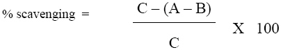
African Journal of Traditional, Complimentary and Alternative Medicines, Vol. 5, No. 3, 2008, pg. 230-237 Biological Activities Of Asparagus racemosus Buppachart Potduang, Maneerat Meeploy, Rattanasiri Giwanon, Yaowaluck Benmart, Montree Kaewduang and Winai Supatanakul Pharmaceuticals and Natural Products Department, Thailand Institute of Scientific and Technological Research, Technopolis, Thanon Liab Klong 5, Klong Luang, Pathumthani 12120, Thailand. E-mail: buppachart@hotmail.com; buppachart@tistr.or.th Code Number: tc08034 Abstract Cytotoxic, antioxidant, tyrosinase inhibitory, antimicrobial activities of the crude ethanol extract of dry powdered roots of Asparagus racemosus(Liliaceae) were investigated. The LC50to brine shrimp was 2189.49mg/ml; the EC50 for DPPH radical scavenging was 381.91mg/ml; the IC50 for tyrosinase inhibition was 7.98 mg/ml. The extract was active at 5-20mg/ml against various pathogenic microbial (16 species, 18 strains) using the agar dilution assay, with the minimum inhibitory concentration (MIC) between 10-20mg/ml for enteropathogens, the MIC between 5-20 mg/ml for dermatopathogens, and MIC = 10 mg/ml for a pneumonia causing bacteria Klebsiella pneumoniae. TLC and HPLC finger printing showed the presence of steroids-terpenes, alkaloids and flavonoids. Key words: Asparagus racemosus, Antioxidant, Antityrosinase, Antimicrobial, Phytochemistry Introduction Asparagus racemosus Willd. (Liliaceae), locally known in Thailand as ‘Rag Samsib’,is a woody climber growing to 1-2 m in height. The Thai local name ‘Rag Samsib’refers to its finger-like andclustered roots. The leaves are like pine-needles, small and uniform. The inflorescence has tiny white flowers, in small spikes (Vichien, 2003).The plant is common at low altitudesinshade and in tropical climates throughout Asia, Australia and Africa. In India, the plant is calledShatavariin Hindi. The root has long been used in Ayurvedaas a tonic remedy to promote fertility and reducing menopausal symptoms. It is also used for dry coughs and gastric ulcers (Winston, 2004). Recent research indicates Shatavari enhances immune function, increases corticosteroid production, and promotes cell regeneration (Rege et al., 1999). In Thailand the root is claimed as a galactagogue, antidysenteric, antipeptic, antipruritic, antirheumatic, tonic andlongevity enhancer. This study investigated various biological activities of the crude ethanol root extract of A. racemosus cultivated in Thailand. TLC fingerprints and HPLC fingerprint of the root powder were performed. Materials and Methods Plant material Fresh roots of A. racemosus were collected in May 2003 from Nakhon Rachasima Province, Thailand. Thesample was dried in a hot air oven at 40-50oC, and then pulverized into powder. The specimen (no. LRS-0110)was authenticated by the Research Botanist Officer and kept at the Lamtakhong Plant Research Station,TISTR. Preparation of crude ethanol extract The root powder was repeatedly macerated with 95%ethanol in a percolator. The combined filtrate was evaporated to dryness under reduced pressure at 40-50°C. The resulting crude ethanol extract was then stored at 10-15oC. Cytotoxicity to brine-shrimp The brine-shrimp micro-plate assaywas amodified version of Solis et al. (1992) used to determine the inhibitory activity on Artemia sp. in 0.25% Tween 80-artificial seawater, as described by Potduang et al. (2007). The sample solution was added into 6 wells, each containing 5 newly hatched brine shrimps to make overall 30 brine shrimps in contact with the sample for 24hr. The dead organisms were counted under a binocular microscope (4x). Plot %Lethality vs Log concentration. Substituted y = 50 in the resulted linear equation to obtain the x value. The antilog x was then the LC50 (conc. of 50% lethality) value (Ballantyne et al., 1995). Thymol and kojic acid were used as reference standards. Antioxidant activity Scavenging of DPPH radical The DPPH radicalscavenging micro-plate assay modified from Hatano et al. (1989), as described by Potduang et al. 2007, was used. Equal volumes of absolute ethanol solutions of the extract and 0.06mM DPPH (2,2-diphenyl-1-picrylhydrazyl from Sigma, Germany) were mixed for 30 min in a micro-well plate, and absorbance measured at 517nm in a micro-plate reader (TECAN, Sunrise remote). All samples were run in triplicate. The % scavenging activity of test samples was determined as follows:
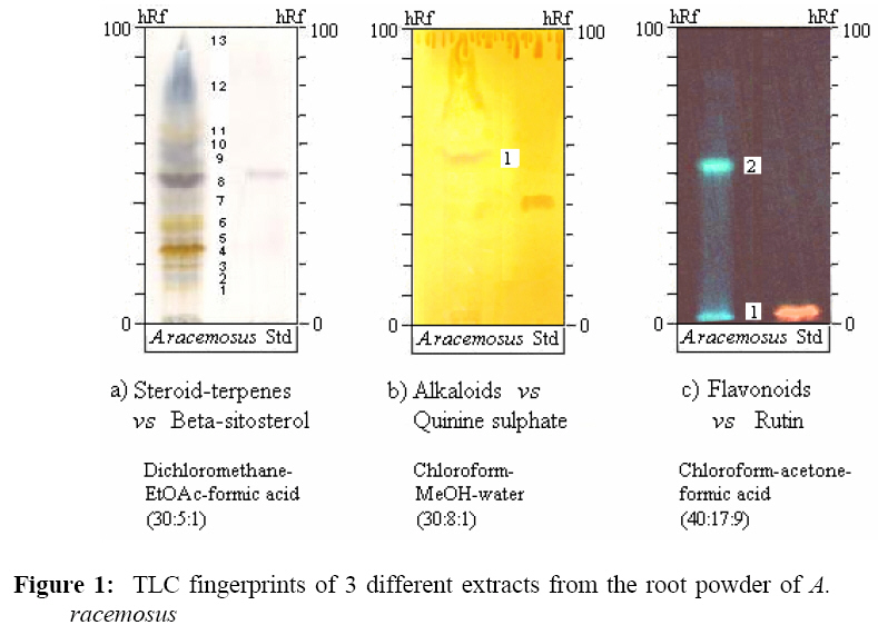
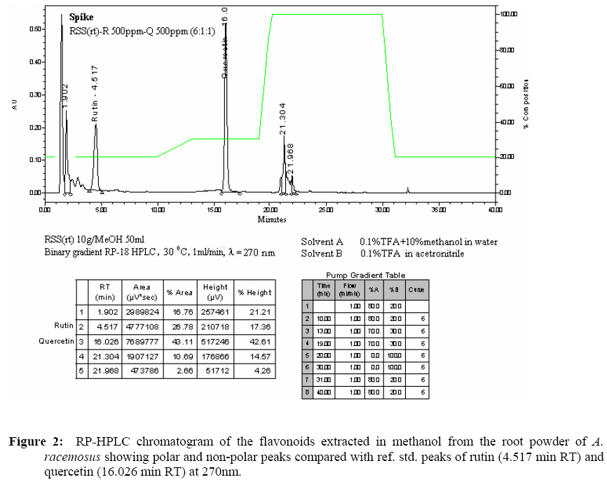
Where A, B and C represent the absorbances of DPPH in the reactionmixture, the blank, and the control respectively.Plot %Scavenging vs Log concentration. Substituted y = 50 in the resulted linear equation to obtain the x value. The antilog x was then the EC50(conc. of 50% scavenging)value (Ballantyne et al., 1995). BHT, BHA and vitamin C were used as reference standards. Tyrosinaseinhibition The dopachrome micro-plate assay modified fromIidaet al. (1995) was used to investigate the tyrosinase inhibition of the 20% ethanol derived extract,as described by Potduang et al. 2007. The 50 ml sample solution was mixed with 50 ml of mushroom tyrosinase buffer solution (314.8U/ml, Fluka) and 150 ml of 0.02 M sodium phosphate buffer (pH 6.8), and allowed to stand for 10 min. Added was 50 ml of 0.34 mM L-Dopa (Sigma Chemical) buffer solution as substrate, mixed and then incubated for 2 min. The absorbance was measured at 492nm by a micro-plate reader (TECAN, Sunrise remote). All samples were run in triplicate. The absorbance differences before and after the 2 min-incubation were used to calculate the percentage inhibition of tyrosinase as follows:
Where the absorbance difference A represents the control (L-Dopa mixed with enzyme in buffer); B represents the blank (L-Dopa in buffer); C represents the reaction mixture; and D represents the blank of C (L-Dopa mixed with test sample in buffer). Plot %Tyrosinase inhibition vs Log concentration. Substituted y = 50 in the resulted linear equation to obtain the x value. The antilog x was then the IC50 (conc. of 50% inhibition) value (Ballantyne et al., 1995). A well-known tyrosinase inhibitor, kojic acid, was used as the reference standard. Anti-microbial activity The agar dilution method (Washington and Sutter, 1980) was used to test the activities against pathogenic microorganisms, using specific assay media and broths as described by Potduang et al. 2007. The media were Mueller Hinton Agar (MHA; Difco Laboratories) for aerobes; WC Agar (Wilkins and Chalgren, 1976) for anaerobes; and Saboraud Dextrose Agar (SDA; Difco Laboratories) for yeasts. The isolates suspension was adjusted to McFarland 0.5 turbidity standard. Spot inoculated the 5-20 mg/ml dilution plates of the crude extract and incubated at 37oC (overnight for aerobes; 3 days for anaerobes; 48 hr for yeast). The minimum inhibitory concentrations (MICs) of the extract were determined. TLC fingerprints Thin-layer chromatography (TLC) of 3 different extracts containing steroids-terpenes, alkaloids or flavonoids from the root powder were performed on 0.25 mm thick TLC plates (Merck Silica gel 60 F254-precoated) using suitable developing solvents and special detection reagents (Merck, 1980; Wagner and Bladt, 1996). These 3 easily extracted groups possess various biological activities. 1. TLC fingerprint of the steroids-terpenes extract. The extract was prepared by stirring 5 g of the root powder with hexane (3x50 ml) for 30min. The filtrate was concentrated to dryness under reduced pressure, and then dissolved in 0.5 ml chloroform. The extract (2 ml)was applied onto a TLC plate to perform 10 cm chromatography with suitable solvent system. The developed plate was sprayed with vanillin-sulfuric acid reagent, then heated until the spots attain maximum colour intensity of steroids-terpenes compared to ref. std. 1:1 w/v b-sitosterol (Sigma, USA) in chloroform. 2. TLC fingerprint of the alkaloids extract. The extract was prepared by stirring20 g of the root powder with100 ml 0.1N sulphuric acid for 20 min. The filtrate was alkalinized to pH 8-9 with 5% ammonium hydroxide. The free alkaloids were extracted by partitioning with chloroform (3x80 ml). The combined chloroform extracts were dried over anhydrous sodium sulphate before evaporated to dryness under reduced pressure. The dried extract was dissolved in 0.2 ml methanol before applying 10 ml onto a TLC plate to perform 10 cm chromatography with a suitable solvent system. The developed plate was sprayed with Dragendroff’s reagent to visualize orange-brown zones of alkaloids compared to ref. std. 1:1 w/v quinine sulphate (BDH, England) in methanol. 3. TLC fingerprint of the flavonoids extract. The extract was prepared by stirring0.5 g of the root powder with 5 ml methanol on a dry block heat bath (60°C, 5 min), allowed to cool, filtered, evaporated to dryness under reduced pressure. Dissolved the dried extract in 0.2 ml methanol, then filtered through a PTFE syringe filter membrane (Orange Scientific, Belgium) before applying 5 ml onto a TLC plate to perform 10 cm chromatography with a suitable solvent system. The developed plate was sprayed with natural products-polyethylene glycol (NP/PEG) reagent to achieve fluorescing zones of flavonoids under UV-365nm compared to ref. std. 1:1 w/v rutin (Fluka, Switzerland) in methanol. HPLC fingerprint A methanol extract containing flavonoids was prepared by shaking5 g of the root powder with 25 ml of methanol at 1,500 rpm for 2 min. The filtration was made through a Whatman paper no.41, and then added methanol to make the filtrate to 25 ml in a volumetric flask. The sample solution was then filtered through a 0.45 mnylon syringe filter membrane before subjected to binary gradient RP-18, 30°C, 1 ml/min flow rate, HPLC analysis with 270nm UV detector. Solvent A was water with 0.1% TFA + 10% methanol, and solvent B was acetronitrile with 0.1% TFA. Standard addition of rutin (Merck, Germany) and quercetin (Fluka, Switzerland) was applied to HPLC chromatogram. Results The crude ethanol root extract of A. racemosus was9.01%. The extractexhibited anLC50 of 2189.49 mg/ml on brine shrimp cytotoxicity, and gave EC50 of 381.91 mg/ml on DPPH radical scavenging. The derived 20% ethanol extract gave the IC50 of 7.98 mg/ml on mushroom tyrosinase inhibition (Tables 1-4). The agar dilution assay indicated that the crude extract at5-20mg/ml, was active against various disease causing microorganisms (16 species, 18 strains). The minimum inhibitory concentrations (MICs) were 10-20 mg/ml against enteropathogens: Enterococcus faecalis,Salmonella velterans, Shigella dysenteriae, Staphylococcus aureus, Escherichia coli, Bacteroides spp., Clostridium spp., Peptococcus spp., Lactobacillus spp.andStreptococcus mutans. The MICs were 5-20 mg/ml against dermatopathogens as Staphylococcus epidermidis, Propionibacterium acnes, Candida albicans, Pseudomonas aeruginosaand Streptococcus spp. The MIC was 10 mg/ml against a pneumonia causing bacteria Klebsiella pneumoniae(Table 5). TLC investigation showed the presence of steroids-terpenes, alkaloids and flavonoids(Table 6, Figure 1). RP-HPLC fingerprint, under a suitable 40min-program linear gradient, showed 1 peak more polar than ref. std. rutin (4.517 min RT) and 2 peaks more non-polar than ref. std. quercetin (16.026 min RT) at 270nm, as shown in Figure 2. Discussion The concentration of 50% activity of the A. racemosus root extract were calculated from the following computerized linear equations: y = 138.86x – 413.84 on brine shrimp cytotoxicity; y = 52.359x – 85.189 on DPPH radical scavenging; and y = 46.767x + 7.8191 on tyrosinase inhibition. Where x was obtained by substituting y = 50, the antilog x gave the value of either the LC50, EC50 or IC50, respectively. The roots had mild cytotoxicity (brine shrimp inhibition approx.0.8% of kojic acid and 0.6% of thymol), mild DPPH radical scavenging activity (approx.1% of BHT, BHA and 0.3% of vitamin C), and non-significance melanin biosynthesis inhibitors (anti-tyrosinase activity approximately 0.03% of kojic acid). The MICs of 5-20 mg/ml against various pathogenic microbial (16 species, 18 strains) indicated that A. racemosus root extract has a wide spectrum activity. RP-HPLC chromatogram of the flavonoid extract and zoning patterns of steroids-terpenes, alkaloids and flavonoids on the TLC fingerprints were specific enough to be used for the identification of A. racemosus root powder. Table 1: Biological effects of the ethanol extract fromthe roots of A. racemosus
Table 2: Inhibitory effect onbrine-shrimp of the ethanol extract from the roots of A. racemosus
Table 3: In vitro DPPH radical scavenging effect of the ethanol extract from the roots of A. racemosus
Table 4: Inhibitory effect onmushroom tyrosinase of the 20% ethanol fraction from the ethanol extract from the rootsof A. racemosus
Table 5: Minimal inhibitory concentrations (MICs) of the ethanol extract from the roots of A. racemosus on various pathogenic microorganisms
+ = cultured growth, 1assay media with acetone, 2assay media Table 6: hRf values of chief constituents detected on TLC of 3 different extracts fromthe root powder of A. racemosus.
Conclusion The root of A. racemosus is a potential broad spectrum antibiotic. Acknowledgements We thank the Pharmaceuticals and Natural Products Department Thailand Institute of Scientific and Technological Research (TISTR) for providing the fund and good laboratory facilities, and to Mr. Parinya Wilairatana, Ex-Director of the Lamtakhong Plant Research Station TISTR for providing the plant materials. References
© Copyright 2008 - African. Journal. Traditional, Complementary and Alternative Medicines The following images related to this document are available:Photo images[tc08034f2.jpg] [tc08034f1.jpg] | ||||||||||||||||||||||||||||||||||||||||||||||||||||||||||||||||||||||||||||||||||||||||||||||||||||||||||||||||||||||||||||||||||||||||||||||||||||||||||||||||||||||||||||||||||||||||||||||||||||||||||||||||||||||||||||||||||||||||||||||||||||||||||||||||||||||||||||||||||||||
| |||||||||

