 |
| About Bioline | All Journals | Testimonials | Membership | News |
|
||||||
|
||||||
African Journal of Traditional, Complimentary and Alternative Medicines, Vol. 5, No. 4, 2008, pg. 363 - 369 Research PaperAntimicrobial activity of various extracts of Ocimum basilicum l. And observation of the inhibition effect on bacterial cells by use of scanning electron microscopy Ilhan Kaya1, Nazife Yiğit 2, Mehlika Benli*3 1Yuzuncuyil University, Faculty of
Agriculture, Department of Plant Protection, Van, Turkey Code Number: tc08050 Abstract The antimicrobial activities of chloroform, acetone and two different concentrations of methanol extracts of Ocimum basilicum L. were studied. These extracts were tested in vitro against 10 bacteria and 4 yeasts strains by the disc diffusion method. The results indicated that the methanol extracts of O. basilucum exhibited the antimicrobial activity against tested microorganisms. While the chloroform and acetone extracts had no effect, the methanol extracts showed inhibition zones against strains of Pseudomonas aeruginosa, Shigella sp., Listeria monocytogenes, Staphylococcus aureus andtwo different strains of Escherichia coli. The cells of microorganisms, which were treated and untreated with plant extracts, were observed by using the scanning electron microscope. It was observed that the treated cells were damaged. Key words:Antimicrobial activity, Disc diffusion method, Ocimum basilicum, Plant extract, SEM, Spices. Introduction Some medical plants have been used for a wide variety of purposes such as food preservation, pharmaceutical, alternative medicine, and natural therapies for many thousands of years. It is generally considered that compounds produced naturally, rather than synthetically, will be biodegraded more easily and therefore be more environmentally acceptable. Thus, natural antioxidants, antibacterial, cytotoxic, antiviral, fungicidal agents and nutrients have gained popularity in recent years, and their use and positive image among consumers are spreading. In recent years, multiple drug resistance in both human and plant pathogenic microorganisms have been developed due to the indiscriminate use of commercial antimicrobial drugs commonly used in the treatment of infectious diseases (Davis, 1994; Service, 1995).In order to find new therapeutic agents, plants that have antimicrobial activity have attracted attention (Kalemba and Kunika, 2003; Juliani and Simson, 2002; Falerio et al., 2003). The Labiatae family is one of the most employed medicinal plants as a worldwide source of spices and also as a consolidated source of extracts with strong antibacterial and antioxidant properties. Within this family, the genus Ocimum provides various species, and Ocimum basilicum is one of the more than 60 Ocimum species. These Ocimum plants, plant parts, extracts and essential oils are used as spices and flavours for various food products as well as effective drugs for many applications in folk medicine especially in Africa and Asia (Sacchetti et al., 2004; Jirovetz et al., 2003). Ocimum basilicum, commonly known as basil, is naturally distributed in the East Anatolia region in Turkey. The leaves of these plants are used in traditional cuisine as spices. Basil, which is a popular culinary herb, and its essential oils have been used extensively for many years in flavouring of meats and sausages. Basil oil has also found a wide application in perfumery, as well as in dental and oral products. In addition, because the public nowadays prefers natural food additives, naturally derived antimicrobial agents such as basil, are becoming more important in antimicrobial packaging as they present a perceived lower risk to consumers (Sappakul et al., 2003). Leaves and flowering parts of O. basilicum are traditionally used as antispasmodic, aromatic, carminative, digestive, galactogogue, stomachic, and tonic agents (Chiej, 1984; Lust, 1983; Duke and Ayensu, 1985). They have also been used as a folk remedy to treat various ailments such as; feverish illnesses, poor digestion, nausea, abdominal cramps, gastro-enteritis, migraine, insomnia, depression, gonorrhoea, dysentery, and chronic diarrhoea exhaustion (Chopra et al., 1986). Externally, they have been applied for the treatment of acne, loss of smell, insect stings, snake bites, and skin infections (Martin and Ernst, 2004). Medicinal plants have constituted an important source of active biomolecules for centuries. Although medicinal plants in Turkey are quite large in variety, ethnobotanical and pharmaceutical studies on these plants have been made only in recent years (Ates and Erdogrul, 2003; Kivcak et al., 2002; Erturk et al., 2003; Ozdemir et al., 2004; Dulger and Gonuz, 2004a; Dulger and Gonuz, 2004b). The objective of the present study was to evaluate the potential antimicrobial activities of O. basilicum plant extracted with acetone, chloroform and two different concentrations of methanol. Materials and Methods Plant material Ocimum basilicum L. (voucher No. 1281) was selected as a test plant. Fresh plants of O. basilicum were collected from Van (Turkey) during June-September of 2004. These plants were identified and preserved at Yuzuncuyil University, Faculty of Agriculture, Department of Plant Protection, and then all plants were air-dried. The leaves were taken and used in this study to determine its antimicrobial activity against the test microorganisms. Preparation of plant extracts To prepare plant extracts three different solvents were used. Dried leaves of plants were mechanically ground, and 2 g of plant was extracted with 20 ml of acetone or chloroform or methanol, then it was gently heated. Upon heating, it was rinsed for 24 h at room temperature. Then the extracts were filtered using Whatman filter paper No. 1 and the filtrates were then evaporated in incubator at 30 ºC. The dried extracts of acetone or chloroform were resuspended with 10ml distilled water. The dried extracts of methanol were resuspended with two different volumes (10 ml or 5 ml) of distilled water. The extract of suspension with 10ml distilled water was further diluted than the extract of suspension with 5ml distilled water (Ates and Erdogrul, 2003; Kivcak et al., 2002; Ozdemir et al., 2004). Test microorganisms In vitro antimicrobial studies were carried out on ten bacteria strains (Enterococcus gallinarum CDC-NJ-4, Enterococcus faecalis ATCC 29212, Bacillus subtilis RSHI, Escherichia coli ATCC 25922, Shigella sp. RSHI, Escherichia coli RSHI, Streptococcus pyogenes ATCC 19615, Staphylococcus aureus ATCC 29213, Listeria monocytogenes ATCC 7644, Pseudomonas aeruginosa ATCC 27853), and on four yeast strains (Saccharomyces cerevisiae Pakmaya, Candida albicans ATCC 845981, Candida crusei ATCC 6258, Candida albicans ATCC 90028). E. coli, B. subtilis and Shigella species were used in this study were pathogens from isolates from Refik Saydam Hifzissihha Institute (RSHI) (Ankara, Turkey). All strains were grown on nutrient broth (Merck). Determination of antimicrobial activity by disc diffusion method The disc diffusion method was used to determine the antimicrobial activities by the disc diffusion method. Fresh cultures of microorganisms that were grown for 24 h were used and diluted 10-1 with sterile physiological saline solution (0.85% NaCl). 100 µl of test microorganisms containing 2.0x10⁶ colony forming units (cfu/ml) for bacteria were inoculated on the surface of Muller Hinton Agar (Merck) plates. The three sterile discs with a diameter of 6 mm were placed onto each agar plate containing microorganisms. Then 30 µl of extracts were dropped onto discs under sterile conditions and were incubated at +37 º ± 0.1 ºC for 24 h. After incubation, the diameters of inhibition zones were measured in millimetres on all plates. All experiments were repeated three times (Phadke and Kulkarni, 1989).Amikacin (30 µg/ml) (Eczacibasi), vancomycin (30 µg/ml) (Mayne), penicillin (10 U/ml) (I.E.Ulagay), gentamicin (10 µg/disc) (I.E.Ulagay), rifamicin (5 µg/ml) (Aventis), tetracycline (30 µg/ml) (SIGMA), ampicillin (10 µg/ml) (SELVA), chloramphenicol (30 µg/ml) (SIGMA) and erythromycin (15 µg/ml) (SIGMA) were used as positive controls. Positive control discs were tested on the same microorganisms under the same conditions. The stock antibiotics mentioned above were prepared in appropriate amounts (µg/ml) and 20 µl were dropped on discs (6 mm), and for negative control; acetone, methanol and chloroform were used. Scanning electron microscopy The bacteria that were susceptible to the plant extracts were prepared for Scanning Electron Microscope (SEM). Small agar pieces were cut out from the inhibition zone and they were fixed in 3 % (v/v) glutaraldehyde buffered with 0.1 M sodium phosphate buffer (pH 7.2) for an hr at room temperature and then washed four times in sodium phosphate buffer. The pieces were then post-fixed in 1 % (w/v) osmium tetroxide (OsO4) for an hr and then washed four times in the buffer. They were dehydrated in a graded alcohol series. The last stages of dehydration were performed with propylene oxide (CH3CH.CH2.O). The specimens were dried and were mounted onto stubs using double-sided carbon tape, and then were coated with a thin layer of gold by a Polaron SC 502 sputter coater. They were examined in a Jeol JSM 6060 LV Scanning Electron Microscope (Hayat, 1981). Results and Discussion Disc diffusion assay The antimicrobial activities of O. basilicum (acetone, methanol, and chloroform) extracts against the microorganisms were examined in the present study, and their potency, were assessed by the presence or absence of inhibition zones and zone diameter. The results are given in Table 1. Also, the inhibition zones formed by positive control of antibiotic discs are presented in Table 2. As it can clearly be seen in Table 1, the methanol extract has stronger and broader spectrum of antimicrobial activities compared with acetone and chloroform extracts. The results showed that two concentrations of methanol extracts have different antimicrobial effects. The methanol extract suspended with 10 ml deionized water exhibited inhibition effects against 2 bacterial strains; namely P. aeruginosa and Shigella sp. The methanol extract suspended with 5 ml deionized water show inhibitory effects against 6 bacterial strains; namely P. aeruginosa, Shigella sp., L. monocytogenes, S. aureus andtwo different strains of E. coli. Two different concentrations of methanol extracts were used in our study. It was not surprising that more condensation of methanol extracts resulted in more effective antimicrobial activity. If more concentrations of extracts are used, antimicrobial effect will increase. On the other hand, the chloroform and acetone extracts of basil showed antimicrobial activity against only one bacterial strain, P. aeruginosa. Besides, the results showed that three different extracts exhibited inhibitory effects against bacterial strains but had no effect against yeast strains. Ethanol, methanol, and hexane extracts from Ocimum basilicum L. were investigated for their in vitro antimicrobial properties by Adiguzel and colleagues (Adiguzel et al., 2005). Their result showed that none of the three extracts tested have antifungal activities, but these extracts have anticandidal and antibacterial effects. Among these three extracts, while hexane was observed to be more effective in a wider spectrum when compared to methanol, no effects were observed in the extracts prepared with ethanol. In our study, methanol extracts have showed the most effective result among the extracts tested. When Adiguzel and his colleagues’ findings (2005) are compared to ours, similar results with respect to common bacteria tested were obtained, in our study. While no effects on Candidas were observed in our study, anticandidal effect on Candida albicans was reported in Adiguzel and his colleagues’ study (2005). In the other study, the extract of O. basilicum had antibacterial activity against only P. aeruginosa (Nascimento et al., 2000). In our study, effective antibacterial activity on the four tested extracts of O. basilicum on P. aeruginosa was observed. In another study, the extract of O. basilicum had antimicrobial properties against E. coli, Salmonella paratyphi and Shigella dysenterea (Omoregbe et al., 1996). The antibacterial activity of different extracts from the leaves of O. gratissimum, another species of Ocimum,were tested against pathogenic bacteria that cause diarrhea and effective results on all the four bacteria studied were obtained (Adebolu and Oladimeji, 2005). Even though they are different species of Ocimum, it has been observed that these two plants have the same effects on S. aureus and E. coli strains. Even though the type of plants the antimicrobial effects of which are tested are the same, when the results are compared, there are differences in terms of the bacteria against which they are effective; and in cases where they are effective on the same bacteria, they perform different activities in terms of quality and quantity. It is considered that these differences depend on the individual different chemical contents of the plants and thus, such differences are caused by some factors such as the structure of the soil, daily and seasonal changes that occur during the collection of the plant material, physiological growth period of the plant, part of the plant studied, extraction process, solvent material and the types of bacteria used (Izzo et al., 1995; Martinez et al., 1996; Tunon et al., 1995). When the antimicrobial activity of O.basilicum methanol extract is compared with the standard Table 1: The antimicrobial activity of O. basilicum extracts against 14 microorganisms.
Table 2. The inhibition zone formed by standard antibiotic disks at same conditions. *RSHI: Microbiology Laboratory Culture Collection of Refik Saydam Hifzisihha Institute (-): No inhibition zone (resistant).
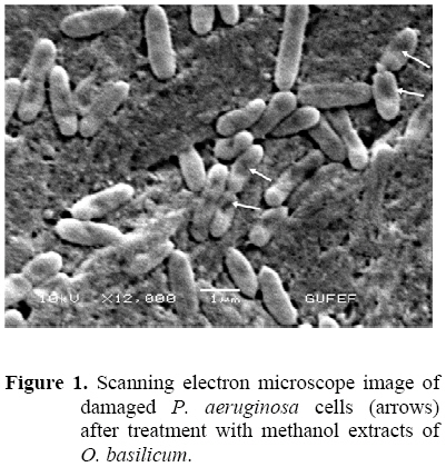
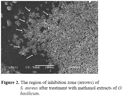
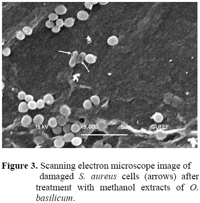
antibiotics that were tested, as it can be seen in Table 2, Shigella sp., P. aeruginosa, Escherichia coli ATCC 25922, Escherichia coli were resistant to a variety of standard antibiotics tested but methanol extract of Ocimum had antimicrobial activities on these microorganisms. On the other hand, L. monocytogenes and S. aureus were sensitive to a variety of antibiotics as well as O. basilicum methanol extract. Consequently, O. basilicum has an inhibitory activity on these bacteria that is as effective as antibiotics. It would be possible to produce new effective antibiotics using the effective material to be refined from this plant. Scanning electron microscope observations To observe morphological alteration after the cell was treated with plant extracts we used scanning electron microscope. When the bacterial cells treated with plant extracts were compared with untreated cells, the treated cells appeared to be shrinking and there was a degradation of the cell walls (Figures 1, 2, 3). As the case is in our study, Burt and Reinders (2003) found that oregano and thyme essential oil exhibit strong antimicrobial properties against E. coli O157:H7 and observed cells were damaged when treated with essential oil using SEM and the damage in the cells was similar. Burt (2004) tried to explain the mechanism of action for essential oil components in bacterial cells. The mechanism of action was thought to be the degradation of the cell wall, damage to cytoplasmic membrane proteins, the binding of proteins, leakage of cell contents, and coagulation of cytoplasm and depletion of the proton motive force. All these findings indicate that O.basilicumextracts possess antibacterial activity and they cause lysis and eradicate bacteria by degrading bacterial cell walls. By the findings and purification of the active agent that is present in the extract of O.basilicum, it will be possible to discover new natural drugs serving as chemotherapeutic agents for treatment with nosocomial pathogens and could control antibiotic-resistant bacteria. This study will be a base to our further investigations on advanced purification and effect mechanism of its active compounds. Conclusions The methanol (suspended with 5 ml deionized water) extracts of O.basilicum showed antimicrobial activity against the test microorganisms and caused degradation of the cell walls of sensitive bacteria. References
© Copyright 2008 - African. Journal. Traditional, Complementary and Alternative Medicines The following images related to this document are available:Photo images[tc08050f3.jpg] [tc08050f2.jpg] [tc08050f1.jpg] | |||||||||||||||||||||||||||||||||||||||||||||||||||||||||||||||||||||||||||||||||||||||||||||||||||||||||||||||||||||||||||||||||||||||||||||||||||||||||||||||||||||||||||||||||||||||||||||||||||||||||||||||||||||||||||||||||||||||||||||||||
| |||||||||
