 |
| About Bioline | All Journals | Testimonials | Membership | News |
|
||||||
|
||||||
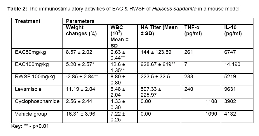
African Journal of Traditional, Complimentary and Alternative Medicines, Vol. 5, No. 4, 2008, pg. 394 - 398 Research PaperToxicity and immunomodulatory activity of fractions of Hibiscus sabdariffa linn (Family Malvaceae) in animal models Titilayo Fakeye Department of Clinical Pharmacy & Pharmacy Admin, University of Ibadan, Nigeria E-mail: titifakeye@yahoo.com Code Number: tc08055 Abstract This study evaluated immunomodulatory properties and the sub-acute toxicity profile of two fractions of the aqueousalcoholic extract of the dried calyx of Hibiscus sabdariffa in experimental animals. Immunomodulatory activity was evaluated using red blood cell-induced immunostimulation. The fractions were not found to be toxic after 7-day administration, though there was severe weight loss with the residual water-soluble fraction (RWSF) and weight gain with the ethyl acetate soluble fraction (EAC). The EAC exhibited a significant dose-dependent immunostimulation (p<0.05)higher than thatobserved for levamisole (positive control). The residual water-soluble fraction exhibited immunostimulatory activity at 100mg/kg body weight. The two fractions caused a significant reduction in production of tissue necrosis factor – alpha and an increase in interleukin 10 (IL-10). Keywords: Hibiscus sabdariffa, plant extract and fractions, sub-acute toxicity, immuno-modulatory activity,cytokine production Introduction The extracts and some of the constituents of dried calyx of Hibiscus sabdariffa Linn (family Malvaceae) have been reported to possess impressive antioxidant and antihypertensive activities in animal models and human, (Tseng et al., 1997; Tseng et al., 1998; Ali et al.., 2003; Lin et al., 2003; Odigie et al.., 2003; Herrera-Arellano et al., 2004; Amin and Hamza, 2005), and in vitro antimicrobial and in vivo anti-inflammatory activity (Ogundipe et al., 1998). The extracts were found at low doses to cause inhibition of serum lipids with anti-atherosclerotic activity (Chen et al., 2003) and induction of apoptosis (Hou et al., 2005; Chang et al, 2005; Lin et al., 2005). Chronic oral administration of the water and alcoholic extracts of Hibiscus sabdariffa had been found toxic in a previous study affecting liver function enzymes, serum creatinine and causing muscular dystrophy and significant weight loss in mice at 300mg and 2000mg/kg body weight doses, while the alcoholic and water extracts possess significant immunostimulatory and anxiolytic activities, and cause significant increase in ketamine-induced sleep in mice (Fakeye et al., In press, 2008). This study was therefore designed to evaluate the immunomodulatory properties of two fractions of the crude aqueousalcoholic extract of the dried calyx of Hibiscus sabdariffa. The sub-acute toxicity profile of the doses for the immunomodulatory activity was also evaluated. Methodology The dried flowers of Hibiscus sabdariffa L (family Malvaceae) were purchased at the local market and further dried at 40 °C until a constant weight was obtained. The material was authenticated at the Forestry Research Institute of Nigeria (FRIN), Ibadan. A herbarium species (reference number FHI 107622) was submitted for future reference.The dried calyx was pulverized to obtain a coarsely powdered material. One liter 50% ethanol (water/ethanol - 50:50) was used to infuse 100g of the powdered plant material for four hrs. The extract obtained was decanted and the material was re-extracted with another one L. The extracts obtained were pooled, filtered and dried in vacuo using a rotary evaporator. The extract was further partitioned successively into dichloromethane (Merck KGaA, Darmstadt, Germany), ethyl acetate (Thomas Baker Chemicals PVT Ltd, Mumbai, India) and butanol (Thomas Baker Chemicals PVT Ltd, Mumbai, India). The fractions obtained were dried in vacuo and the yield was noted. Sub-acute toxicity tests Male swiss albino mice obtained from the animal house of Central Institute of Medicinal and Aromatic Plants (CIMAP), Lucknow, with weight ranging from 18 – 24 g (16.25 ± 9.85 g) were kept in laboratory cages. Feed and water were allowed ad libitum and the animals were maintained in standard environmental conditions (temperature 27 ± 1.5 0C; humidity 73 ± 2.3%) throughout the period of the study. Different concentrations of residual water-soluble fraction (RWSF) and the ethyl acetate-soluble fractions (EAC) were prepared in 20% Tween 80. To three groups of four male swiss albino mice each were administered 50, 100 mg/kg of the ethyl acetate-soluble fraction or 100 mg/kg body weight of the residual water-soluble fraction daily for seven days with oral feeding needle. Another group of four male mice were administered 0.8 ml of 20% Tween 80 as control. The weight of each animal was monitored over a 7-day observation period. The animals were closely monitored the first 24 hr post administration of the fractions. Thereafter, a daily observation was kept for seven days. Blood (0.7 ml) was obtained through the medial canthus of the mice seven days after administration of the fractions. Erythrocyte, total leukocyte and differential leukocyte count were determined using standard methods. Plasma was obtained by centrifuging heparinised blood at 2500 rpm for 10 minutes. The plasma obtained was analysed for glucose, triglycerides and creatinine using a RA-50 standardised Clinical Chemistry System (RA232C, Serial No 30650, Bayer Diagnostics Mfg. Ltd., Swords, Co. Dublin, Ireland). All experimental protocols were in compliance with CIMAP institutional Ethical Committee Guidelines as well as internationally accepted principles for laboratory animal use and care as found in US guidelines (NIH publication #85-23, revised in 1985 Immunomodulatory tests This activity was evaluated using red blood cell-induced immunostimulation in swiss albino mice weighing 18-24 g (16.25 ± 9.85 g). Residual water soluble fraction (RSWF) at 100 mg/kg body weight and ethyl acetate-soluble fraction (EAC) at 50 and 100 mg/kg body weight were administered to groups of mice each with oral gavage needle daily for 28 days. Levamisole (0.6818 mg/kg) administeredorally was used as positive control, while the vehicle-control group was administered 0.5 ml of water daily. The negative control group was administered intraperitoneal cyclophosphamide (200 mg/kg) on day 5. On day 7, 200 µl of 10% of freshly prepared New Zealand rabbit red blood cell (RRBC) suspension in normal saline was administered intra-peritoneally. This was repeated 14 days later as a booster dose. On day 28, all the animals were bled and the serum collected was stored at -20 oC for further studies. Body weights of the animals were monitored weekly. To evaluate the ability of the fractions to enhance or diminish the formation of antibodies to the RRBC, haemaglutination test was performed by adding 100 µl of rabbit RBC (6 X 10³ cells per ml) to 25 µl of the serial two fold dilutions of the serum in Alsever’s solution in U-bottom microtitre plates. This was shaken and allowed to stand for 4 hr at 25 oC. Rabbit RBC-setting patterns were then read. The haemaglutination (HA) titer was expressed as the reciprocal of the highest dilution of the serum showing definite agglutination formation as opposed to smooth dot in the center of the well. Cytokine determination Previous studies have showed that extracts (Chen et al., 2003) and some of the fractions of the dried calyx possess anti-inflammatory activity in carrageenan-induced rat paw model, and were able to reduce production of tissue necrosis factor-α (TNF-α) and increase interleukin-10 (IL-10) production (implicated as pro-inflammatory and anti-inflammatory interleukins respectively). Enzyme-linked immunosorbant assay (ELISA) for the quantitative measure of mouse TNF-α, and IL-10 (Endogen(R) Mouse ELISA kit, Pierce Biotechnology, Inc, Rockford) was used to evaluate the effects of the fractions on the production of the two cytokines. Sera from animals given the same treatment were pooled. A 50 µl of the pooled sera (from animals which had received the same treatment) or standard concentrations of lyophilized recombinant mouse tissue necrosis factor- alpha was added to biotin-labelled detecting antibody reagent in TNF-α precoated wells. Incubation was done for 2 hr at 37 oC. Thereafter, a 100 µl dilution of 1:400 streptavidin conjugated with highly purified horseradish peroxidase was added to each well and further incubated for 30 mins. For determination of IL-10, 50 µl assay buffer was added to each endogen mouse interleukin-10 pre-coated well. The same amount of standards (containing known concentrations of lyophilized recombinant mouse IL-10) or samples from the pooled sera was added to the wells and incubated at 37 oC for 3 hr. After washing the plates, 50 µl of biotin-labelled detecting antibody reagent was added to each well, incubated for 1 hr, after which 100 µl of 1:400 diluted streptavidin-horse radish peroxidase was added and incubated for 30 mins. To both IL-10 and TNF-α plates, 100 µl of premixed 3,3/,5,5/-tetremethylbenzidine substrate solution was added to each well and developed in the dark for 30 mins. The blue color obtained was stopped with 100 µl of 0.18M sulphuric acid. The difference in absorbance was measured using a spectrophotometer plate reader (Versamax, Molecular Devices, USA) at 450 nm and 550 nm. The difference in absorbance was read off the calibration curve obtained from the standard concentrations. The concentration of each cytokine was determined in picogram per milliliter of serum. Results and Discussion Fractionation of the crude aqueous alcoholic extract gave a yield of 5% and 18.5% for ethyl acetate-soluble and residual water-soluble fractions respectively. The fractions were generally found non-toxic at the dose used to evaluate the immunomodulatory properties in the animals. However, the residual water-soluble fraction, RWSF, caused a severe weight loss when compared with the control animals. Table 1 shows the toxic effects of the fractions on some biochemical and hematological parameters. The ethyl acetate-soluble fraction, EAC, was found to cause an increase in the total leucocyte count at 100 mg/kg (p<0.05) but with a reduction at 50 mg/kg. The two fractions caused a significant reduction in basophils,the white blood cells responsible for antigen-antibody response. The RWSF caused a reduction in neutrophil count which deals with defense against bacterial infections. There was no change with the lymphocyte count when compared with the control group even though results obtained showed that the fractions enhanced the antigen-antibody reaction to the RRBC antibodies. The EAC soluble fraction exhibited a significant dose-dependent activity in cytokine production with more than a 6-fold increase in IL-10 production activity with a double increase in dose (100 mg/kg versus 50 mg/kg; Table 2). The EAC at 100 mg/kg caused a profound reduction in the production of TNF-α while the RWSF (100 mg/kg) and EAC at 50 mg/kg compared with levamisole, the positive control, in the production of TNF-α. Concentrations of the TNF-α in the serum of the experimental animals, though not exhibiting dose-dependence in any of the test groups, was lowest with EAC 100 mg/kg. Previous studies have implicated high levels of TNF-α in clinical diagnosis of atherosclerosis. The noticeable reduction in production of TNF-α in this study by EAC and RWSF confirm the activity of extracts of Hibiscus sabdariffa reducing atherosclerosis in an animal model (Chen et al., 2003). Interleukin 10 (IL-10) was highest in the serum of animals on EAC 100 mg/kg. The interplay of the reduction in concentration of TNF-α and increase in IL-10 has been linked with stress, inflammation and tumors. IL-10 has been known to down regulate factors present in inflammation Table 1: Biochemical and hematological effects of sub-acute administration of fractions of Hibiscus sabdariffa in mice
Key: CRT – Creatinine; TRIG
– Triglycerides; GLUC - Glucose and tumors. An extract or chemical compound that could increase anti-inflammatory interleukin, for example, IL-10, and reduce the formation of a pro-inflammatory factor such as TNF-α, as EAC has done, may be found to be of use as a source of drug candidate for preventing or reducing formation of atherosclerotic plaques and may also help in reduction of stress and possess antitumour activity. The low level and high level of TNF-alpha and IL-10 respectively confirmed that the fractions tested may also stimulate immunomodulation through the activities of cytokines. Conclusion Multiple doses of the fractions of the aqueous alcoholic extracts of the dried calyx of Hibiscus sabdariffa were not found to be toxic at the doses found to possess immunostimulatory activities. The fractions possess impressive immunostimulatory activities with a profound increase in production of an anti-inflammatory cytokine, IL-10 and a great reduction in production of tissue necrosis factor – alpha. The two fractions, EAC and RWSF, showed good possibilities of being developed into drug entities that may be used to stimulate immunity as an adjunct to therapy in immunosuppressed disease conditions. References
© Copyright 2008 - African. Journal. Traditional, Complementary and Alternative Medicines The following images related to this document are available:Photo images[tc08055t2.jpg] [tc08055t1.jpg] | ||||||||||||||||||||||||||||||||||||||||||||||||||||||||||||||||||
| |||||||||
