 |
| About Bioline | All Journals | Testimonials | Membership | News |
|
||||||
|
||||||
African Journal of Traditional, Complimentary and Alternative Medicines, Vol. 6, No. 1, 2009, pp. 1-8 Protective effect of Withaferin-A on micronucleus frequency and detoxication agents during experimental oral carcinogenesis Kuppusamy Panjamurthy , Shanmugam Manoharan, Subramanian Balakrishnan, Kathiresan Suresh, Madhavan R. Nirmal, Namasivayam Senthiland Linsa Marry Alias 1Department of Biochemistry & Biotechnology,
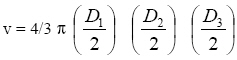
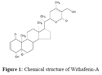
Faculty of Science, Code Number: tc09001 AbstractOur aim was to investigate the effect of Withaferin-A on bone marrow micronucleus frequency and buccal mucosa detoxication agents during 7, 12–dimethylbenz[a]anthracene (DMBA) induced hamster buccal pouch carcinogenesis. Oral squamous cell carcinoma was developed in hamsters’ buccal pouches by painting 0.5% DMBA in liquid paraffin, three times per week for 14 weeks. We observed 100% tumor formation in DMBA painted hamsters. Elevated frequency of bone marrow micronucleated polychromatic erythrocytes (MnPCEs) and decrease in buccal mucosa phase II detoxication agents were noticed in tumor bearing hamsters. Oral administration of Withaferin-A significantly reduced the micronucleus frequency and brought back the status of phase II detoxication agents in DMBA painted hamsters. Our study thus demonstrated the protective effect of Withaferin-A on DMBA-induced micronucleus frequency in the bone marrow of golden Syrian hamsters. Also, Withaferin-A maintained the status of buccal mucosa detoxication agents during DMBA-induced hamster buccal pouch carcinogenesis. Key words: Withaferin-A, DMBA, Oral cancer, Micronucleus, Detoxication Abbreviations: DMBA - 7,12–dimethylbenz[a]anthracene; MnPCEs - micronucleated polychromatic erythrocytes; PCEs - polychromatic erythrocytes; NCEs - Normochromatic erythrocytes; GST - Glutathione-S-transferase; GSSG – Oxidized glutathione; GSH-reduced glutathione. Introduction Cancer of the oral cavity results due to repeated exposure of carcinogens to the entire epithelial cells of the oral cavity. 7, 12–dimethylbenz[a]anthracene (DMBA) is the most widely used organ specific carcinogen to induce oral carcinogenesis in the buccal pouches of golden Syrian hamster. The pre-cancerous and cancerous lesions induced by this potent carcinogen are morphologically, histologically and biochemically similar to that of human oral carcinoma. Also, DMBA induced oral cancer expresses similar molecular changes that are expressed in human oral carcinoma (Miyata et al., 2001; Hodgson, 2000; Senthil et al., 2007). Cytogenetic markers such as micronuclei frequency are relatively rapid, facile and sensitive indicators of genetic damage. Micronuclei could be formed from either lagging anaphase chromosome or anaphase bridges that can be detected in the cytoplasm of a daughter cell after cell division (Morita et al., 1997). Bone marrow micronucleus test has been widely used as a tool to indicate carcinogen-induced DNA damage as well as to assess the antigenotoxic effect of natural and synthetic chemopreventive agents (Miyamoto et al., 2007). Several studies have reported that phytochemicals inhibit neoplastic transformation by inducing phase II defoliation agents. Glutathione-S-Transferase (GST) detoxifies carcinogens either by destroying their reactive centers or facilitating their excretion by conjugation process (Dasgupta et al., 2004). Withania somnifera (Solanaceae), a shrub commonly found on the Indian subcontinent, has been used for centuries as a traditional medicine for various human ailments. Experimental studies have reported that W. somnifera have antitumor and immunomodulatory activities (Davis and Kuttan, 2000, Davis and Kuttan, 2001). Withaferin-A, a highly oxygenated steroidal lactone, is the principal withanolide in Indian W. somnifera and its related Solanaceae species. Diverse pharmacological activities reported for Withaferin-A includes anti-inflammatory, antitumor and antioxidant properties (Sharada et al., 1996; Bhattacharya et al., 1997). The chemical structure of Withaferin-A is given in Figure 1. To the best of our knowledge, we have found no scientific literature on modifying effects of Withaferin-A on bone marrow micronucleus frequency and buccal mucosa detoxicating agents during DMBA induced hamster buccal pouch carcinogenesis. The present study was thus designed to assess the protective role of Withaferin-A on bone marrow micronucleus frequency and buccal mucosa detoxicating agents during DMBA induced hamster buccal pouch carcinogenesis. Materials and Methods Animals Male golden Syrian hamsters, 8-10 weeks old weighing 80-120g, were purchased from National Institute of Nutrition, Hyderabad, India, and maintained in Central Animal House, Rajah Muthiah Medical College and Hospital, Annamalai University. The hamsters were housed four or five in a polypropylene cage and provided standard pellet diet and water ad libitum. The hamsters were maintained under controlled conditions of temperature and humidity with a 12h light/dark cycle. The hamsters were maintained as per the principles and guidelines of the ethical committee for animal care of Annamalai University in accordance with Indian National Law on animal care and use. The experimental design was approved by the Annamalai University animal ethical committee [Reg. No: 160/1999/CPCSEA], Annamalai University, Annamalainagar, India. Chemicals 7,12-dimethylbenz[a]anthracene (DMBA), Colchicine, Giemsa and May-Grunwald’s stain were purchased from Sigma-Aldrich Chemical Pvt. Ltd., Bangalore, India and all other reagents used were of analytical grade. Isolation of Withaferin-A from Withania somnifera root Withaferin-A (WA) was extracted and isolated from commercially available Withania somnifera root powder by the method of Subramanian and Sethi (1969). The crude extract was prepared with 95% ethanol and further fractionation was carried out using petroleum ether, ether and chloroform, in that order. The ether and chloroform fractions were subjected to column chromatography (neutral alumina) and thin layer chromatography (silica gel). The final product, Wihaferin-A, a steroidal lactone (4β, 27 dihydroxy-1-oxo-5β, 6β, epoxy witha 2-24 dienolide) was obtained as a creamy white crystalline substance, which has Rf = 0.4 and molecular weight 470. The identity of the isolated Withaferin-A was done by Mass spectral analysis and its identity was confirmed by comparing with the authentic Withaferin-A, purchased from Calbiochem, Darmstadt, Germany. The yield and purity of the isolated Withaferin-A was found to be 0.11% and >90% respectively. For experimental studies, the obtained Withaferin-A was first dissolved in a few drops of absolute ethanol followed by dilution with 30% polyethylene glycol-400 (PEG-400) in phosphate-buffered saline (PBS). Experimental design A total number of 40 hamsters were randomized into four groups of 10 hamsters each. Group 1 hamsters were served as control and were painted with liquid paraffin thrice a week for 14 weeks on their left buccal pouches. Groups 2 and 3 hamsters were painted with 0.5% DMBA in liquid paraffin thrice a week for 14 weeks on their left buccal pouches. DMBA painting in hamsters’ buccal pouches was done using number 4 painting brush, which leaves approximately 0.4mg DMBA for each application (Shklar, 1999). Group 2 hamsters received no other treatment. Group 3 hamsters were orally given Withaferin-A at a dose of 20mg/kg bw, starting one week before the exposure to the carcinogen and continued on days alternate to DMBA painting, until the hamsters were sacrificed. Group 4 hamsters received oral administration of Withaferin-A alone throughout the experimental period. The experiment was terminated at the end of 14 weeks and all hamsters were sacrificed by cervical dislocation. The numbers of tumors in the hamsters’ buccal pouches were counted. The diameter of each tumor was measured with a caliper. The tumor volume was calculated by the formula,
where, D1, D2 and D3 are the three diameters (mm) of the tumor. Tumor burden was calculated by multiplying tumor volume and the number of tumors/hamster. Bone marrow micronucleus testBone marrow micronucleus test was carried out according to the method of Schmid (1975). The femur bones removed from the hamsters were cleaned and the content was flushed into tube containing 1 ml of calf serum and was centrifuged at 500 g for 10 mins. The pellet obtained was suspended with few drops of fresh serum and slides were prepared and air-dried for 18 hours. After drying, the slides were stained with May Grunwald stain followed by Giemsa stain. The frequency of MnPCEs in each group was calculated by scoring 2500 polychromatic erythrocytes (PCEs) per hamster. Biochemical estimations Biochemical studies were conducted on buccal mucosa of control and experimental hamsters in each group. Tissue samples from hamsters were washed with ice cold saline and homogenized using appropriate buffer [Glutathione-S-Transferase (GST) – 0.3 M phosphate buffer, PH 6.5; GSH – 0.4 M phosphate buffer, pH – 7.0] in an all glass homogenizer with teflon pestle and used for biochemical estimations. The reduced glutathione level in the buccal mucosa was determined by the method of Beutler and Kelley (1963). The technique involves protein precipitation by meta-phosphoric acid and spectrophotometric assay at 412nm of the yellow derivative obtained by the reaction of the supernatant with 5-5’dithiobis-2-nitrobenzoic acid. The activity of glutathione-S-transferase (GST) in buccal mucosa tissue homogenate was assayed by the method of Habig et al (1994). GST activity was measured by incubating the tissue homogenate with the substrate 1-chloro 2,4 dinitrobenzene (CDNB). The absorbance was followed for 5 mins at 540nm after the reaction was started by the addition of reduced glutathione. The level of oxidized glutathione (GSSG) was estimated by the method of Anderson (1985). This method is based on oxidation of reduced nicotinamide adenine dinucleotide phosphate by glutathione, which was read at 340nm. Statistical Analysis The data are expressed as mean ± SD. Statistical comparisons were performed by one way analysis of variance (ANOVA), followed by Duncan’s Multiple Range Test (DMRT). The results were considered statistically significant if the p values were 0.05 or less. Results Table 1 shows the tumor incidence, tumor volume and tumor burden of control and experimental hamsters in each group. We have observed 100% tumor formation with mean tumor volume (384.10 mm³) and tumor burden (1344.35 mm³) in DMBA alone painted hamsters (Group 2). Oral administration of Withaferin-A at a dose of 20mg/kg bw for 14 weeks completely prevented the tumor incidence, tumor volume and tumor burden in DMBA painted hamsters (Group 3). No tumors were observed in control hamsters painted with liquid paraffin alone (Group 1) as well as Withaferin-A alone administered hamsters (Group 4). The frequency of MnPCEs in control and experimental hamsters in each group are given in Table 2. Hamsters painted with DMBA alone (group 2) for 14 weeks showed higher frequency of MnPCEs as compared to control hamsters. The frequencies of MnPCEs were significantly reduced in DMBA painted hamsters treated with Withaferin-A Orally (group 3) for 14 weeks. Oral administration of Withaferin-A alone (group 4) to hamsters displayed no significant differences in MnPCEs as compared to control hamsters (group 1) Table 3 shows the status of GSH, GSSG and GST in the buccal mucosa of control and experimental hamsters in each group. The status of GSH and GST were significantly increased whereas GSSG was decreased in tumor bearing hamsters (Group 2) as compared to control hamsters. Oral administration of Withaferin-A to DMBA painted hamsters (Group 3) significantly reverted to near normal concentrations. Hamsters painted with Withaferin-A alone (Group 4) showed no significant difference in the status of GSH, GSSG and GST as compared to control hamsters (Group 1). Table 1. Incidence of oral neoplasms in control and experimental hamsters in each group (n = 10)
Tumor volume was measured using the formula v = 4/3 p
where D1, D2 and D3 are the three diameters (mm) of the tumor. Tumor burden was calculated by multiplying tumor volume and the number of tumors/hamster. ( ) indicated total number of hamsters bearing tumors. Values are given as mean ± SD. Table 2. Effect of Withaferin-A on DMBA induced bone marrow micronuclei formation
Values are expressed as mean
± SD (n =10; 2500 PCEs were scored per hamster). Table 3: Activities of detoxication agents in buccal mucosa of control and experimental hamsters in each group (n = 10)
Values are expressed as mean ± SD for 10 hamsters in each group. Values not sharing a common superscript letter differ significantly at P<0.05 (DMRT). Discussion Chemopreventive agents exert their tumor suppressive effect by preventing metabolic activation of carcinogens, stimulating detoxication of the carcinogens, blocking the interaction of ultimate carcinogen with cellular macromolecules or by suppressing the clonal expression of the neoplastic cells (Aruna and Sivaramakrishnan, 1990).In the present study, we have investigated the modifying effects of Withaferin-A on bone marrow micronucleus frequency and buccal mucosa detoxication agents during DMBA-induced hamster buccal pouch carcinogenesis. Withaferin-A completely prevented the tumor formation in DMBA painted hamsters, which is probably due to its suppressive effect on cell proliferation. Genotoxic agents such as carcinogens can enhance the error rate in the genome reduplication and cause mutation in the DNA of an organism. DMBA exposure result in a marked increase in tumor burden and tumor volume in rodent models and pronounced mutagenic response in several in vivo and in vitromutation assay system (Chang et al., 1996). DMBA on metabolic activation produces dihydrodiol-epoxides, which mediate neoplastic transformation by causing chromosomal abnormalities and mutations in key growth regulatory genes (Miyata et al., 2001; Hodgson, 2000; Senthil et al., 2007). DMBA can oxidize both DNA bases and deoxyribose sugar through its active metobolite diol epoxides (Miyata et al., 2001). N-ras mutation, Ha-ras mutation and A-T transversion in H-ras codon 61 were demonstrated in DMBA induced experimental oral carcinogenesis (Osaka et al., 1996; Chang et al., 1996). Several studies have also documented increased MnPCEs frequency during DMBA-induced hamster buccal pouch carcinogenesis (Bhuvanesvari et al., 2005; Chandramohan et al., 2006). Our results lend credence to these observations. Oral administration of Withaferin-A to DMBA painted hamsters significantly reduced the frequency of MnPCEs. Some studies have demonstrated that Withaferin-A has potent anti-inflammatory, antioxidant and antitumor properties (Yang et al., 2007; Bargagna-Mohan et al., 2007; Bhattacharya et al., 1997). Our results indicate that Withaferin-A might have protected DNA damage by inhibiting the metabolic activation of DMBA. Phase II detoxication agents play a vital role in detoxication of pro- and ultimate carcinogens and thereby reducing DNA damage (Senthil et al., 2007). Altered activities of liver and buccal mucosa phase II detoxication agents are well documented in experimental oral carcinogenesis (Schwartz and Shklar, 1996; Chandramohan et al., 2006). Our results corroborate these observations. The status of phase II detoxication agents in the buccal pouch reverted to near normal in DMBA painted hamsters, after treatment with Withaferin-A. Our results indicate that Withaferin-A probably modulated the activities of phase II detoxication agents to prevent the mutagenic and carcinogenic effect of the carcinogen, DMBA. Conclusion Our study thus demonstrates the protective effect of Withaferin-A on DMBA-induced micronucleus frequency in the bone marrow of golden Syrian hamsters. Also, Withaferin-A maintained the status of buccal mucosa detoxication agents during DMBA induced hamster buccal pouch carcinogenesis. Acknowledgements Financialsupport from Indian Council of Medical Research (ICMR), New Delhi to Mr. K. Panjamurthy, in the form of Senior Research Fellowship (SRF) is gratefully acknowledged. We thank Dr. S. Narasimhan and Dr. R. Mohan Kumar, Asthagiri Herbal Research Fundation, Chennai for their valuable assistance in isolating Withaferin-A from W. somnifera root powder. References
© Copyright 2009 - African. Journal. Traditional, Complementary and Alternative Medicines The following images related to this document are available:Photo images[tc09001f1.jpg] |
| |||||||||

