 |
| About Bioline | All Journals | Testimonials | Membership | News |
|
||||||
|
||||||
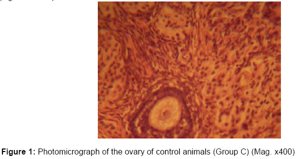
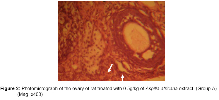
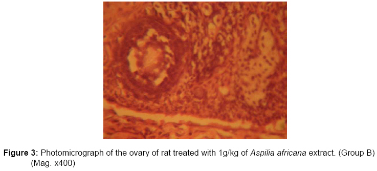
African Journal of Traditional, Complimentary and Alternative Medicines, Vol. 6, No. 1, 2009, pp. 57-61 Histological studies of the effects of oral administration of Aspilia africana (Asteraceae) leaf extract on the ovaries of female wistar rats A.O. Eweka Department of Anatomy, School of Basic Medical Sciences, University of Benin, Edo state, Nigeria.+E-mail: andreweweka@yahoo.com Code Number: tc09008 Abstract Histological studies of the effects of oral administration of extract of Aspilia africana, used in ethno medical practice in Africa for the management of various ailments, on the ovarian tissues of matured female Wistar rats were studied. The rats (n=24), average weight 182g were randomly assigned into two treatment (n=16) and a control (n=8) group. The rats in the treatment groups received 0.5g/kg and 1g/kg of aqueous extract of Aspilia africana orally through orogastric tube for fourteen days, while the control rats received equal volume of distilled water without the extract of Aspilia added. The rats were fed with growers’ mash and were given water liberally. The rats were sacrificed on day fifteen of the experiment. The ovary was carefully dissected out and quickly fixed in 10% formal saline for routine histological study after H&E method. The histological findings indicated that the treated sections of the ovary showed cellular hypertrophy of the theca folliculi, distortion of the basement membrane, degenerative and atrophic changes in the oocyte and zona granulosa. There were marked vacuolations appearing in the stroma cells. These findings indicate that Aspilia africana consumption may probably have adverse effects on the ovaries by its deleterious effects on the oocytes and stroma cells of ovary of adult Wistar rats. It is recommended that further studies aimed at corroborating these observations be conducted. Key words: Aspilia africana, Histological effect, theca folliculi, oocyte, cellular hypertrophy, vacuolations, ovaries and Wistar rats. Introduction In ethno-medical practice, plant materials have been used as sources of medical compounds and had played a dominant role in the maintenance of human health in most rural communities in developing countries in time past. Like any therapeutic agent, when overdosed or incorrectly used they also have the potential to induce adverse effects. The historic role of medicinal herbs in the treatment and prevention of disease, and their role as catalysts in the development of pharmacology do not, however, assure their safety for uncontrolled use by an uninformed public (Matthews et al., 1999). There has been minimal research to address possible adverse reproductive, immunologic, or neurological effects or even systemic toxicity and/or carcinogenicity that might be associated with high doses or prolonged use of these products (Miller, 1998). This concern frequently expressed at the International Workshop to Evaluate Research Needs on the Use and Safety of Medicinal Herbs (1998) could not be assumed safe because they are “natural”. In Benin City, Nigeria, many plants are used in herbal medicine to treat diseases and heal injuries. Such medicinal plants include Aspilia africana (Asteraceae); (Pers) C.D. Adams, a perennial herb varying in height from 60cm to about 1.5m depending on rainfall. It is a common weed of field crops in West Africa and sometimes found in fallow land, especially the forest zones (Akobundu, 1987). It is ligneous at the base, its fruit quadrangular akenes and leaves opposite and hairy. The plant is a weed grazed by cattle and sheep and is mostly used in the western state of Nigeria as food for rabbits and hares (Burkill, 1985). A. africana is widely used in ethno medical practice in Africa for its ability to stop bleeding, even from a severed artery, as well as promote rapid healing of wounds and sores and for the management of problems related to cardiovascular diseases (Dimo et al., 2002). It has also been established that Aspillia africana has an anticoagulant activities when applied topically to wounds (Hanna and Niemetz, 1987). Infusion of a liquid made from the leaves is taken by children and can also be mixed with clay as a medicine for stomach trouble (Okwu and Josiah, 2006). It has been reported that the plant is effective against malaria (P. falciparum) infection Okokon et al (2006). It has been classified among substances with a low potential for toxicity, with an LD50 averaging 6.6g/Kg body weight (Taziebou et al., 2007). The methanolic and aqueous extracts of the leaves of A. africana have exhibited differential anti-infective activities on both Gram-positive and Gram-negative bacterial species (Macfoy and Cline, 1990; Adeniyi and Odufowora, 2000). A. africana has many other additional uses such as palliative properties as documented by Okoli CO et al that the leaves of A. africana possess constituents capable of arresting wound bleeding, inhibiting the growth of microbial wound contaminants and accelerating wound healing which suggest good potentials for use in wound care (Okoli et al., 2007a, 2007b), alleviating menstrual cramps and dysmenorrheal (which are not documented) probably because empirical studies had not been carried out on them to authenticate their efficacy. In some communities in Nigeria, women boil and filter the leave of A. africana, which they drink to prevent conception. It is, therefore, suggestive that A. africana may have some contraceptive or anti-fertility properties, The ovary is a paired, egg-producing reproductive organ found in female organisms. The ovaries also functions in the production of various steroid and peptide hormones like estrogen and progesterone which sub serve many functions in the reproductive system (Wikipedia, 2007). About 15% of cases of female infertility investigation will show no abnormality. In these cases abnormalities are likely to be present but not detected by current methods, ^American Society for Reproductive Medicine (FAQ) 2006. This work was performed in order to investigate potential histological effects of A. africana leaf extracts on the of adult female Wistar rats. Materials and Methods Plant Materials Fresh leaves of Aspilia africana (voucher number UBAD-Aa001) were collected in November, 2006 at Oluku Town in Ovia North-East local government area of Edo State. The plant was identified and authenticated at the Botany Department of the University of Benin, Benin City. The harvested fresh leaves were sun dried and ground into a fine powder. The dried material (300g) was macerated in 6 liters of distilled water for 48hrs at 4oC in a refrigerator. The extract was sieved and the juice was filtered using Whatman No 1 filter paper. The filtrate was placed in a stainless-steel tray, and concentrated in an air-circulating oven at 42oC until totally dry. The resultant extract (8g) was placed into small glass dishes and stored at 28oC in an incubator for further studies. Animals: Twenty four, (24) adult female Wistar rats with an average weight of 182g were randomly assigned into three groups A, B and C (n=8 in each group). Groups A and B (n=16) served as treatment groups while Group C (n=8) was the control. The rats were obtained and maintained in the Animal Holdings of the Department of Anatomy, School of Basic Medical Sciences, University of Benin, Benin city, Nigeria. They were fed with growers’ mash obtained from Edo feed and flour mill limited, Ewu, Edo state and given water liberally. The rats gained maximum acclimatization (2 weeks) before actual commencement of the experiment. The consent and approval for the methodology and other ethical issues concerning the work were obtained from the University of Benin Research Ethics Committee. Aspilia africana administration: The rats in the treatment groups (A and B) were given 0.5g/kg/day and 1g/kg/day extract of A. africana orally through an orogastric tube, respectively on a daily basis. The control group (C) received an equal volume of distilled water without the extract of A. africana added for fourteen days. The rats were sacrificed on the fifteenth day of the experiment. The ovaries were quickly dissected and fixed in 10% formal saline solution for routine histological assessment. The 0.5g/kg and 1g/kg extract of A. africana doses were chosen and extrapolated in this experiment based on the indiscriminate use of the plant here in Nigeria and on previous work conducted with this plant (Taziebou et al., 2007). ; Macfoy and Cline, 1990;, Adeniyi and Odufowora, 2000; Okoli et al., 2007a, 2007b not in references list; Benoit et al., 2005). Histological study: Ovarian tissues were dehydrated in an ascending grade of alcohol (ethanol 70%), cleared in xylene and embedded in paraffin wax after the method of Drury and Wallington (1980). Serial sections of 7 microns thick were obtained using a rotatory microtome. The deparaffinized sections were stained routinely with haematoxylin and eosin. Photomicrographs of the desired results were obtained using digital research photographic microscope in the University of Benin research laboratory. Results The ovaries of the control group showed normal histological features, illustrating a well defined zonal granulosa surrounding the oocyte and compact theca folliculi and the presence of some primordial follicles (Figure 1). The ovaries of the treated groups showed some cellular hypertrophy of the theca folliculi, complete distortion/destruction of the basement membrane separating the theca folliculi from the zona granulosa. Degenerative and atrophic changes were observed in the oocyte and zona granulosa; these were more pronounced in those that received 1g/kg/ of A. africana extract. There were marked vacuolations appearing in the stroma cells (Figures 2and 3). Discussion The results of the haematoxylin and eosin staining (H & E) reactions showed some cellular hypertrophy of the theca folliculi, complete distortion/destruction of the basement membrane separating the theca folliculi from the zona granulosa. Degenerative and atrophic changes were observed in the oocyte and zona granulosa; these were more pronounced in those that received 1g/kg of A. africana. There were marked vacuolations appearing in the stroma cell. The increase in cellular hypertrophy of the theca folliculi in the treatment groups as reported in this study may have been as a result of cellular proliferation; its mechanism is not yet clear. The vacuolation probably indicates the presence of mucous. Degenerative and atrophic changes which were observed in the oocyte and zona granulosa were more pronounced in the group treated with higher dose (1g/kg) of A. africana extract. It may be inferred from the present results that the higher dose and prolonged administration of A. africana extract resulted in degenerative and atrophic changes observed in the ovaries. The actual mechanism by which A. africana extract induced cellular degeneration observed in this experiment needs further investigation. Degenerative changes have been reported to result in cell death, which is of two types, namely apoptotic and necrotic cell death. These two types differ morphologically and biochemically (Wyllie, 1980). Pathological or accidental cell death is regarded as necrotic and could result from extrinsic insults to the cell such as osmotic, thermal, toxic and traumatic effects (Farber et al., 1981). In this experiment A. africana extract could have acted as toxins to the oocyte and follicular cells of the ovaries. The process of cellular necrosis involves disruption of membrane’s structural and functional integrity which was also a landmark of this experiment. In cellular necrosis, the rate of progression depends on the severity of the environmental insults. The greater the severity of insults, the more rapid the progression of cellular injury (Ito et al., 1975). The principle holds true for toxicological insults to the brain and other organs (Martins, 1984). It may be inferred from the present results that prolonged intake of A. africana extract resulted in a dose-dependent toxic effects on the ovaries. Conclusion and Recommendation In conclusion, the study revealed that A. africana extract causes some cellular hypertrophy, complete distortion/destruction of the basement membrane, degenerative and atrophic changes, and vacuolations in the cells of the ovaries. With these results, it is probable that the functions of the ovary may be adversely affected, and A. africana could be one of the factors causing female infertility by its varied use in the management of other medical conditions by alternative medical practitioners and rural dwellers. It is recommended that further studies be carried out to examine these findings. References
© Copyright 2009 - African. Journal. Traditional, Complementary and Alternative Medicines The following images related to this document are available:Photo images[tc09008f3.jpg] [tc09008f2.jpg] [tc09008f1.jpg] |
| |||||||||
