 |
| About Bioline | All Journals | Testimonials | Membership | News |
|
||||||
|
||||||
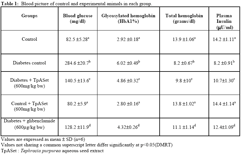
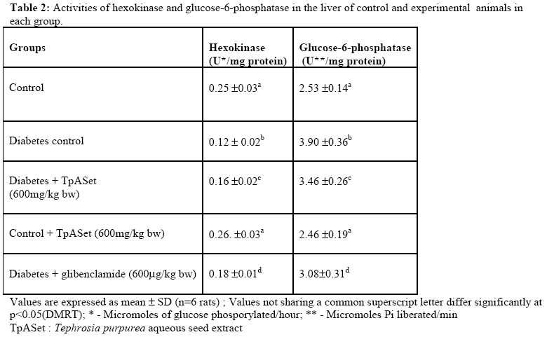
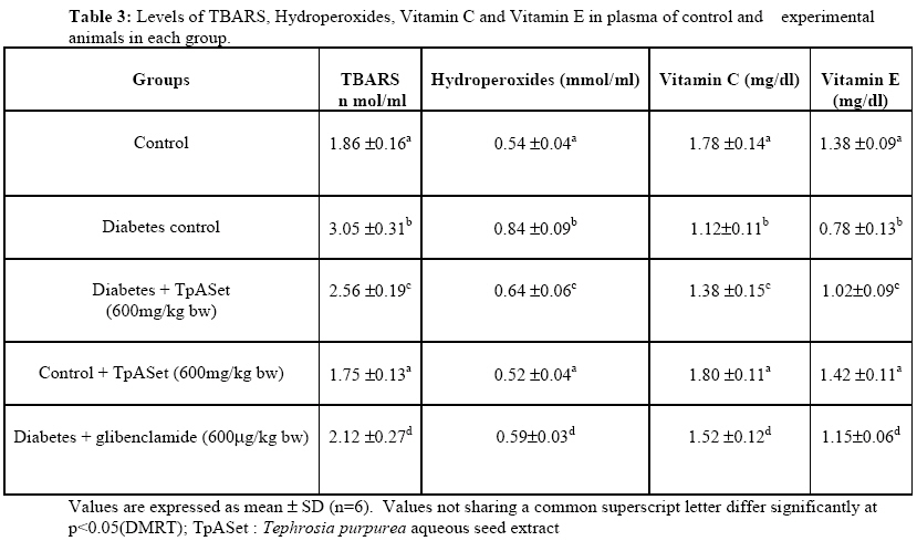
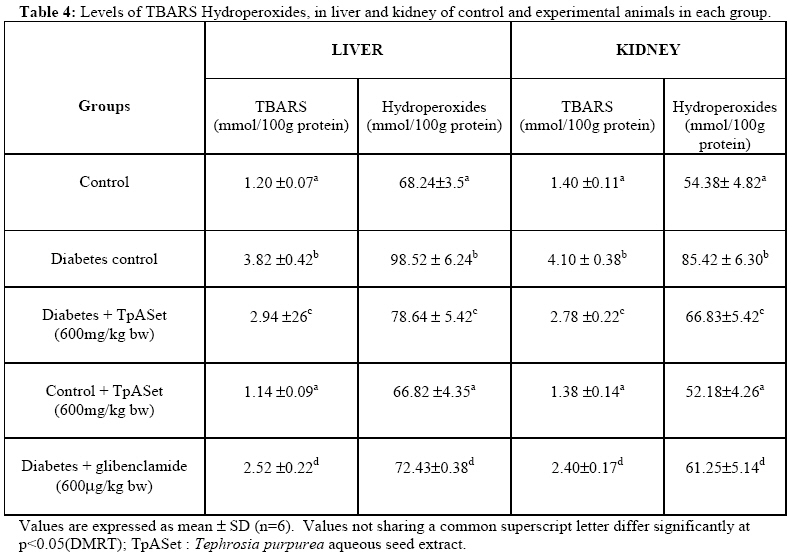
African Journal of Traditional, Complimentary and Alternative Medicines, Vol. 6, No. 1, 2009, pp. 78-86 Research Paper Effects of Tephrosia purpurea aqueous seed extract on blood glucose and antioxidant enzyme activities in streptozotocin induced diabetic rats Pavana Pb, Sethupathy Sa, Santha Ka, Manoharan Sb* aDivision of Biochemistry, Rajah Muthaiah Medical College and Hospital, Annamalai University, Annamalainagar. B. Department of Biochemistry, Faculty of Science, Annamalai University, Annamalainagar-608002 Tamilnadu. INDIA. *Email:manshisak@yahoo.com Code Number: tc09011 Abstract The aim of the present study was to evaluate the effects of aqueous seed extract of Tephrosia purpurea (TpASet) on blood glucose and antioxidant status in streptozotocin induced diabetic rats. Hyperglycemia associated with an altered hexokinase and glucose-6-phosphatase activities, elevated lipid peroxidation, disturbed enzymatic [Superoxide dismutase (SOD), catalase (CAT) and glutathione peroxidase (GPx)] and non enzymatic [Glutathione, vitamin C and vitamin E] antioxidant status were observed in streptozotocin induced diabetic rats. Oral administration of “TpASet” at a dose of 600mg/kg body weight showed significant improvement in above mentioned parameters. Our results clearly indicate that “TpASet” has potent antihyperglycemic and antioxidant effects in streptozotocin-induced diabetic rats and therefore further studies are warranted to isolate and characterize the bioactive principles from “TpASet”. Key words: Streptozotocin; Tephrosia purpurea; Antioxidants; Lipid peroxidation Introduction Diabetes mellitus is a clinically and genetically heterogeneous group of disorders, characterized by abnormally high blood glucose concentration, Hyperglycemia is due to deficiency of insulin secretion or resistance of the body cells to the action of insulin, often associated with carbohydrate, protein and lipid metabolism. These metabolic disturbances result in acute and long term diabetic complications, which are responsible for premature death and disability. According to World health organization, approximately more than 100 million peoples were reported to have diabetes mellitus worldwide (Zimmet, 1999). India is one of the leading countries with high number of people with diabetes mellitus and it is estimated that around 57 million peoples will be suffering from diabetes mellitus by the year 2025 (Aravind et al., 2002). Oxygen free radicals have been implicated in the pathogenesis of various disorders including diabetes mellitus. Oxygen free radicals are formed by auto oxidation of glucose and non enzymatic glycation of proteins. It is well documented that diabetes is associated with increased oxidative stress as evidenced by the increased accumulation of lipid peroxides (Maritim et al., 2003). Under physiological conditions antioxidant defense system protects the body against adverse effects of free radical generation. In diabetes mellitus hyperglycemia may depress the natural antioxidant system. Elevated generation free radicals resulting in the consumption of antioxidant defense components may lead to disruption of cellular functions and oxidative damage to membranes and may enhance susceptibility to lipid peroxidation. Several reports indicate that modified oxidative stress is due to chronic hyperglycemia (Bhor et al., 2004). Enzymatic antioxidants form the first line of antioxidant defense mechanism to protect the organism from reactive oxygen species mediated oxidative damage (Santhakumari et al., 2003). Enhanced oxidative stress has been well documented in both experimental and human diabetes mellitus (Baynes, 1991). Streptozotocin induced diabetes mellitus is an ideal model to study the beneficial effects of investigating agents. World health organization has suggested that medicinal plants may provide valuable therapeutics agents in modern medicine and in traditional system, especially in areas where the modern drugs are unavailable (World Health Organization, 1994). Oral hypoglycemic agents such as metformin and sulphonylureas have to be given through out life time and produce side effects like hypoglycemia (Dixit and Joshi, 1985). Folk medicine in India describes several kinds of indigenous herbs and plants belonging to various families to be recommended for the treatment of diabetes mellitus. There are several reports which strongly recommended for hypoglycemic and antihyperglycemic effects of different medicinal plants (Bordia et al., 1997); Upadhya et al., 2004). Tephrosia purpurea, commonly known as “Sarapunkha” in Sanskrit and “Purple tephrosia” in English, is widely used in Ayurveda and Siddha medicines to treat various disorders like jaundice, kidney disorders and to reduce thirst in diabetes mellitus (Joshi, 2000; Kritikar and Basu, 1956). However, we have not found any studies on the antihyperglycemic and antioxidants effects of T. Purpurea in experimental diabetes mellitus. Thus, in the present study, we have evaluated the antihyperglycemic and antioxidant effects of T. purpurea in streptozotocin induced diabetic rats. Materials and Methods Plant material Seeds of T. purpurea were collected in and around Chidambaram, Tamil Nadu and it was botanically authenticated. A voucher specimen (AU05102) was deposited in the Department of Botany, Annamali University, Annamalai nagar, Tamilnadu. Preparation of plant extract About 100g of fine powder of T. purpurea seeds was suspended in 250 ml of water for 2 hrs and then heated at 60-65oC for 30 min. The extract was preserved and the process was repeated three times with the residual powder, each time collecting the extract. The collected extract was pooled and passed through fine cotton cloth. Filtrate upon evaporation at 40-50oC yielded 13% semisolid extract. This was stored at 0-4oC until used. Animals Albino Wistar male rats 7 to 8 weeks old and weighing 150-200g were used for the present study. The animals were obtained from central animal house, Rajah Muthiah Institute of Health Sciences, Annamalai University, India and were housed in the central animal house with 12 light and 12 h dark cycles. The animals were randomized into experimental and control groups and housed 4 or 5 in a polypropylene cages. Standard pellets obtained from Mysore Snack Feed Ltd, Mysore, India, were used as a basal diet during the experiment. The control and experimental animals were provided food and drinking water ad libitum. We got ethical committee clearance for our animal experimental design (House registration number 160/1999/CPCSEA and approval date 25-08-2003). Induction of diabetes mellitus Diabetes mellitus was induced in Wistar rats by single intraperitoneal injection of streptozotocin (50mg/kg bw) dissolved in 0.1M citrate buffer (pH 4.5) to overnight fasted Albino Wistar rats (Chang, 2000). The diabetes was assessed in streptozotocin induced rats by determining the blood glucose concentration, 48 hours after injection of streptozotocin. The rats with blood glucose level above 250 mg/dl were selected for the experimental studies. In the experiment, a total number of 30 rats (18 diabetic rats, 6 normal rats 6 normal rats treated with “TpASet” alone) were used. The rats were divided into 5 groups of six each. Group I : Served as control rats
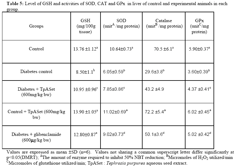
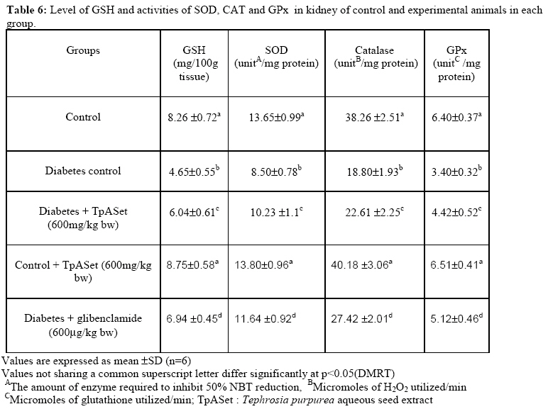
and did not receive any other treatment After the experimental period, all animals were sacrificed by cervical dislocation and biochemical studies were conducted on plasma, liver and kidney of control and experimental animals in each group. Blood glucose (Sasaki et al., 1972) glycosylated hemoglobin (Sudhakar Nayak. and Pattabiraman, 1981), total hemoglobin (Drabkin and Austin, 1932) and plasma insulin (Anderson et al., 1993) were estimated. TBARS in plasma (Yagi, 1987) and tissues (Ohkawa et al., 1979) were assayed. Hydroperoxides (Jiang et al., 1992) was estimated. Superoxide dismutases (Kakkar et al., 1984), catalase (Sinha, 1972) and glutathione peroxidase (Rotruck et al., 1984) were estimated and reduced glutathione (Beutler and Kelly, 1963), vitamin C (Omaye et al., 1979) vitamin E (Desai et al., 1984) were estimated. Hexokinase (Brandstrup et al., 1957) and glucose-6-phosphatase (Koida and Oda, 1959) activities in liver were estimated. Analysis The data were expressed as mean±SD. Statistical comparisons were performed by one way analysis of variance [ANOVA] followed by Duncan’s multiple range test [DMRT]. The results were considered statistically significant if the p values were 0.05 or less. Results Table 1 shows the blood picture of control and experimental animals in each group. The levels of blood glucose and glycosylated hemoglobin were significantly increased (p<0.05) whereas the levels of plasma insulin and total hemoglobin were significantly decreased (p<0.05) in streptozotocin induced diabetic animals as compared to control animals. However, the above said parameters were significantly improved (p<0.05 ) in diabetic rats treated with “TpASet” and glibenclamide. Table 2 indicates the activities of hexokinase and glucose-6-phosphatase in the liver of control and experimental animals in each group. A significant decrease (p<0.05) in hexokinase and increase (p<0.05) in glucose-6-phosphatase activities were noticed in the liver of diabetic animals as compared to control animals. Oral administration of “TpASet” to diabetic animals reverted the enzyme activities to near normal concentrations. Table 3 shows the levels of TBARS, hydroperoxides, vitamin C and vitamin E in plasma of control and experimental animals in each group. The levels of TBARS and hydroperoxides were significantly (p<0.05) increased whereas vitamin C and E levels were decreased in diabetic animals. The levels of TBARS and hydroperoxides, vitamin C and E levels were significantly (p<0.05) improved in diabetic animals treated with “TpASet Table 4 presents the levels TBARS and hydroperoxides in liver and kidney of control and experimental animals in each group. The levels of TBARS and hydroperoxides were significantly (p<.0.05) increased in diabetic animals compared to control animals. Oral administrations of “TpASet” at a dose of 600mg/kg bw reverted the levels of TBARS and hydroperoxides in diabetic animals. Table 5 and 6 indicate the concentration of GSH and the activities of enzymatic antioxidants such as SOD, CAT and GPX in the liver and kidney of control and experimental animals in each group. Activities of these enzymes and GSH level significantly (p<0.05) decreased in diabetic animals compared with control. The activities of these enzymes were significantly (p<0.05) improved in diabetic animals treated with “TpASet”. Rats administered with “TpASet” alone did not produce any significant alteration in plasma and tissue antioxidant parameters. Discussion Hyperglycemia is associated with the development of micro and macrovascular complications. Streptozotocin, a naturally occurring nitrosamide, damages pancreatic β-cells possibly by generating excess reactive oxygen species and thus widely used for the induction of diabetes mellitus in experimental models. Streptozotocin generated lipid peroxidation and DNA breaks in pancreatic islet cells have been demonstrated (Takasu et al., 1991). Oxidative stress is an imbalance between the formation of free radicals and the rate at which they are scavenged by enzymatic and non enzymatic antioxidants in diabetes. Lipid peroxidation is one of the characteristic features of diabetes mellitus. In diabetes red blood cells were more susceptible to lipid peroxidation. Measurement of plasma thiobarbituric acid reactive substances (TBARS) was used as an index of lipidperoxidation and it helps to assess the extent of tissue damage (Gutteridege, 1995). Several studies have reported an increase in TBARS and hydroperoxides in plasma, liver and kidney in experimental diabetes mellitus. (Ananthan et al., 2004; Venkateswaran and Pari, 2002) Our results corroborate these observations. Glutathione is a tripeptide, intracellular antioxidant and protects the cellular system from adverse effects of lipid peroxidation. In diabetic rats we have found that lower levels of GSH in tissues which indicate increased utilization due to oxidative stress. Previous studies reported that hyperglycemia induces polyol pathway that consumes NADPH which is necessary for GPx redox cycle (Subbiah Rajasekaran et al., 2005). GPx is a selenium containing antioxidant which plays crucial role in scavenging toxic free radicals by using GSH as substrate. Decreased GPX activity in diabetic rats is probably due to insufficiency of GSH. Vitamin E is one of the most important free radical scavenging chain-breaking antioxidant in the cell membrane (Parks and Traber, 2000). In diabetes, disturbances in ascorbic acid metabolism might have a great role in the pathogenesis of diabetic complications (Gumieniczek et al., 2002). In the present study we noticed decreased plasma vitamin C and E levels in diabetic rats which represent increased utilization by elevated oxidative stress or decreased GSH level since GSH is required for recycling of vitamin C. Bode et al (1993) reported that reduced activity glucose-6-phosphate dehydrogenase in diabetes, which produces NADPH to regenerate ascorbate from dehydrogenase. SOD protects from oxygen free radicals by catalyzing the removal of superoxide radical, which damage the membrane and biological structures. Catalase was shown to be responsible for the detoxification of H2O2, (Mahboob et al., 2005). In the present study we observed decreased SOD and catalase activities in diabetic rats. This may result in number deleterious effects due to accumulation of superoxide radical and H2O2. In the current study, we observed that administration of“TpASet” to diabetic rats at a dose of 600mg/kg body weight for 45 days resulted in decrease in the blood glucose concentration and also increased insulin level which could be due to the stimulation of insulin secretion from remnant pancreatic β-cells which in-turn enhance glucose utilization by peripheral tissues. Glycosylated hemoglobin reflects the mean blood glucose concentration. Hence HbAIC is now considered as the most reliable marker of glycemic control in diabetes mellitus (Latha and Pari, 2004). In diabetes, high blood glucose reacts with hemoglobin and thus increased glycosylated hemoglobin are formed. We observed decreased hemoglobin and increased glycosylated hemoglobin levels in diabetic rats. Increased hemoglobin in “TpASet” treated diabetic rats indicated decreased blood glucose level and glycosylated hemoglobin. Decrease in hexokinase and increase in glucose-6-phosphatase activities were noticed in the liver of diabetic animals as compared to control animals. Oral administration of “TpASet” to diabetic animals which significantly improved hexokinase and glucose-6-phosphatase activities, which suggest its stimulatory effects on glycolysis and inhibitory action on gluconeogenesis in diabetes mellitus. The observed increase in antioxidant activities and decline in TBARS and hydroperoxides in “TpASet” treated diabetic rats suggesting its potent antilipid peroxidative and antioxidant effects. Furthermore the plant drug was found to be as effective as that of the reference drug glibenclamide. Although the plant drug has considerable blood glucose lowering effect, the effect was much lesser than that of glibenclamide, the reference drug. Conclusion In conclusion, the results of the present study showed that the aqueous seed extract of Tephrosia purpurea have blood glucose lowering effects and it also had potent antioxidant properties, which may contribute towards preventing peroxidative damage. References
© Copyright 2009 - African. Journal. Traditional, Complementary and Alternative Medicines The following images related to this document are available:Photo images[tc09011t1.jpg] [tc09011t6.jpg] [tc09011t4.jpg] [tc09011t3.jpg] [tc09011t2.jpg] [tc09011t5.jpg] |
| |||||||||
