 |
| About Bioline | All Journals | Testimonials | Membership | News |
|
||||||
|
||||||
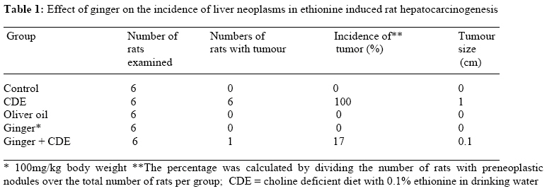
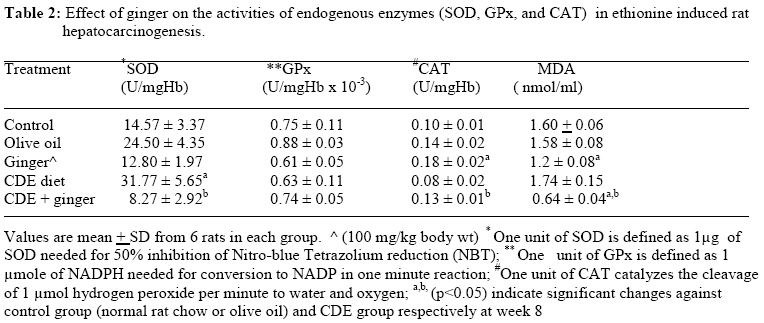
African Journal of Traditional, Complimentary and Alternative Medicines, Vol. 6, No. 1, 2009, pp. 87-93 Research Paper Chemopreventive efficacy of ginger (Zingiber officinale) in ethionine induced rat hepatocarcinogenesis 1Yasmin Anum Mohd Yusof*, 1Norliza Ahmad, 2Srijit Das, 1Suhaniza Sulaiman, 3Nor Azian Murad 1Department of Biochemistry, 2Department of Anatomy, Faculty of Medicine, Universiti Kebangsaan Malaysia, Medical Centre, Jalan Raja Muda Abdul Aziz, 50300 Kuala Lumpur, 3Center of Lipids, Engineering and Applied Research, Universiti Teknologi Malaysia, Kuala Lumpur, Malaysia. Email: anum@medic.ukm.my Code Number: tc09012 Abstract Ginger (Zingiber officinale Rosco) is widely used in foods as a spice all around the world. It has been reported to have antioxidant and anticarcinogenic properties. We investigated the effect of ginger in ethionine induced rat hepatocarcinogenesis. Male Wistar rats were divided into 5 groups: group 1 and 2 served as controls and they received normal rat chow and olive oil respectively. Group 3 was fed with ginger oleoresin dissolved in olive oil at 100 mg/kg body wt. Group 4 was fed with choline deficient diet and 0.1% ethionine in drinking water (CDE diet), and group 5 received ginger with CDE diet. Blood samples were taken from the orbital sinus at 0 and 8 weeks of experiment for the determination of antioxidant enzymes, superoxide dismutase (SOD), glutathione peroxidase (GPx), catalase and lipid peroxidation end product, malondialdehyde (MDA). Rats were also killed at 8 weeks for the observation of liver tumor formation. CDE diet induced the formation of liver nodules in rats and increased SOD activity. However, it had no effect on catalase, GPx and MDA levels when compared to both controls at 8 weeks of experiment. When CDE rats were treated with ginger, the formation of liver tumour, SOD activity and MDA level reduced, catalase activity was increased but no change was observed for GPx activity when compared to CDE group. In conclusion, ginger supplementation suppressed liver carcinogenesis by scavenging the free radical formation, and by reducing lipid peroxidation. Keywords: Choline deficient diet, Ethionine, Liver cancer, Ginger, Antioxidants, Lipid peroxidation. Introduction Hepatocellular carcinoma (HCC) is one of the most common cancers in the world with an annual incidence of 250,000 to 1,000,000 cases worldwide (Haydon and Hayes, 1995). The cancer registry from Ministry of Health Malaysia reported that liver cancer ranked the 11th among 15 most common cancers in Malaysia (Malalysian National Cancer Registry, 2004). Hepatitis B (HBV) and Hepatitis C virus (HCV) are the risk factors attributed to 80% of HCC cases globally (Chen et al., 1997). Other risk factors include toxin (aflatoxin) and exposure to certain chemical carcinogens such as diethylnitrosamine (DEN), polyaromatic hydrocarbon (PAH) and acetyl aminofluorene (AAF) (USA, 2004; Chandrasoma and Taylor, 1998). Similar to pathogenesis of other cancer, liver cancer has also been implicated as a consequences of oxidative stress due to continuous production of reactive oxygen species (ROS) which form covalent adduct with DNA bases, causing its damage and leads to mutagenicity (Packer and Colman, 1999; Halliwell, 1987). Currently there have been many studies focusing on the search for chemopreventive agents from herbs and natural products in combating cancer. Zingiber officinale Roscoe known as ginger is widely used as food condiments in many parts of the world. It has been extensively studied for its pharmacological and biological activities such as antiviral, antiulcer, antitumour, anti thrombotic, antiemesis and as anti cholesterolemic agents (Vimala et al., 1999; Sharma and Gupta, 1998; Bhandari et al., 1998; Sharma et al., 1997; Katiyar et al., 1996; Al-Yahya et al., 1989; Denyer et al., 1994). The antioxidative capacities of ginger and its active phenolic components 6-gingerol, shogaol and zingerone have been reported by several researchers. Ginger supplementation increased SOD activities in erythrocytes of rats and [6]-gingerol inhibited phospholipid peroxidation induced by FeCl3-ascorbate system and xanthine oxidase which is responsible for reactive oxygen species generation (Aeschbach et al., 1994; Masuda et al., 1995; Chang et al., 1994). The evidence thus implicated that ginger and its phenolic omponents were able to scavenge free radicals like superoxide and hydroxyl radicals (Cao et al., 1993; Reddy and Lokesh, 1992). Inhibition of carcinogenesis by ginger extract and its polyphenols have been documented in several studies. In skin cancer study, pre-application of ginger extract and its active ingredient 6-gingerol at the back of mice afforded significant inhibition of the skin tumour promoter, 12-O-tetradecanoylphorbol-13-acetate (TPA) and 7,12-dimethylbenz[a]anthracene-induced pamillomagenesis (Katiyar et al., 1996; Park et al., 1998). A recent in vivo study by Manju and Nalini (2005) reported that ginger supplementation suppressed colon carcinogenesis induced by a procarcinogen, dimethylhydrazine (DMH). The antitumourigenic effects of ginger has been implicated to the phenolic contents of ginger extract such as 6-gingerol, shogaol and zingerone (Manju and Nalini, 2005). To our knowledge no single study has reported the anti liver tumour effect of ginger extract. In the present work we determined the anticancer effect of ginger extract in liver cancer-induced rats by looking at the cancer incidence, enzymic antioxidant status and lipid peroxidation. We used the choline-deficient diet in combination with ethionine as a model diet to induce liver cancer in rats (Copeland and Salmon, 1946; Salmon and Copeland, 1954; Poirer, 2002). Ethionine , a methionine antagonist, will be metabolized to its active ethyl radical metabolite which interacts with DNA, inhibiting methylation and inducing mutagenesis (Poirer, 2002; Farber, 1963). Materials and Methods Chemicals Locally grown ginger (Z. officinale) was dried and finely ground. Ethanolic crude extract of ginger was kindly supplied by Noor Azian Murad, Center of Lipids, Engineering and Applied Research, Universiti Teknologi Malaysia. Animals Male Wistar albino rats aged 3-4 months and weighing 200-250 g were supplied by the Animal Care Unit of Universiti Kebangsaan Malaysia (UKM), Kuala Lumpur, Malaysia. The study was approved by the the Animal Ethics of Faculty of Medicine, UKM. The animals kept in a polycarbonate cage were provided with food and water ad libitum. They were maintained under standard conditions of temperature and humidity with an alternating light and dark cycle. Rats were randomized into 5 groups of 6 animals each. The first group and the second group served as the control group and were fed with normal rat chow (Gold Coin, Malaysia) and olive oil respectively. Rats in group 3 received ginger extract at 100 mg/kg body weight by gavage method. Rats in group 4 were fed with choline deficient diet ( ICN Biochemicals, USA) plus 0.1 % ethinonie (Sigma Chemical Co., USA ) in drinking water, known as CDE diet. Rats in group 5 received ginger as in group 3 plus CDE diet. Blood sample was obtained from all rats via the orbital sinus at 0 and 8 weeks for the analysis of endogenous antioxidant enzymes (SOD, GPx and catalase) and lipid peroxidation product, MDA. All rats were killed at 8 weeks for the observation of liver tumour incidence. The rats were cut open to expose the liver and gross tumours were counted. Preparation of blood samples Blood obtained from the orbital sinus was collected in test tubes with heparin to prevent blood coagulation, and the plasma was separated. The blood samples were then rinsed with the same volume of 0.9% NaCl (normal saline) and centrifuged at 3000 rpm for 10 min at 4oC. The upper buffy coat layer was removed and the packed red blood cells were washed twice with cold 0.9% NaCl until it became clear. The lower layer, termed as hemolysate was then used for the antioxidant enzyme assays and MDA determination. Biochemical determinations Superoxide (SOD) activity was determined by the method of Beyer and Fridovich (1987) at 560 nm spectrophotometrically. One unit of enzyme activity is defined as the amount of the enzyme that causes 50% inhibition of the reduction of formazan observed in the blank. Catalase activity was determined by the method of Aebi [1984] while glutathione peroxidase (GPx) measurement was assayed using the method of Paglia and Valentine (1967). Plasma lipid peroxidation was determined by the formation of malondialdehyde (MDA)-thiobarbituric acid reactive substrates (TBARS) adduct according to the method of Ledwozyw et al. (1986). The released malondialdehyde served as the index of lipid peroxidation. 1.1.1.3-tetraethoxypropane (Sigma, USA) was used as the standard. Statistical analysis Data was analysed using SPSS 9 package. Results were expressed as mean ± SEM of 6 rats in each group. Statistical evaluations were assayed using the one-way analysis of variance [ANOVA]. Results were regarded statistically significant at p < 0.05. Results Table 1 summarizes the incidence of liver neoplasms in rats on a low lipotrope diet with ethionine in drinking water and the effect of ginger on cancer incidence. There were no tumors in both controls (normal rat chow and olive oil), and in ginger treated rats. However, in CDE rats the tumor incidence was 100% and the average size of the tumor was 0.5-1 cm. Treatment of ginger to CDE rats managed to lower the tumor incidence to 17%. Table 2 represents the activities of enzymic antioxidants (SOD, GPx, CAT) and lipid peroxidation level (MDA) in hemolysate and plasma of control and experimental groups respectively. The activity of SOD enzyme increased significantly (p<0.05) in CDE treated rats compared to both control groups (normal rat chow and olive oil) at 8 weeks of experiment. However no significant changes were observed in ginger treated group when compared to the control group. On the other hand, there was a significant decrease of SOD activity in CDE group treated with ginger extract when compared with CDE group alone. There were no significant changes in GPx enzyme activities in all groups when compared to control group at 8 weeks of experiment. As for the activity of CAT, with the exception of ginger group, there were no significant changes observed in its activity in all groups at week 8 when compared with the control group. A significant increase in catalase activity however was observed in CDE group treated with ginger extract when compared with CDE group alone. Although there were no significant changes observed in MDA levels in CDE treated rats when compared to the control group, ginger supplementation however decreased MDA levels significantly in CDE-treated group when compared to the control group and to the CDE group alone. Discussion Ginger is among the most frequently and heavily consumed dietary condiments throughout the world. Ginger oleoresin and its polyphenols [6]-gingerol, shogaol, and zingerone have been studied extensively for their chemopreventive and antioxidative effects in carcinogenesis (Vimala et al., 1999; Katiyar et al., 1996; Park et al., 1998; Manju and Nalini, 2005; Chung et al., 2001; Mohd Yusof et al., 2002). Reactive oxygen species (ROS) such as superoxide radicals, hydroxyl radicals, hydrogen peroxide are produced in living systems under normal conditions and the body handles free radicals formed by scavenging them through the endogenous enzymic (SOD, GPx, catalase) and non enzymic antioxidants (glutathione, bilirubin, vitamins). However, enhanced concentrations of ROS and circulating lipid peroxidation product (MDA) produced have been implicated with carcinogenesis (Manju and Nalini, 2005; Chung et al., 2001; Ledwozyw et al., 1986; Cerutti, 1994). Superoxide anions or hydroxyl radicals thus generated caused oxidative damage of DNA and RNA leading to mutagenesis. Cell membranes surrounding the cells are also the prime target to free radicals attacks causing lipid peroxidation (Cerutti, 1994). Based on the available evidence, our body does not only require endogenous protective antioxidants to scavenge free radicals but it also requires exogenous antioxidants such as vitamins A, C, E , flavonoids and polyphenols derived from plants. Plant foods might have critically important components for cancer prevention because of their capacity to inhibit cell growth, induce apoptosis and scavenge free radicals (Surh, 1999; Lambert et al., 2005). Reports regarding status of enzymic antioxidants in carcinogenesis study have been highly variable. While some reports have noted increased enzymic antioxidant activities in rats and mice induced with carcinogens (L’Abbe et al., 1989; Manju et al., 2005), others have reported decreased or no changes of enzymic antioxidant status in carcinogenesis induced animals (Karimov et al., 2003; Chandra Mohan et al., 2003). The results of our study clearly exhibited the anti liver tumor promoting activity of ginger extract as evidenced by the reduced number of tumors in CDE treated rats. Since tumour promotion is closely related to oxidative stress, a compound that exhibits antioxidative properties is expected to act as an antitumour promoter. Antioxidative capacity of ginger is clearly seen when increased SOD activity as a result of superoxide free radical generation in liver cancer induced rats was abrogated by ginger oleoresin supplementation. Ginger was able to attenuate the harmful effects of ethionine by scavenging the increased superoxide anions generated during hepatocarcinogenesis induced by the CDE diet and thus sparing the endogenous SOD activity. The enhanced level of SOD activity observed during hepatocarcinogenesis may also be due to an active rate of cell proliferation that confers a selective growth advantage on cancer cells (Kulalko and Pence, 1992). Increased expression of SOD activities have been reported in many human cancers including brain tumors, breast and colorectal cancers and other malignant tumours (Cobbs et al., 1996; Janssen et al., 1998; Seth et al., 2003; Skrzydlewska et al., 2001). The release of serum SOD from rapidly multiplying tumour cells may be an effort to protect themselves from oxidative damage (Seth et al., 2003). Our study showed that ginger supplementation was also able to inhibit lipid peroxidation in CDE-treated rats. Manju and Nalini (2005) have also established in their laboratory that ginger inhibited lipid peroxidation and scavenged ROS. The high level of MDA during carcinogenesis implicates peroxidation of phospholipids in cell membrane causing cellular damage, and reduced level of MDA in CDE-treated rats by ginger extract indicates the protective effect of ginger. Ginger enchanced further the catalytic activity of catalase in CDE-treated rats perhaps due to its ability to scavenge free radicals and toxic carcinogenic electrophiles, thus sparing the concentration of the endogenous antioxidant enzymes (Chandra et al., 2003). GPx did not show any antioxidative property as shown in our study, and the absence of antioxidative activity in CDE treated rats could probably be due to the compensational relationship between catalase and GPx (L’Abbe et al., 1989; Manju et al., 2005), since both enzymes reacted with the same substrate, namely H2O2. Our study thus confirmed earlier findings that had shown both ginger extract and its phenolic components such as [6]-paradol and [6]-gingerol have strong potential antioxidant and antitumour activites. Ginger extract has been tested for the antitumor effect on rodent skins and models of intestinal carcinoma (Surh,1999; Hu et al., 2002; Lee et al., 2007). It has been observed that the pungent ginger ingredients such as [6]-gingerol and [6]-paradol have potential in vitro antitumour activity against different cell lines (Lee and Surh,1998; Chung et al., 2001: Wang et al.,2003).Ginger extract has been shown by our group in a previous study to exhibit antiproliferative and apoptotic activities against hepatoma cell line, HepG2 (Mohd Yusof et al., 2002). Research studies have depicted that some of the food constituents like phytochemicals, can easily modulate the complex multistage process of carcinogenesis through alteration of the genes and cell apoptosis (Fleischer et al., 2006). Ginger has been proven to suppress apoptosis (Manju et al.,2005). In the present study, ginger may have acted as an antitumour agent by reducing oxidative stress and thereby suppressing the growth of cancer cells. Conclusion Based on the results of the present study, ginger supplementation suppressed liver carcinogenesis by scavenging the free radical formation, and by reducing lipid peroxidation Further human studies may be needed to support our results but a strong evidence exists on the role of ginger as an anticancer agent. Acknowledgement This study was funded by the Research and Development Grant from the Malaysian Ministry of Science, Technology and Innovation. The authors wish to thank Ms Nor Ashikeen Mokti for expert technical assistance. References
© Copyright 2009 - African. Journal. Traditional, Complementary and Alternative Medicines The following images related to this document are available:Photo images[tc09012t2.jpg] [tc09012t1.jpg] |
| |||||||||
