 |
| About Bioline | All Journals | Testimonials | Membership | News |
|
||||||
|
||||||
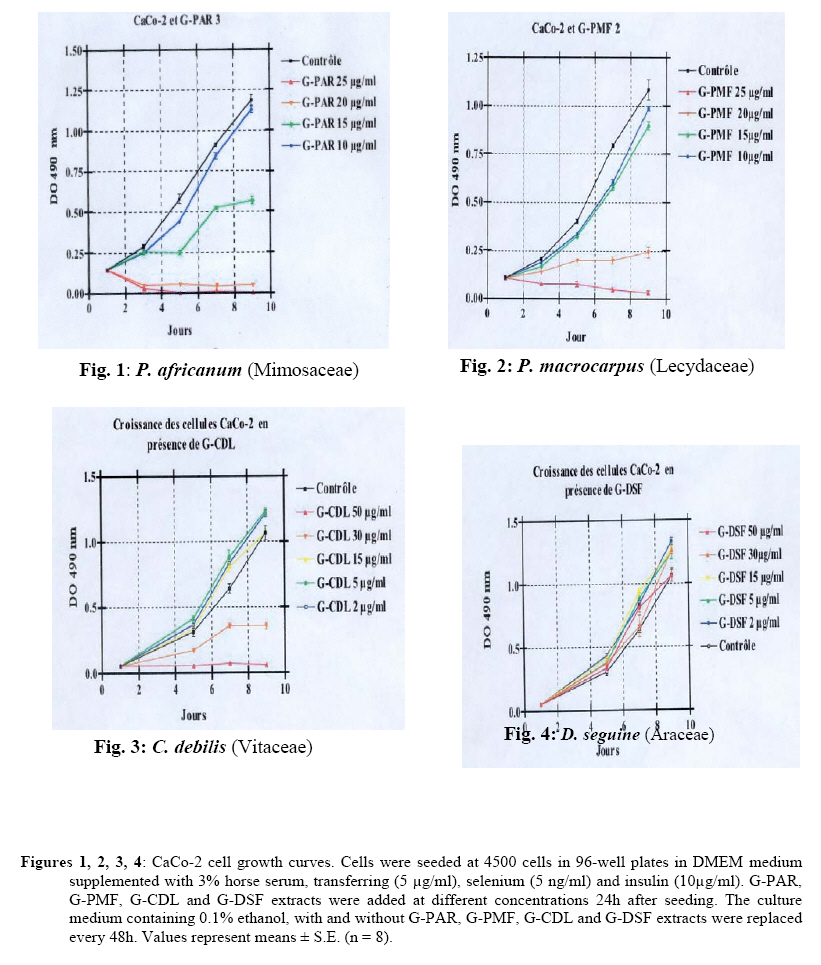
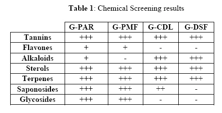
African Journal of Traditional, Complementary and Alternative Medicines, Vol. 6, No. 2, 2009, pp. 112-117 ANTIPROLIFERATIVE EFFECT OF ALCOHOLIC EXTRACTS OF SOME GABONESE MEDICINAL PLANTS ON HUMAN COLONIC CANCER CELLS Mengome Line-Edwigea*, Feuya tchouya Guy Raymondb, Eba Françoisc and Nsi-Emvo Edouardb aLaboratoire de Phytochimie, Institut de Pharmacopée et de Médecine
Traditionnelle (IPHAMETRA)/
CENAREST, BP 1935 Libreville / Gabon; Code Number: tc09015 Abstract Extracts from Piptadeniastrum africanum Brenan (Mimosaceae), Petersianthus macrocarpus (Breauv) L. (Lecydaceae), Dieffenbachia seguine Planch (Vitaceae) and Dieffenbachia seguine Jacq. (Araceae) were tested in vitro for their antiproliferative activity on human colon cancer cell line (CaCo-2). The highest antiproliferative activities were obtained with the alcoholic extracts of the roots of Piptadeniastrum africanum (G-PAR), the leaves of Petersianthus macrocarpus (G-PMF) and the stem of Dieffenbachia seguine (G-CDL), with 50% inhibition concentrations (IC50) of 15 µg/ml, 17 µg/ml and 25 µg/ml respectively. Only one extract (leaves of Dieffenbachia seguine (G-DSF)) exhibited weak antiproliferative activity with 50% inhibition concentration (IC50) higher than 50 µg/ml. Keywords: Dieffenbachia seguine; Dieffenbachia seguine; Petersianthus macrocarpus; Piptadeniastrum africanum; antiproliferative activity; CaCo-2 cells. Introduction In the past, it was thought that the prevalence of cancer was higher in the developed than in developing countries. Nowadays, contrary to the literature stating their scarcity in black Africa, (Attia et al., 1991 ; Nguéma–Mvé et al., 1995), the colon cancer is more and more frequent in African poor countries. It represents 32% of digestive cancers in Cameroon, 1% and 5% of cancers in Gabon and in Congo respectively (Ndjitoyap- Ndam et al., 1990 ; Takongmo et al., 2000) and 25% of the whole digestive cancers or half of colorectal cancers in some countries (Benhamiche, 1998). Many therapeutic strategies were used to fight this cancer. The surgical treatment of the colon cancer is influenced by a great mortality and the risk of local repetition (Fabre et al., 2000; Ele et al., 2006). The radiotherapy and the chemotherapy with drugs such as 5-fluorouracile (5Fu) (Carnesecchi et al., 2002a) can expose the patients to many troublesome side effects. The forest covers 85% of Gabon, and many plants are traditionally used as anticancer agents. The assessment of the anticancer activities of compounds from plants, fruits and vegetables in vitro and in vivo would represent a therapeutic alternative to fight cancer (Metz et al., 2000). Some recent work showed that resveratrol, a natural polyphenolic compound from grapes and wine, exerted a variety of pharmacological effects. Hence, the treatment of human colonic cancer cell line CaCo-2 with 25 µM resveratrol caused a 70% growth inhibition (Schneider et al., 2000; 2001). Other studies showed that, geraniol, a monoterpene from plant essential oils, inhibits not only the growth of CaCo-2 cells, but also the growth of CaCo-2 cells treated with 5Fu (Carnesecchi et al., 2001; 2002a ; 2002b). Some studies on pharmacological properties of chemicals from Sorghum revealed that the consumption of this cereal significantly reduces the risk of chronic diseases as cancer, cardiovascular diseases and obesity (Awika and Rooney, 2004). In the present study, the in vitro growth inhibition activities of alcoholic extracts of P. africanum (Mimosaceae), P. macrocarpus (Lecydaceae), C. debilis (Vitaceae) and D. seguine (Araceae) on human colonic cancer cell line CaCo-2 was investigated. Decoctions of these plants were said to be used by oral and enema in Gabonese traditional medicine in the treatment of haemorrhoids, paralysis and ulcerative incurable wounds. As there is no word in local language to describe cancer, we assimilated ulcerative wounds to cancers and this encouraged us to submit the extracts of these plants to the in vitro growth inhibition activities on the human colonic cancer cell line CaCo-2. Methodology Plant material Plants were selected on the basis of their ethnomedical studies. Leaves of Petersianthus macrocarpus (Breauv) L. (Lecydaceae); leaves of Dieffenbachia seguine Jacq. (Araceae); stem of Dieffenbachia seguine Planch (Vitaceae) and roots of Piptadeniastrum africanum Brenan (Mimosaceae) were harvested in Cap Esterias region, neighbourhood of Libreville, Gabon Republic, during the dry season in June 2004. Plants were authenticated in the Gabon National Herbarium, Libreville, for Dieffenbachia seguine (2658); Dieffenbachia seguine (2759); Petersianthus macrocarpus (1614) and Piptadeniastrum africanum (2876), where different voucher specimens were conserved. Extraction procedures Plant materials were dried and pulverised. Dry and powdered stem of Dieffenbachia seguine, leaves of Petersianthus macrocarpus, leaves of Dieffenbachia seguine, and the roots of Piptadeniastrum africanum were each extracted with ethanol. Hence, dried and ground plant material (1 kg Dieffenbachia seguine, 2.5 kg Dieffenbachia seguine, 1 kg Petersianthus macrocarpus and 4 kg Piptadeniastrum africanum) were macerated with ethanol for 48 h and 96 h. The extracts were filtered and the filtrates were freeze-dried to yield (7.2% w/w Dieffenbachia seguine, 3.8% w/w Dieffenbachia seguine, 4.2% w/w Petersianthus macrocarpus, 1% w/w Piptadeniastrum africanum). Cell culture CaCo-2 cells, obtained from the European Collection of Animal Cell Culture (CERDIC, Sophia Antipolis, France), were cultured in 75-cm2 Falcon flasks containing Dulbecco’s modified Eagle’s medium (DMEM) at 25 mM glucose supplemented with 10% heat-inactivated horse serum, 100 U/ml penicillin and 100µ g/ml streptomycin. Cells were incubated at 37 °C in a humidified atmosphere of 5% CO2, and subcultured after trypsinization (0.5% trypsin/2.6 mM EDTA). They were used up to 30-40 passages. In the experiments, cells were seeded at 6 x 10⁵ cells on culture dishes (100 mm in diameter), or at 4500 cells/well in 96-well plates. Cells were grown in DMEM supplemented with 3% horse serum, transferrin (5 µg/ml), selenium (5 ng/ml) and insulin (10 µg/ml) (TSI-defined medium; Gibco BRL, Life Technologies SARL, France). Plant extracts were dissolved in absolute ethanol and added 24 h after seeding to the culture medium (final concentration of ethanol, 0.1%). In all experimental settings, the culture medium and plant extracts were replaced every 48 h. Cells were harvested after various times, washed three times with phosphate-buffered saline (PBS; pH 7.2) and kept frozen at -70 °C until the assays were performed. Cell growth The cells were seeded in 96-well plates and incubated for different times. Cell growth was stopped by the addition of 50 µl trichloroacetic acid (50% v/v) and the protein content of each well was determined by staining with sulforhodamine B (Skehan et al., 1990). The absorbance was determined at 490 nm. The relationship between cell number (protein content/well) and absorbance is linear from 0 to 200 000 cells. Chemical screening procedures Alkaloids: 0.5 g of each extract was agitated with 5 ml aqueous hydrochloric acid on a steam bath. 1 ml filtrate was treated with Mayer reagent another 1 ml treated similarly with Dragendorff reagent. The appearance of a precipitate with one of these reagents was considered as a preliminary indicator of the presence of alkaloids. For confirmation, a little quantity of the extract was treated once with a solution of ammonia (40%) and twice with chloroform then resubmitted to Mayer and Dragendorff tests (Bruneton, 1999). Sterols and terpenes: 0.5 g of extract was dissolved in 0.5 ml chloroform. 0.5 ml acetic anhydride was added in the solution, cooled in ice. Few drops of sulphuric acid were then added carefully in the solution. A change in colour from violet to blue indicated the presence of sterols while a green or purplish red indicated the presence of triterpenes (Bruneton, 1999). Tannins: a drop of the extract was settled on a TLC silica gel plate. The elution was performed with a chloroform/acetic acid/formic acid (5/4/1) mixture. The plate was sprayed with a 10 ml solution of methanol/nitrous acid (95%/5%), then heated in an oven at 80 °C for 10 min. The presence of tannins was revealed by the appearance of blue spots (Bruneton, 1999). Flavones: a little quantity of the extract was dissolved in 2 ml methanol (50%). Few magnesium ships and few drops of concentrated hydrochloric acid were added in the solution. The appearance of a red-orange or violate colour showed the presence of flavones aglycones (Bruneton, 1999). Glycosides: a little quantity of extract was dissolved in an ethanol/a-naphthol (99%/1%) solution contained in a test tube, then allowed to run on the tube wall few drops of concentrated sulphuric acid. Glycosides presence was detected by the emergence of a red ring at the interface (Bruneton, 1999). Saponosides: 1% decoction of each sample was distributed gradually in 10 test tubes for a final volume of 10 ml. After two successive shakings, the test tubes were left to stand for 15 mins and then, the height of the foam was measured in each tube. The tube in which the height of the foam was 1 cm which indicated the value of the foam index (Bruneton, 1999). For all the families tested, according to the precipitation or colour intensity of each tube, following evaluations were given: (+++); (++); (+). Results Plants samples and CaCo-2 cell growth Dose-dependent effect of the plants extracts were studied on cell proliferation at concentrations from 2 to 50 µg/ml (Dieffenbachia seguine (stem) and Dieffenbachia seguine (leaves)); 10 to 25 µg/ml (Petersianthus macrocarpus (leaves) and Piptadeniastrum africanum (roots)). The Plant extracts demonstrated antiproliferative activities on the growth of CaCo-2 cells. The IC50 values obtained for the antiproliferative effects of the extracts on the growth of the CaCo-2 cells were 15 µg/ml for G-PAR (Figure 1); 17 µg/ml for G-PMF (Figure 2); 25µ g/ml for G-CDL (Figure 3). Only minor inhibition of the growth of the CaCo-2 cells were observed in the presence of G-DSF (IC50 > 50 µg/ml) (Figure 4). The phytochemical analysis (Table 1) revealed the presence of many chemical groups in the extracts that could be responsible of their activities. Discussion The chemical screening of the stem of C. debilis (G-CDL), the leaves of D. seguine (G-DSF), the leaves of P. macrocarpus (G-PMF) and the roots of P. africanum (G-PAR) showed the presence of tannins, flavones, alkaloids, steroids, terpenoids, saponosides and glycosides. We realised that G-PAR, G-PMF and GCDL, rich in saponosides inhibited the growth of CaCo-2 cells with 50% growth inhibition concentrations of 15µ g/ml, 17 µg/ml and 25 µg/ml respectively (Table 1, Figures 1; 2 and 3). In contrast, G-DSF extract containing also saponosides possessed very weak antiproliferative activity on CaCo-2 cells with IC50 higher than 50µg/ml (Table 1, Figure 4). These results did not show the importance of saponosides in the fight against cancer. On the other hand, saponosides had exerted an antiplasmodial activity on the Plasmodium falciparum strain (W2) (Lamidi et al., 1996). The antioxidant activities of the polyphenols have been revealed (Awika and Rooney, 2004). Polyphenolic compounds showed pharmacological effects on many chronic diseases such as cancer (Liu, 2004). It was established that the polyphenolic tannins of a cereal (Sorghum) had carcinogenic effects (Morton, 1970; 1972 ; Oterdoon et al., 1985). Although some recent studies showed that a natural polyphenolic component of grapes, wine and green tea extracts, resveratrol, exhibited antiproliferative activity on the growth of human colonic cancer cell line CaCo-2 in vitro, and anticancer activities in cancerous rats, inhibiting or activating some target molecules implied in the progression of the cell cycle (Metz et al., 2000; Schneider et al., 2000), in this study, it was not possible to correlate the anticancer activities of our extracts with the presence of tannins, since even the less active extract (G-DSF) contains tannins (Table 1). May be, the effect was related to the concentration of tannins. Subsequent studies, including the quantitative analysis of the chemicals from the studied plants should be made before any conclusion is made. It could also be interesting to speculate that the anticancer activities of our extracts could be due to a synergy between their various chemical constituents. This would be in agreement with the results of Rui Hai Lin in 2004, revealing that the combination of phytochemical agents exerts most of the total antioxidant activity. The healings observed in the treatment of ulcerative incurable wounds in the Gabonese traditional medicine with these plants suggested that the anticancer activities of the extracts should not be simply related to the probable cytotoxic effect of their polyphenolic compounds (tannins). For clarifications, future studies including the cytotoxicity analysis of extracts will be carried out. Conclusion In this study, the in vitro growth inhibition activities of alcoholic extracts of P. africanum (Mimosaceae), P. macrocarpus (Lecydaceae), C. debilis (Vitaceae) and D. seguine (Araceae) against the human colonic cancer cell line CaCo-2 was studied. No study on the anticancer effects of the Gabonese medicinal plants have been yet carried out. We showed for the first time that the ethanolic extracts G-PAR; G-PMF; GCDL and G-DSF possessed anticancer activities. This work showed that ethanolic extracts G-PAR, G-PMF and G-CDL strongly inhibited the growth of the human colonic cancer cell line CaCo-2 in vitro. Our work authenticated the therapeutic indications of the traditional preparations containing G-PAR, G-PMF and G-CDL. Acknowledgements The authors are grateful to Dr. Francis Raul for supplying of CaCo-2 colonic cancer cell line, Mr. Raoul Niangadouma and Mr. Edouard Mintsa Obiang for the determining botanical data. References
© Copyright 2009 - African. Journal. Traditional, Complementary and Alternative Medicines The following images related to this document are available:Photo images[tc09015f1-4.jpg] [tc09015t1.jpg] |
| |||||||||
