 |
| About Bioline | All Journals | Testimonials | Membership | News |
|
||||||
|
||||||
African Journal of Traditional, Complementary and Alternative Medicines, Vol. 6, No. 2, 2009, pp. 131-138 Research Paper PROTECTIVE ACTIVITY OF THE STEM BARK AQUEOUS EXTRACT OF MUSANGA CECROPIOIDES IN CARBON TETRACHLORIDE- AND ACETAMINOPHENINDUCED ACUTE HEPATOTOXICITY IN RATS Adeneye, Adejuwon Adewale Department of Pharmacology, Faculty of Basic Medical Sciences, Lagos State University College
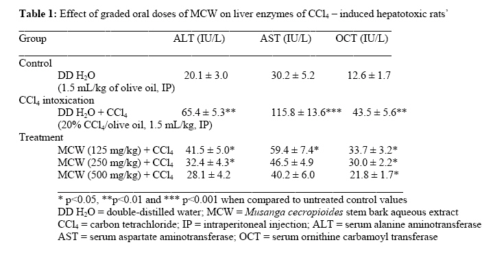
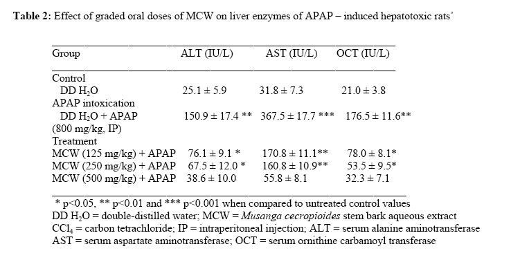
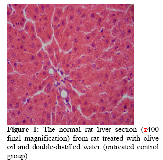
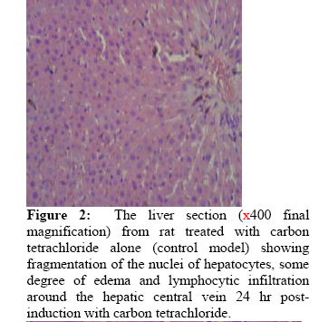
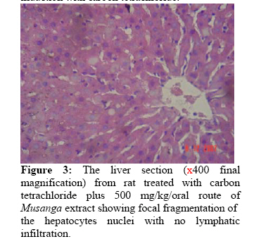
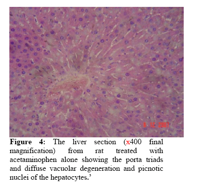
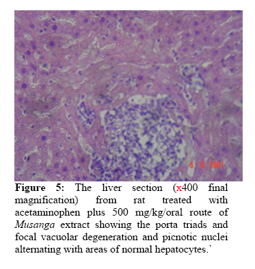
of Medicine, PMB 21266, Ikeja, Lagos, Nigeria Code Number: tc09018 Abstract The hepatoprotective activities and the mechanisms of actions of Musanga cecropioides stem bark aqueous extract (MCW) were investigated on acute hepatocellular injuries induced by intraperitoneal (IP) carbon tetrachloride (CCl4) (20% CCl4/olive oil, 1.5 mL/kg) and 800 mg/kg/IP of acetaminophen (APAP) in normal saline, in male Wistar rats. Among the Yorubas (South-West Nigeria), cold decoction of MCW is used as a natural antidote for oral gastric poisonings, infective hepatitis and other liver diseases. Its hepatoprotective activities were monitored by assaying for the serum aminotransferases, ornithine carbamoyl transferase and the toxicant-induced histopathological lesions in rat livers 24 hours postinduction. These enzymes are markers of acute hepatic injuries and their elevations are indications of acute liver injuries. Pretreatment of rats with graded doses (125 - 500 mg/kg) of MCW significantly attenuated the acute elevation of the liver enzymes and the hepatotoxin-induced histopathological lesions in the rat livers. The presence of two active natural antioxidants (flavonoids and alkaloids) in high concentrations in MCW may account for the hepatoprotective activities observed in this study. These results, thus, support the folkloric use of MCW for treatment of hepatic injuries resulting from acute gastric poisonings, infective hepatitis or other liver diseases. Key words: Musanga cecropioides, Hepatoprotective activities, Carbon tetrachloride, Acetaminophen, Wistar rats Introduction The use of herbal remedies these days has become more popular partly because of scientific validation of some of their medicinal values (Odetola and Akojenu, 2000). For example, the clinical benefits of St. John’s Wort extract (Hypericum perforatum L. extract) in the treatment of mild to moderate depression, viral and bacterial infections, leukemia and inflammatory conditions are well established. Oil of St. John’s Wort is also used externally for the treatment of sores, burns, myalgia and bruises (Erdelmeier and Koch, 2006); African ginger (Zingiber officinale) is used in the treatment of infective hepatitis, asthma, rheumatism and immunosuppression (Gill, 1992). Musanga cecropioides R.Br. Apud Tedlie (family: Cecropiaceae) is an erect, rapidly growing tree of the deciduous tropical West African rainforest. It grows up to 20 m tall with umbrella-shaped crown, straight and cylindrical trunk (up to 2 m in girth) and stilt root of up to 3 m above ground level (Adeneye et al., 2006a). The plant is highly valued by the local herbalists due to its diverse medicinal properties such as oxytocic, contraceptive, galactogogue, antihypertensive, antidiabetic, analgesic and diuretic (Adeneye et al., 2006a). Recently, the oral hypotensive (Adeneye et al., 2006b), hypoglycemic and antidiabetic activities (Adeneye et al., 2007) of the aqueous stem bark aqueous extract of the plant were investigated and validated. Amongst the Yorubas (South-West, Nigeria), hot infusion of the plant stem bark is also used for treatment of fever, jaundice, acute gastric poisonings and liver diseases. However, despite its extensive use, there are no scientific evidences to justify its use in folk medicines. Hepatitis and other liver diseases constitute a major cause of mortality and morbidity in Nigeria, and these can be prevented with proper treatments and well taken prophylactic measures (Adesunkanmi et al., 2002). However, treatment and prophylaxis options for these conditions are limited. Therefore, it is important to make continuous effort to develop more effective therapeutic strategies or prophylactic modalities to eradicate or stem the disease. Reasons attributed to liver disease being a leading cause of medical emergency in Nigeria includes poorly treated or untreated infective hepatitis, late presentation at hospitals for treatment, alcoholism and drug misuse (Adesunkanmi et al., 2002). The aim of the current study is to validate the folkloric claim of the hepatoprotective efficacy of MCW in the treatment of liver diseases resulting from acute gastric poisonings and to elucidate its possible mechanism(s) of actions using carbon tetrachloride and acetaminophen experimental models of acute liver injury in rats. Carbon tetrachloride is known to generate lipid radicals that initiate the chain reactions of lipid peroxidation within and outside the hepatic cells causing liver damage. Acetaminophen, on the other hand, is also known to produce a highly reactive electrophilic metabolite which conjugates with cellular glutathione (GSH), producing marked GSH depletion, lipid peroxidation and hepatocyte injury. Materials and Methods Plant Materials One (1) kg of the fresh stem bark sheaths of the Musanga cecropioides tree was collected from the deciduous forest within the complex of the Permanent Site of Olabisi Onabanjo University, Ogun State, Nigeria, during the first week of April, 2006. Plant identification, authentication and deposition of the voucher specimen for referencing had been done earlier (Adeneye et al., 2006a). The sheaths were washed with normal saline, sorted and air-dried at room temperature and protected from direct sunlight and heat for 2 weeks until completely air-dried. These were then pulverized using the laboratory hammer-mill and the powdered samples were stored in air- and water-proof containers until required for extraction. Preparation of Aqueous Extract The powdered plant material (100 g) was extracted for 6-8 hours with 1L of double distilled water using Soxhlet extractor. The filtrate obtained was completely dried using a digital aeration oven (Genlab Ltd., UK) preset at 50 ºC. The percent yield was 12.5 ± 0.5 %. The deep-brown crude extract obtained was stored in tight-fitted capped containers and stored at 4 ºC. The crude extract was dissolved in double distilled water to make a concentration of 100 mg/ml from which different doses (125, 250 and 500 mg/kg body weight/oral route) was re-constituted. Experimental Animals Animal experiment was conducted after approval was given by the Ad hoc Animal Ethical Committee of the Lagos State University College of Medicine, Ikeja, Lagos State, Nigeria. Experimental procedures involving the animals and their care were conducted in conformity with the institutional guidelines that are in compliance with the national and international laws and the Guidelines for Care and Use of Laboratory Animals in Biomedical Research as adopted and promulgated by the Canadian Council of Animal Care (1984) and United States National Institutes of Health (1985). Inbred male Wistar rats, 6-8 weeks old and weighing 120 - 150 g, were used for the study. They were fed with a standard laboratory diet and drinking water made readily available. The experimental animals were maintained at standard laboratory conditions (12:12 hour dark/light periodicity, 23 ± 2 ºC temperature and 55 ± 5 % humidity). Prior to experimentation, the rats were fasted for 18 - 20 hours. Chemicals and reagents The following reagents were used: carbon tetrachloride (CCl4) (Merck, Darmstadt, Germany), olive oil (Roberts Lab. Ltd., Belton, England), normal saline (Unique Pharmaceuticals, Sango- Otta, Nigeria), Paracetamol (Emzor Pharmaceuticals Industries Ltd., Isolo-Lagos, Nigeria), Alanine aminotransferase (ALT) (Boehringer, Germany), Aspartate aminotransferase (AST) (Boehringer, Germany), Ornithine carbamoly transferase (OCT) (Boehringer, Germany). Other reagents were all of analytical grade. CCl4 -induced Hepatotoxicity in rats The fasted rats were randomly divided into 5 groups of six rats each such that the difference within and between groups does not exceed ± 20 % of the average weight. Of the five groups used for this study, Group I was used as the untreated control group, Group II served as the model control and was treated with CCl4 intraperitoneallly, and Groups III-V were pre-treated with different doses of MCW (125, 250 and 500 mg/kg/oral of the extract, respectively) 1 hour before the intraperitoneal injection of CCl4 (1.5 mL/kg body weight as a 20% olive oil solution). Twenty-four hours post-CCl4 treatment, the rats were anesthetized with inhalational halothane and blood samples were withdrawn through cardiac puncture. APAP-induced hepatotoxicity in rats The protocol adopted in the case of pretreatment with acetaminophen was similar to that described for CCl4 except that CCl4 was replaced with 800 mg/kg body weight of APAP in normal saline, which was injected intraperitoneally. Assay of marker enzymes of liver damage The whole blood collected was centrifuged at 3000 revolutions/minute using a table-top centrifuge (Shanghai Surgical Instrument Factory, Shanghai, China) at 37 ºC for 15 min to separate the sera. Serum alanine (ALT) and aspartate (AST) aminotransferases and ornithine carbamoyl transferase were assayed using Boehringer Mannheim diagnostic kits. Histopathological studies of the rat liver The livers of the rats were carefully dissected out and freed from the supporting adipose and ligamental tissues. After rinsing the liver in normal saline, different sections were taken from each lobe of the liver. The tissue was fixed in 10% formo-saline, dehydrated with 100% ethanol solution and embedded in paraffin. It was then processed into 5 μm thick sections stained with hematoxylin-eosin and observed under a photomicroscope (Model N - 400ME, CEL-TECH Diagnostics, Hamburg, Germany). Statistical analysis Data were expressed as mean ± S.E.M. (standard error of mean) of six observations and statistically assessed by two-way analysis of variance using statistical software program SYSTAT 10.2. Further analysis among the treatment groups, was statistically evaluated by Student-Newman-Keuls test. Statistical significance was considered significant at p<0.05, p<0.01, p<0.001. Results Effect of MCW on CCl4-induced hepatocellular injury In Table 1, it can be seen that IP administration of CCl4 induced severe hepatocellular damage as evidenced by the significant (p<0.05) elevation of the serum AST, ALT and OCT. However, these elevations were attenuated in MCW-pretreated rats, in dose related fashion as the values of the liver enzymes fall with increasing treatment doses of MCW although the values of OCT at 500 mg/kg body weight of the extract were still statistically higher than that of untreated control values. Effect of MCW on APAP-induced hepatocellular injury Table 2 shows that high doses of APAP (800 mg/kg) given intraperitoneally also cause severe hepatocellular injury as indicated by the marked elevation of ALT, AST and OCT. However, these increases were significantly (p<0.05) reduced when the rats were pretreated with MCW 1 hour prior to APAP induction. These reductions were in dose-dependent fashion and that striking reduction was at the highest dose. Histopathological changes in MCW-pretreated and CCl4- and APAP-induced hepatotoxic rat livers’ Figures 1, 2, 3, 4, and 5 depict the histopathological changes seen in the normal, CCl4-induced, MCWpretreated- CCl4-induced, APAP-induced and MCW-pretreated-APAP-induced rat livers, respectively. As seen in the figures, the histopathological lesions varied from one group to the other. Figures 2 and 4 show the typical CCl4- and APAP-induced histopathological lesions which were characterized by diffuse hepatocyte necrosis and lymphocytic infiltration , while figures 3 and 5 show histological improvements in the rat liver protected with MCW before induction of CCl4- and APAP hepatotoxicities respectively. Discussion It is well established that acute hepatocellular injury can be induced by various hepatotoxins, including carbon tetrachloride and high dose acetaminophen. CCl4 is a highly reactive halogenated aromatic hydrocarbon which has been used in experimental models of drug-induced hepatocellular injury (Shah et al., 1979; Letteron et al., 1990; Tsai et al., 1997). CCl4 is metabolized by the cytochrome P450 family in the hepatic endoplasmic reticulum to form highly reactive and unstable trichloromethyl radicals (.CCl3) (Recknagel and Glende, 1973) which binds to the unsaturated fatty acids of membrane lipids covalently, resulting in the formation of chloroform and lipid radicals (Parker et al., 1978). These lipid radicals initiate the chain reactions of lipid peroxidation within and outside the hepatic cells and cause liver damage Also, microsomal auto-oxidation of chloroform results in the formation of highly reactive phosgene, a hepatotoxin. To validate the acute model of CCl4-induced hepatotoxicity, 1.5 mL/kg of CCl4 as 20% olive oil solution was administered to rats intraperitoneally. This dose induced a significant elevation of the liver enzymes (Table 1) when compared to the untreated control values, the normal range being 10 - 35 IU/L for humans (Kee, 2002). These biochemical alterations were corroborated by the histopathological lesions of hepatic necrosis which was characterized by diffuse nuclear fragmentations of hepatocytes with lymphocytic infiltration and some degree of edema, predominantly found around the centrilobular region of the liver. In the present study, pretreatment of rats with graded oral doses of MCW attenuated CCl4-induced hepatocellular injury in dose related fashion, as evidenced by the dose related reduction in the CCl4-induced hepatic aminotransferases and ornithine carbamoyl transferase (OCT) (Table 1). This observation was further corroborated by attenuation of CCl4-hepatocellular distortion on histopathological examination which showed a focal nuclear fragmentation of the hepatocytes with no lymphocytic infiltration (Figure 3). Serum levels of ornithine carbamoyl transferase and type-1 arginase have been documented to be effective and reliable markers of acute and chronic liver injuries as these enzymes are primarily synthesized and confined to hepatocytes. Whenever, the liver is damaged, their serum levels are significantly elevated indicating hepatocellular injury (Murayama et al., 2007). In fact, ornithine carbamoyl transferase is reported to be more sensitive than other liver enzyme markers as its serum level rises first and more significantly than the aminotransferases, although it is less specific than ALT (Tegeris et al., 1969). Thus, the assays of ornithine carbamoyl transferase and type-1 arginase are often complementary to that of ALT and AST in evaluating hepatocellular damage. Acetaminophen-induced hepatotoxicity as an established experimental model of drug-induced hepatotoxicity is well documented in the literature (Mitchell et al., 1973; Comporti et al., 1991). Acetaminophen is known to be metabolized to produce acetaminophen electrophilic metabolite, N-acetylpara- benzoquinonimine (NAPQI), which conjugates with cellular glutathione (GSH) to cause marked GSH depletion (Muriel et al., 1992). In the current study, acetaminophen induced a significant elevation of serum aminotransferases and OCT levels. The associated histopathological studies also showed a diffuse vacuolar degeneration of the hepatocyte cytoplasm as well as picnosis of the hepatocytes 24 hours post-induction with APAP (Figure 4), which reflect high level of lipid peroxidation. Both the degeneration of cytoplasm and picnosis of hepatocyte suggest that lipid peroxidation is an important factor in the etio-pathogenesis of acetaminophen-induced liver cell death. As shown in the result, MCW has protective effects in the liver by counteracting the elevation of the liver aminotransferases induced by APAP- (Table 2) and this is evident in the rats pretreated with the high dose (500 mg/kg/oral route) of MCW which were the least affected by the APAP induction (Table 2) and (Figure 5). Inflammatory process has been documented in the literature to contribute to the development of liver injury induced by hepatotoxins such as APAP and CCl4 (Luster et al., 2001). As inflammatoryresponse, chemokine- and cytokine-releasing leukocytes such as neutrophils and macrophages are often mobilized to hepatic injury site (Blazka et al., 1995; Blazka et al., 1996; Ishida et al., 2002). Also administration of anti-neutrophil serum and any other agents that inhibit leukocyte infiltrations has been documented to diminish ALT elevation in the serum and histopathological changes of the liver in hepatotoxin-treated rats (Smith et al., 1998). Thus, reduced leukocyte infiltration in the hepatocytes of MCW-pretreated rats suggests that MCW has an in vivo hepatic anti-inflammatory activity. Previous phytochemical results showed that MCW contain high concentrations of alkaloids, flavonoids and other biological principles (Adeneye et al., 2006). It has been reported that flavonoids are able to inhibit drug-induced hepatotoxicity in experimental models (Carini et al., 1992; Paya et al., 1993) due to their potent anti-oxidant or free radicals scavenging properties (Fraga et al., 1987; Laughton et al., 1989; Sanz et al., 1994). This active principle may account for the extract pharmacological properties. In addition, literatures have shown alkaloids to elicit hepatoprotective activity by strongly inhibiting lipid peroxidation (reduction of malonaldehyde (MAD) production) and cell membrane disruption (reduction of lactate dehydrogenase (LDH) leakage) induced in isolated hepatocytes (Lanhers et al., 1991). Thus, the presence of these two active natural antioxidants in high concentrations in MCW may account for the hepatoprotective activities observed in this study despite the differences in their mechanisms of inducing hepatocellular injury. This appears that the extract elicits its hepatoprotective function through more than one mechanism of action. Conclusion Overall, results of this study support the folkloric use of the decoction of the plant stem bark in the treatment of patients with suspected liver disease and acute gastric poisonings. Isolation and evaluation of the suspected hepatoprotective principle(s) in the extract would constitute a future research interest. Acknowledgements The author wishes to acknowledge the expertise of Dr. A.S. Benebo, a Consultant Pathologist and Lecturer in the Department of Pathology and Forensic Medicine, Lagos State University College of Medicine, Ikeja, Nigeria and the technical assistance of Mr. J.A. Akinmurele, Assistant Chief Technologist, Pharmacology Department, Lagos State University College of Medicine, Ikeja, Nigeria. References
© Copyright 2009 - African. Journal. Traditional, Complementary and Alternative Medicines The following images related to this document are available:Photo images[tc09018f2.jpg] [tc09018f4.jpg] [tc09018f5.jpg] [tc09018f3.jpg] [tc09018t2.jpg] [tc09018t1.jpg] [tc09018f1.jpg] |
| |||||||||
