 |
| About Bioline | All Journals | Testimonials | Membership | News |
|
||||||
|
||||||
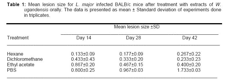
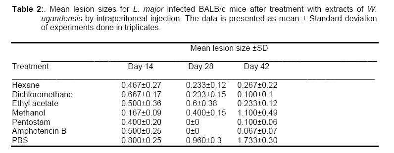
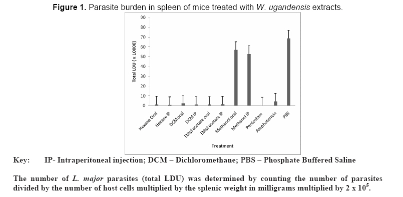
African Journal of Traditional, Complementary and Alternative Medicines, Vol. 6, No. 2, 2009, pp. 207-212 Research Paper In vivo EFFICACY OF ORAL AND INTRAPERITONEAL ADMINISTRATION OF EXTRACTS OF Warburgia ugandensis (CANELLACEAE) IN EXPERIMENTAL TREATMENT OF OLD WORLD CUTANEOUS Leishmaniasis CAUSED BY Leishmania major 1* Peter Kamau Ngure, 3 Zipporah Ng’ang’a, 2 Johnstone Ingonga 2 Geoffrey Rukunga and 2 Willy Kiprotich Tonui. 1Daystar University, Science department, P.O. Box 44400, 00100, Nairobi. Code Number: tc09028 Abstract The antileishmanial activity of extracts of Warburgia ugandensis Spraque (Canellaceae), a known traditional therapy in Kenya was evaluated in vivo. Treatment of infected BALB/c mice with W. ugandensis extracts orally resulted in a reduction of the size of lesions compared to the untreated control. The lesion sizes differed significantly for the four extracts (p=0.039) compared to the untreated control. For mice treated by intraperitoneal injection, the lesion sizes increased initially for the hexane, dichloromethane and ethyl acetate extracts and healed by day 42. The lesion sizes for mice treated with methanol increased steadily from 2.47mm to 3.57mm. The parasitic burden was significantly higher (p<0.001) in mice treated with methanol extracts and PBS compared to those treated with hexane, dichloromethane and ethyl acetate. This study demonstrated the antileishmanial potential of extracts of W. ugandensis. Key words: Warburgia ugandensis; Leishmaniasis; BALB/c. List of abbreviations Introduction The Leishmaniases are diseases caused by obligate intracellular, kinetoplastid protozoa of the genus Leishmania (Trypanosomatidae) (Roberts, 2006). Although it is not a household name like malaria, the diseases caused by infection with Leishmania continue to have a major impact on much of the world’s population and are currently considered to be an emerging illness with high morbidity and mortality in the tropics and subtropics (Handman, 2001; Santos et al., 2008). Neglected by researchers and funding agencies, Leishmaniases are endemic in 88 countries of the World in which 350 million people who are considered at risk of infection live (WHO, 2007). There are 2 million new cases of Leishmaniasis annually and 14 million infected people worldwide (WHO, 2007). An increase in the incidence of Leishmaniasis can be associated with urban development, destruction of forests, environmental changes, migrations of people to areas where the disease is endemic and wars which contributes to the spread due to displacement of people (Ashford, 2000; Patz et al., 2000; Kolaczinski et al., 2007). Proven therapies against human Leishmaniasis include pentavalent antimonials (sodium stibogluconate and meglumine antimoniate), amphotericin B, pentamidine, miltefosine and paromomycin (Berman, 1996, 1997). These drugs are unsatisfactory because of their limited efficacy, frequent side effects, and increasing drug resistance, therefore new, safer, and more efficacious drugs are urgently required (Croft and Seifert, 2005). Moreover, there is no effective vaccine against Leishmaniasis (Handman, 2001). In this regard, medicinal plants offer prospects for discovering new compounds with therapeutic properties. Warburgia ugandensis Sprague (Canellaceae), the East African greenheart is one of the most highly utilized medicinal plants in tropical and subtropical Africa and is now highly endangered in the wild (Kioko et al., 2005). It is rated as the second highest priority medicinal plant species in Kenya (Kariuki and Simiyu, 2005). According to the World Agroforestry Centre, (http://www Worldagroforetsrycentre.org/sea/products/AFDbases/af/asp/SpeciesInfo) the dried bark of the tree is commonly chewed and the juice swallowed as a remedy for stomach ache, constipation, toothache, venereal diseases, cough, fever, muscle pains, weak joints and general body pains. The leaf decoction baths are used as a cure for skin diseases while the bark, roots, or leaves can be boiled in water and drunk to treat malaria, although this causes violent vomiting. Warburgia ugandensis which is known as “soket” in Tugen tribe is used by traditional healers to treat visceral Leishmaniasis (VL) and cutaneous Leishmaniasis (CL) (W. Tonui, Kenya Medical Research Institute, Nairobi, personal Communication). The stem barks are taken orally in boiled water or soup. Previous studies on W. ugandensis have shown good antibacterial, antifungal, antiviral activity and trypanocidal effects. Crude extracts and purified compounds of W. ugandensis showed activity against Mycobacterium tuberculosis H37Rv and M. Bovis BCG Pasteur (Madikane et al., 2007), Candida albicans and measles virus (Olila et al., 2001, 2002). Similarly, the activity of W. ugandensis against trypanosomes has been demonstrated (Kioy et al., 1998; Olila et al., 2001). A recent study demonstrated that hexane, dichloromethane and ethyl acetate extracts of W. ugandensis had good antileishmanial activity in vitro (Ngure et al., 2008, unpublished report). The objective of the present study was to determine the effect of extracts of W. ugandensis on Leishmania major infected BALB/c mice. Materials and Methods Source of W. ugandensis Warburgia ugandensis stem barks (CTMDR 007) were collected from Sesia village in Kabarnet, Baringo District, in the Rift Valley Province in Kenya. Botanical identification was done using standard identification keys by the National Museums of Kenya botanists. Voucher specimens were kept in the Kenya Medical Research Institute laboratory in Nairobi. Sample preparation and extraction of compounds of W. ugandensis The stem barks were cut into small pieces and air-dried for three weeks under a shed. The dried specimens were shred using an electrical mill in readiness for extraction. The sample preparation and extraction procedure were carried out as described by Harbone (1994). Cold sequential extraction was carried out on plant material with analar grade organic solvents of increasing polarity, which included n-hexane, dichloromethane, ethyl acetate and methanol. Six hundred millilitres of n-hexane was added to 300gms of the shred specimen and flasks placed on a shaker and soaked for 48 hrs. The residue was filtered using a Buchner funnel under vacuum until the sample dried. The sample was soaked further with 600ml of n-hexane for 24 hrs until the filtrate remained clear. The filtrate was then concentrated under vacuum by rotary evaporation at 30-350C (Harborne, 1994). The concentrate was transferred to a sample bottle and dried under vacuum; the weight of the dry extract was recorded and stored at -200C until required for bioassay. The process was repeated sequentially for dichloromethane, ethyl acetate and methanol. Testing for antileishmanial activity of extracts of W. ugandensis Experimental animals Eight week old BALB/c mice were obtained from the Kenya Medical Research Institute’s (KEMRI) animal house facility. The experiments using mice were done in compliance with the Animal Care and Use Committee (ACUC) guidelines of KEMRI. Leishmania parasites Metacyclic promastigotes of L. major strain (Strain IDU/KE/83 =NLB-144) were used. Parasites were maintained as previously described (Titus et al., 1984) and metacyclics were isolated from stationary phase cultures by negative selection using peanut agglutinin (Tonui et al., 2004). Briefly, the promastigotes were cultured in Schneider’s Drosophila medium supplemented with 20% foetal bovine serum (FBS), glutamine (2mM), penicillin G (100 U/ml), and streptomycin (100 μg/ml). Stationary-phase promastigotes were obtained from 5 to 7-day-old cultures. Preparation of drugs Stock solutions of the fractions were made in culture media for anti-Leishmanial assay and resterilized by passing through 0.22 μm micro-filters under sterile conditions in a laminar flow hood. The extracts that were insoluble in water or media were first dissolved in 1% dimethyl sulphoxide to avoid solvent carry over, (Dorin et al., 2001). All the prepared drugs were stored at 40C and retrieved only during use. Infection of mice, treatment and determination of parasite numbers in cutaneous lesions Female BALB/c mice were infected in the hind footpad with 1 x10⁶ L. major metacyclic promastigotes. In all experiments, treatment was initiated 4 or 5 weeks after infection had established as determined by the presence of lesions (Fournet et al., 1996). Two days before administration of the drug, the mice were randomly divided into groups of ten. Infected untreated mice were used as controls. Experimental mice were treated with the standard drug pentostam, in regimens of 20mg of Sb per body weight daily for 30 days intraperitoneally while others were treated with amphotericin B. The extracts were injected intraperitoneally or administered orally using a cannula daily for 30 days. The untreated group received PBS. Lesion development was followed by measuring thickness of the infected footpad as compared to the thickness of the contralateral footpad prior to infection using a vernier caliper. All mice were sacrificed during the 12th week, spleens were weighed and impression smears made. These were fixed in absolute methanol and stained with Giemsa before examination for parasites to determine whether or not visceralization of the parasite had taken place. Statistical analysis All experiments were carried out in triplicate. The mean and standard deviation of at least three experiments was determined. Statistical analysis of the differences between mean values obtained for the experimental groups was done by analysis of variance (ANOVA) and Student’s t test. P values of 0.05 or less were considered significant. Results and discussion Three hundred grams of the stem barks of W. ugandensis yielded 10.5 g for n-hexane extract, 12.1g for dichloromethane, 9.3g for ethyl acetate and 9.9g for the methanol extract. This study examined the antileishmanial activity of extracts of W. ugandensis in vivo. The administration of hexane, dichloromethane, and ethyl acetate extracts led to the recovery of BALB/c mice infected with L. major. The results are in agreement with the claims made by Kenyan traditional practitioners that W. ugandensis stem barks have antileishmanial effects (W. Tonui, 2006, unpublished report). There was no significant difference (p=0.697) in body weight change for mice that were treated orally using a cannula compared to those that were treated by intraperitoneal injection. There was also no significant difference (p=0.462) in change of body weight for mice that were treated with pentostam and amphotericin B compared with those that were treated orally with the hexane, dichloromethane, ethyl acetate and methanol extracts. Similarly, there was no significant difference (p=0.260) between mice treated with pentostam and amphotericin B and those treated intraperitoneally with the four extracts. The administration of the extracts did not have any adverse effects on mice considering that there were no observable changes in mice. Since so far there is no documented report on in vivo studies using W. ugandensis extracts it was necessary to ascertain that the test samples did not affect laboratory mice. Footpad swelling developed in mice within two weeks post infection. Treatment of infected BALB/c mice with W. ugandensis extracts resulted in reduced swellings compared to the negative control. The changes in lesion sizes after treatment with extracts of W. ugandensis orally are as shown in Table 1. The lesion sizes increased initially for the extracts reaching a peak by day 28 post infection then reduced in size by day 42. The difference in lesion sizes was not significant in the different days (p=0.590) but differed significantly for the four extracts (p=0.039) and the negative control. Treatment with the four extracts stopped the development of lesions after 40 days. On the other hand, lesions for mice treated with PBS increased steadily from 2.27 mm reaching a peak of 4.0 mm by the end of the12th week. The number of L. major parasites (total LDU) was determined by counting the number of parasites divided by the number of host cells multiplied by the splenic weight in milligrams multiplied by 2 x 10⁵ . For mice treated by intraperitoneal injection, the lesion sizes increased initially for the hexane, dichloromethane and ethyl acetate reaching a peak on day 28 post infection then reduced in size on day 42. The difference in lesion sizes was significant in the different weeks (p=0.041). However, there was no significant difference in the lesion sizes for the mice treated with the four extracts (p=0.077) by intraperitoneal injection (Table 2). The footpad swelling for mice treated using hexane, dichloromethane and ethyl acetate healed while the lesion sizes for mice treated with methanol increased steadily from 2.47mm to 3.57mm. To establish the extent of parasitism in the spleen, imprints from these organs were microscopically assessed after 12 weeks of infection. The parasitic burden was significantly higher (p<0.001) in the mice treated with methanol extracts and PBS compared to those treated with hexane, dichloromethane and ethyl acetate (Figure 1). The antimicrobial effects of W. ugandensis have previously been demonstrated (Kioy et al., 1998; Rugutt et al., 2006). A study in Kenya showed its activity against soil pathogens namely Fusarium oxysporum, Alternaria passiflorae, and Aspergillus niger (Rugutt et al., 2006). Olila et al. (2001) have demonstrated that this plant has both antibacterial and antifungal activities. However, reports on the antiparasitic effects of this plant are limited since previous studies have indicated no activity against trypanosomes (Kioy et al., 1998) and Giardia lamblia (Johns et al., 1995). To the best of our knowledge, this is the first report on in vivo antileishmanial effects of W. ugandensis. In conclusion, this study demonstrated the antileishmanial potential of extracts of W. ugandensis. Acknowledgements This investigation received financial support from the International Foundation of Sciences through Dr. Willy K. Tonui of the Kenya Medical Research Institute. The technical support of Miss Elizabeth Mumbi and Mr. Albert Kimutai is appreciated. References
© Copyright 2009 - African. Journal. Traditional, Complementary and Alternative Medicines The following images related to this document are available:Photo images[tc09028t1.jpg] [tc09028t2.jpg] [tc09028f1.jpg] |
| |||||||||
