 |
| About Bioline | All Journals | Testimonials | Membership | News |
|
||||||
|
||||||
African Journal of Traditional, Complementary and Alternative Medicines, Vol. 6, No. 3, 2009, pp. 233-240 Research Paper ACUTE TOXICITY STUDY AND ANTIPYRETIC EFFECT OF THE BROWN ALGA Turbinaria conoides (J. AGARDH) KUETZ. S. Sadish Kumar1*; Y. Kumar1; M. S. Y. Khan2; J. Anbu3; K. G. Sam3 1Department of Pharmaceutical Chemistry, I.T.S.Paramedical College
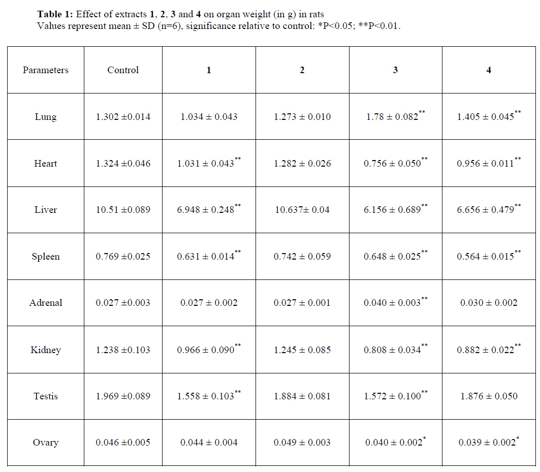
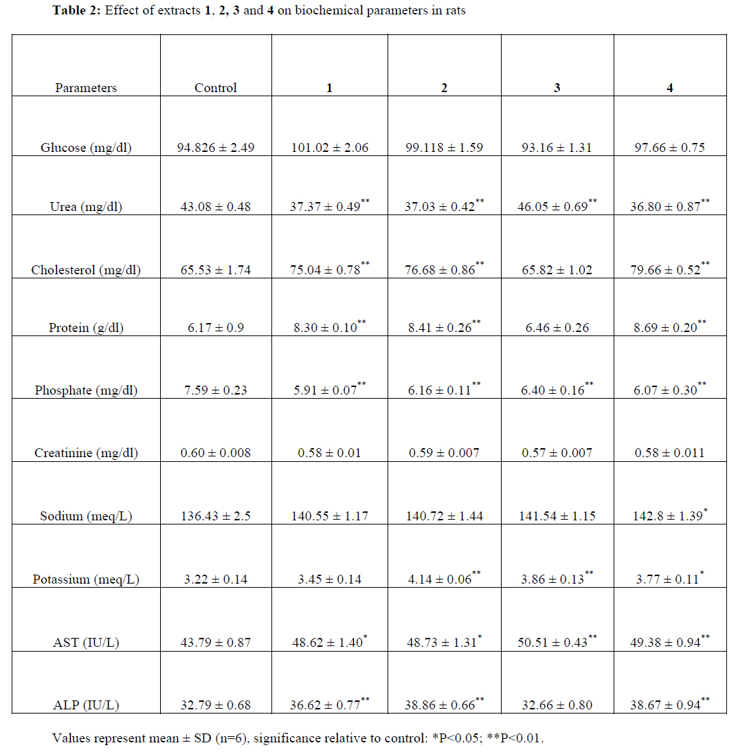
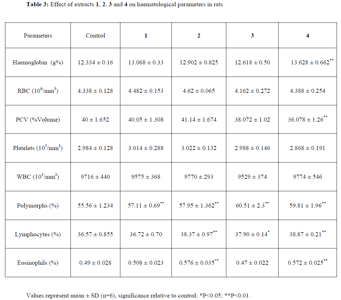
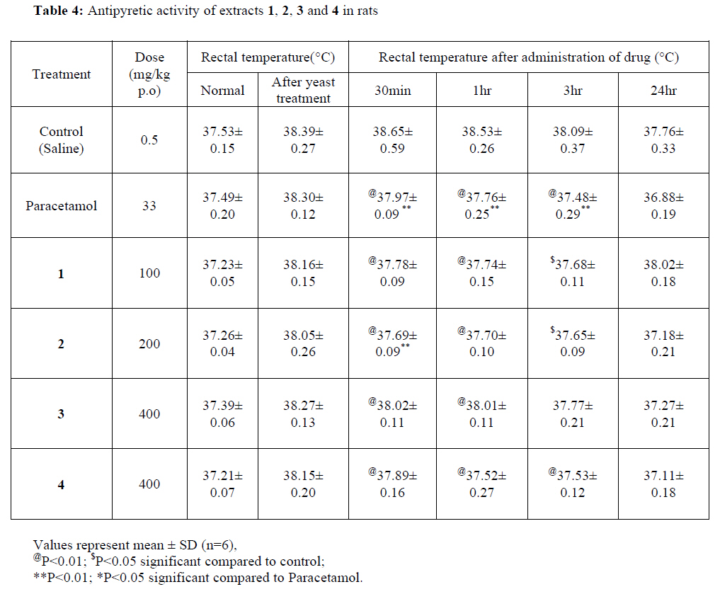
(Pharmacy), Murad Nagar-201 206, Uttar Pradesh, India. Code Number: tc09032 Abstract The active principles of brown alga, Turbinaria conoides (J.Agardh) Kuetz. (Sargassaceae) was extracted with n-hexane, cyclohexane, methanol and ethanol-water (1:1) and investigated for acute toxicity and antipyretic activity. Phytochemical analysis of the extracts revealed the presence of steroids, flavonoids and reducing sugars. Acute toxicity study was performed in Wistar rats after administration of extracts orally. No mortality was observed up to the dose of 5g/kg for methanol and ethanol-water (1:1) extracts whereas n-hexane and cyclohexane extracts were found to be toxic at the dose levels of 1g/kg and 2 g/kg respectively. In biochemical analysis, n-hexane, cyclohexane and ethanol-water (1:1) extracts caused a significant (P<0.01) increase in serum cholesterol, protein and alkaline phosphatase levels. In haematological studies, a significant difference was observed for cyclohexane and ethanol-water (1:1) extracts in polymorphs, lymphocytes and eosinophils when compared to the control. Antipyretic activity of extracts (100-400 mg/kg doses) was carried out on yeast-induced pyrexia in rats. Cyclohexane extract exhibited more significant antipyretic activity (P<0.01) than the other extracts at a dose of 200mg/kg (54.43%), which was comparable to that of paracetamol at a dose of 33 mg/kg. The findings validated the use of this brown alga in traditional cure of children’s fever. Keywords: Acute toxicity study; antipyretic activity; biochemical and haematological studies Turbinaria conoides Introduction Marine organisms are a rich source of structurally novel and biologically active metabolites. Secondary or primary metabolites produced by these organisms may be potential bioactive compounds of interest in the pharmaceutical industry. Recently, researchers have described a wide range of biological activities for algal compounds including anti-HIV, anti-neoplastic, cytotoxic and antipyretic activities. A number of sterols have been isolated from the brown algae belonging to the genus Turbinaria. Turbinaria conoides belongs to the family Sargassaceae. Turbinaria and other members of the family Sargassaceae are inedible, due to the concentration of polyphenolic substances based upon the polymerization of phloroglucinol (Norris and Fenical, 1982; Hay, 1984). Oxygenated steroids of algae have been reported to possess cytotoxic properties (Schroeder et al., 1980; Heltzel et al., 1994; Sheu et al., 1995; Sheu et al., 1996; Sheu et al., 1997a; Sheu et al., 1997b). The ethyl acetate extract of Turbinaria conoides and its oxygenated fucosterols have been reported for their cytotoxicity (Sheu et al., 1999). Traditionally, Turbinaria conoides has been used to cure children’s fever, as human food, fertilizer, insect repellent, antibacterial and pesticide (Erdmann and Bason, 2004). For brown alga to be ratified as a traditional medicine, a scientific approach through experimental and clinical validation of efficacy is required, as it is being done in modern medicine;animal toxicity studies are required to establish the potential adverse effects of Turbinaria conoides. Thus the present study was undertaken to determine the toxicity of the various extracts and to validate the traditional claims as an antipyretic remedy. Materials and Methods Algal material Turbinaria conoides was collected at Salin Munthal, Gulf of Mannar, Bay of Bengal, Ramanathapuram district, Tamil Nadu, India in September, 2005. It was authenticated by K.Eswaran and voucher specimen was deposited at Marine algal research station, Mandapam camp, Tamil Nadu, South India. Preparation of the extracts The algal material was coarsely powdered after air drying at room temperature. The powdered Turbinaria conoides (1 kg) was successively extracted with 2.5 l of n-hexane, cyclohexane, methanol and ethanol-water (1:1) each by maceration with occasional shaking at room temperature for 72 hr. Filtration using Whatman No. 1 filter paper and concentration of the filtrate under reduced pressure provided crude extracts n-hexane 1, cyclohexane 2, methanol 3 and ethanol-water (1:1) 4. Thus obtained residual extracts were kept in desiccator for further investigation. The yields of 1, 2, 3 and 4 were 0.21%, 0.22%, 8.68% and 10.31% w/w respectively. nhexane and cyclohexane extracts showed positive phytochemical tests for steroids; methanol extract for flavonoids and reducing sugars; ethanol-water (1:1) extract for reducing sugars (Khandelwal, 2004). Animal stock Wistar rats were used and fed with standard pellet feeds (Sai Durga feeds Ltd., Bangalore, India) and given water ad libitum. The animals were fasted prior to dosing according to Organization for Economic Cooperation and Development (OECD) guidelines, food but not water was withheld for 3-4 h. The principles of laboratory animal care were approved by the Institutional Animal Ethical Committee (IAEC) and Committee for the Purpose of Control and Supervision of Experiments on Animals (CPCSEA) (Proposal Number: CPCSEA/12/2000/PH-07-10). Acute toxicity Wistar rats weighing about 150-200g were used for acute toxicity study (Jacks et al., 2004; Nwafor et al., 2004; Lorke, 1983). The study was carried out as per the Organization for Economic Co-operation and Development guideline for the evaluation of acute oral toxicity (OECD, 2001). The animals were housed in a cross-ventilated room (12:12hr) and at constant temperature (22±2.5ºC) conditions. The animals were divided into eight groups. Group 1 was administered with Tween-80. Groups 2 – 8 received the suspension of extracts 1, 2, 3 and 4 in Tween-80 (1% w/v) in graded doses (0.10, 0.25, 0.50, 1.0, 2.0, 4.0 and 5.0 g/kg) body weight by oral administration. They were observed continuously and recorded systematically for the physical signs of toxicity including skin changes, mobility, aggressiveness and respiratory movements, sensitivity to sound and pain. Finally, the number of survivors was noted after 24 hr and LD50 was determined (Miller and Tainter, 1944). Gross pathological examination After 24 hrs of observation, the animals were sacrificed by cervical displacement after monitoring for further 13 days, vital organs were isolated, washed with saline and weighed (Benjamin, 1978). Biochemical analysis On the 14th day of experimental period, the blood (~5 ml) was collected from retro-orbital vein using fine capillary tube and centrifuged at 5000rpm for 15min (Varley et al., 1991). The serum was separated and collected into non-heparinized tubes for the following investigation: cholesterol, urea, protein, glucose, aspartate aminotransferase (AST), alkaline phosphatase (ALP) and electrolytes (sodium and potassium). Haematological studies The blood samples before centrifugation that were collected into heparinized tubes were used for the estimation of haemoglobin, red blood cells (RBC), white blood cells (WBC), packed cell volume (PCV), platelets, polymorphs, lymphocytes and eosinophils (Ringler and Dabich, 1979; Dacie and Lewis, 1991). These biochemical and haematological parameters were measured in the Diagnostic Laboratory of the Department of Physiology and Pharmacology, Vel’s College of Pharmacy, Chennai, India. Antipyretic study Either sex of adult Wistar rats weighing 180-230 g (12 weeks) was used. Animals were quarantined in light controlled room (12:12 hr) and at constant temperature (22±2ºC) conditions. 1/10th of the LD50 of each extract was considered for antipyretic study. Animals were distributed into six groups of six each. One group was administered with paracetamol (33 mg/kg) (Mutalik et al., 2003). Control group was given 0.5 ml normal saline. Pyrexia was induced in rats by injecting 20% (w/v) aqueous suspension of Brewer’s yeast intramuscularly. After 18 hr of yeast injection, extracts 1, 2, 3 and 4 in the doses of 100, 200, 400 and 400mg/kg respectively were administered to the other groups orally. The animals rectal temperature and the temperature of each animal was recorded at 0.5, 1, 3 and 24 hr. Percentage reduction in rectal temperature was calculated by considering the total fall in temperature to normal level as 100% (Rao et al., 1997; Dewan et al., 2000). Statistical Analysis The statistical significance between the groups was analyzed by means of an Analysis of variance (ANOVA) with subsequent Dunnet’s multiple comparison tests. Results of the study were expressed as mean± SD. A probability level of less than 0.05 was considered significant. Results and Discussion Oral administration of extracts 3 and 4 did not produce any mortality in rats up to a dose level of 5g/kg, but mild symptoms like tachypnoea and writhing were observed. Thus extracts 3 and 4 of Turbinaria conoides can be considered as substances with low toxicity. The body weight, food and water consumption of the animals treated with the extracts did not show any significant change when compared with the control group. Pathological examinations of the tissues indicated that there were no detectable abnormalities of gross pathological lesions. During the treatment period, 50% of death was recorded in extracts 1 and 2 treated groups given 1g/kg and 2g/kg respectively. The range of toxic symptoms like continuous grooming, aggressive behavior, not responding to stimuli, sedation, abdominal muscle twitch in all the groups and nasal secretion followed by restlessness, gasping and death was generally observed in the animals. As shown in Table 1, there were significant (P<0.05-0.01) differences between the control and treated groups in organ weights except extract 2. For biochemical parameters, extracts 1, 2 and 4 caused a significant (P<0.01) increase in serum protein, ALP and cholesterol levels. Extract 3 did not alter the levels of protein, ALP and cholesterol significantly. Serum glucose and creatinine levels were found to be non-significant, varying when compared with that of the control rats, whereas a significant change in urea (P<0.01) was found for the extracts treated groups. Extracts 1, 2 and 4 showed a significant decrease (P<0.01) in the concentration of urea when compared with control, whereas extract 3 significantly increased the level of urea. There was increased potassium level; however, extracts did not show much change in sodium level (Table 2) Table 3 portrays the haematological status of the rats. Extract 4 gave a significant decrease (P<0.01) in the level of PCV at a dose of 400 mg/kg body weight of extract 4. In contrast a significant increase (P<0.01) was observed in the level of haemoglobin, when compared to control rats. An increase in the values of the polymorphs, lymphocytes and eosinophils was registered (P<0.01) for extracts 2 and 4 when compared to the control. All the other parameters RBC, WBC and platelets in all treated groups remained normal without any significant difference. Table 4 depicts the antipyretic effect of extracts assessed using the Brewer’s yeast induced pyrexia in rats. A remarkable effect (54.43%) was noticed for extract 2 at a dose of 200 mg/kg. The effect became significant (P<0.01) for 30 min after the Brewer’s yeast administration, which was comparable to that of paracetamol. The antipyretic activity could be attributed to the presence of steroids in the extract. Normal rats did not show any decrease in the body temperature on oral administration of extracts. Since antipyretic activity is commonly mentioned as characteristic of drugs or compounds which have an inhibitory effect on prostaglandin biosynthesis (Vane, 1987), the antipyretic activity of cyclohexane extract could be due to the ability of inhibition of prostaglandin biosynthesis. Conclusion Methanol and ethanol-water (1:1) extracts can be considered to be highly non-toxic. Further the results revealed the significant antipyretic activity of cyclohexane extract of Turbinaria conoides when compared to control and standard groups (P<0.01). This potentiality supports the earlier traditional claims as a pediatric antipyretic remedy. Acknowledgements We thank Dr.K.Eswaran for authenticating the Brown alga. We also thank I.T.S.Paramedical College (Pharmacy), Ghaziabad, Uttar Pradesh and Vel’s College of Pharmacy, Chennai, Tamil Nadu for providing necessary facilities to carry out this research work. References
© Copyright 2009 - African. Journal. Traditional, Complementary and Alternative Medicines The following images related to this document are available:Photo images[tc09032t1.jpg] [tc09032t4.jpg] [tc09032t3.jpg] [tc09032t2.jpg] |
| |||||||||
