 |
| About Bioline | All Journals | Testimonials | Membership | News |
|
||||||
|
||||||
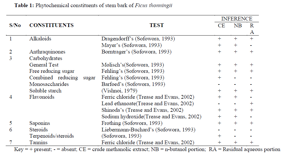
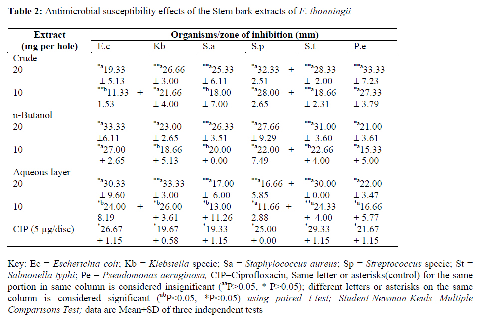
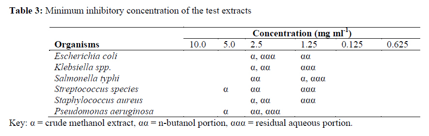
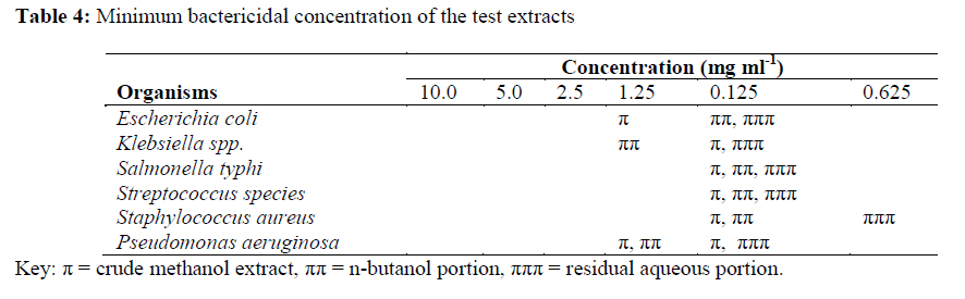
African Journal of Traditional, Complementary and Alternative Medicines, Vol. 6, No. 3, 2009, pp. 275-280 Research Paper QUALITATIVE PHYTOCHEMICAL SCREENING AND in vitro ANTIMICROBIAL EFFECTS OF METHANOL STEM BARK EXTRACT OF Ficus thonningii (moraceae) *Usman H, Abdulrahman, FI and Usman, A Department of Chemistry, Faculty of Science, University of Maiduguri, P.M.B. 1069 Maiduguri, Nigeria. Code Number: tc09039 Abstract The methanolic stem bark extract of Ficus thonningii (moraceae) was subjected to preliminary phytochemical screening and in vitro antimicrobial tests. The phytochemical tests was carried out using standard methods of analysis and these investigations revealed the presence of alkaloids, anthraquinones, carbohydrates, flavonoids, saponins and tannins. The antimicrobial activity of the plant extract was assayed using the agar plate disc diffusion and nutrient broth dilution techniques. Test micro organisms were: Escherichia coli, Klebsiella spp, pseudomonas aeruginosa, Salmonella typhi (Gram-negative), Staphylococcus aureus and Streptococcus spp. (Gram-positive). The extracts inhibited the growth of all the test organisms at different concentrations especially against pseudomonas aeruginosa and Streptococcus spp. which had mean inhibition zone of 33.33±7.33 mm and 32.33±2.51 mm respectively. The results showed the MIC of 10 mg ml-1 against pseudomonas and 1.25 against remaining organisms tested. The MBC against Staphylococcus aureus was 2.5 mg ml-1 and that of Streptococcus spp. was found to be 0.625mg ml-1. The extracts showed varied inhibitory activity against the organisms studied. Key words: Antibacterial, stembark, in vitro, phytochemical, Ficus thonningii, moraceae List of Abbreviations: HCl = Hydrochloric acid, H2 SO4 = tetraoxosulphate (VI) acid, KOH = Potassium hydroxide, MBC=Minimum bactericidal concentration, MIC = Minimum inhibitory concentrations, SD = Standard deviation, Ec = Escherichia coli; Kb = Klebsiella specie; Sa = Staphylococcus aureus; Sp = Streptococcus specie; St = Salmonella typhi; Pe = pseudomonas aeruginosa, CIP = Ciprofloxacin, MOA = Mechanism of action Introduction Traditional and folklore medicines play an important role in health services around the globe. About three quarters of the World’s population relies on plants and its extracts for health care (Premanathan et al., 2000; Gabhe et al., 2006). A good number of our population particularly those living in the villages depend largely on herbal remedies. Most of these herbal remedies have stood the test of time, particularly for the treatment of allergic, metabolic and cardiovascular diseases (Igoli et al., 2005). The genus Ficus of the moraceae family has more than 800 species of which 65 have been recorded in India. They are normally trees with minor leaf veins with out phloem transfer cells and grow to the height of about 10 meters with a thick brown stem (Sharma, 1993). The leaves are large, thick, leathery 25-40 cm long and 30 cm wide. Ficus thonningii contains bioactive substances in leaves, roots, fruits and flowers that are used alone or in combination with other plants for the treatment of diseases like diarrhoea, vomiting and mental illness. In the coastal areas of Nigeria, it is used to treat epilepsy while in the savanna area decoction of the leaves is used to treat mental illness. Also, the ethanolic extract of the leaves is found to exhibit analgesic activity (Igoli et al., 2005). The stem bark of this plant had been reported (Adamu, Pers. Comm.) to be used against diarrhoea, dysentery and wound infections. It is therefore, imperative to screen the said part of the plant against some pathogenic organisms responsible for such diseases. Materials and Methods Plant material The plant material (Voucher number, CHEM/003/2007) (Ficus thonningii Blum) was collected from Gidan madara in Mubi local government area of Adamawa State in April, 2007. The herbarium specimen was identified by Prof. S.S Sanusi of Biological Science Department, University of Maiduguri, Nigeria; where a voucher specimen was deposited for reference. Extraction and preparation of plant extract The stem bark was air-dried at room temperature for seven days and pulverized with mortar and pestle. 350 g of the powdered plant material was weighed and extracted exhaustively with methanol for two (2) weeks using maceration technique. A brownish solid mass which weighed 45.05 g (15.2 % w/w) was obtained and kept asceptically in a desiccator until required for use. About 30 g of the crude extract was completely suspended in 650 ml of distilled water and then filtered. The resulting filtrate was then partitioned with 500 ml n-butanol until the organic layer was visibly clear. The n-butanol and the residual aqueous layer were then concentrated on a reduced pressure to yield a brown-coloured mass and a grey-coloured substance respectively which were also kept in a desiccator until use. The crude stem bark extract and its partitioned portions were screened qualitatively for the phytochemical constituents utilizing standard methods of analysis (Vishnoi, 1979; Sofowora, 1993; Trease and Evans, 2002). Phytochemical Tests Molisch’s test for Carbohydrates: Few drops of Molisch’s reagent was added to each of the portion dissolved in distilled water, this was then followed by addition of 1 ml of conc. H2SO4 by the side of the test tube. The mixture was then allowed to stand for two minutes and then diluted with 5 ml of distilled water. Formation of a red or dull violet colour at the interphase of the two layers was a positive test (Sofowora, 1993). Barfoed’s test monosaccharides: About 0.5 g each portion was dissolved in distilled water and filtered. 1 ml of the filtrate was then mixed with 1 ml of Barfoed’s reagent in a test tube and then heated on a water bath for a period of 2 minutes. Reddish precipitate of cuprous oxide was considered as a positive test (Sofowora, 1993). Fehling’s test for free reducing sugar: About 0.5 g each portion was dissolved in distilled water and filtered. The filtrate was heated with 5 ml of equal volumes of Fehling’s solution A and B. Formation of a red precipitate of cuprous oxide was an indication of the presence of reducing sugars (Sofowora, 1993). Fehling’s test for Combined Reducing Sugars: About 0.5 g each portion was hydrolysed by boiling with 5 ml of dilute hydrochloric acid and the resulting solution neutralised with sodium hydroxide solution. To this, few drops of Fehling’s solution was added and then heated on a water bath for 2 minutes. Appearance of a reddish-brown precipitate of cuprous oxide indicates the presence of combined reducing sugars (Sofowora, 1993). Test for Tannins: About 0.5 g each portion was stirred with about 10 ml of distilled water and then filtered. Few drops of 1% ferric chloride solution were added to 2 ml of the filtrate occurrence of a blue-black, green or blue-green precipitate indicates the presence of tannins (Trease and Evans, 2002). Borntrager’s Test: About 0.2 g of each portion to be tested was shaken with 10 ml of benzene and then filtered. Five millilitres of the 10% ammonia solution was then added to the filtrate and thereafter the shaken. Appearance of a pink, red or violet colour in the ammoniacal (lower) phase was taken as the presence of free anthraquinones (Sofowora, 1993). Liebermann-Burchard test for steroids: To 0.2 g of each portion, 2 ml of acetic acid was added, the solution was cooled well in ice followed by the addition of conc. H2SO4 carefully. Colour development from violet to blue or bluish-green indicated the presence of a steroidal ring i.e. aglycone portion of cardiac glycoside (Sofowora, 1993). Test for terpenoids: A little of each portion was dissolved in ethanol. To it 1 ml of acetic anhydride was added followed by the addition of conc. H2SO4. A change in colour from pink to violet showed the presence of terpenoids (Sofowora, 1993). Test for Saponins: One gram of each portion was boiled with 5 ml of distilled water, filtered. To the filtrate, about ml of distilled water was further added and shaken vigorously for about 5 minutes. Frothing which persisted on warming was taken as an evidence for the presence of saponins (Sofowora, 1993). Shinoda’s test for flavonoids: About 0.5 of each portion was dissolved in ethanol, warmed and then filtered. Three pieces of magnesium chips was then added to the filtrate followed by few drops of conc. HCl. A pink, orange, or red to purple colouration indicates the presence of flavonoids (Trease and Evans, 2002). Ferric chloride test for flavonoids: About 0.5 of each portion was boiled with distilled water and then filtered. To 2 ml of the filtrate, few drops of 10% ferric chloride solution were then added. A green-blue or violet colouration indicated the presence of a phenolic hydroxyl group (Trease and Evans, 2002). Lead ethanoate test for flavonoids: Few quantity of the each portion was dissolved in water and filtered. To 5 ml of each of the filtrate, 3 ml of lead ethanoate solution was then added. Appearance of a buff-coloured precipitate indicates the presence of flavonoids (Trease and Evans, 2002). Sodium hydroxide test for flavonoids: Few quantity of the each portion was dissolved in water and filtered; to this 2 ml of the 10% aqueous sodium hydroxide was later added to produce a yellow colouration. A change in colour from yellow to colourless on addition of dilute hydrochloric acid was an indication for the presence of flavonoids (Trease and Evans, 2002). Test for alkaloids: Few quantity of the each portion was stirred with 5 ml of 1% aqueous HCl on water bath and then filtered. Of the filtrate, 1 ml was taken individually into 2 test tubes. To the first portion, few drops of Dragendorff’s reagent were added; occurrence of orange-red precipitate was taken as positive. To the second 1 ml, Mayer’s reagent was added and appearance of buff-coloured precipitate will be an indication for the presence of alkaloids (Sofowora, 1993). Test for Soluble Starch: Few quantity of each portion was boiled with 1 ml of 5% KOH, cooled and acidified with H2SO4. A yellow colouration was taken as the presence of soluble starch (Vishnoi, 1979). Microorganisms Test micro organisms were: Escherichia coli, Klebsiella spp., pseudomonas aeruginosa, Salmonella typhi, Staphylococcus aureus and Streptococcus spp. All the organisms used in this study were clinical isolates obtained from Department of Medical Microbiology, College of Medical Sciences, University of Maiduguri, Nigeria. Standard antibiotic (Ciprofloxacin 5 μg/disc, Batch no. CT0425B; Expiry: 2011/11) measuring 6 mm in diameter was manufactured by Oxoid® , England. Preparation of sample extract for microbiological assay About 1g of each extract was dissolved in 10 ml (100 mg ml-1) of peptone water to obtain a stock solution; the working solution was prepared thus: The extract was diluted as 1:10 equivalent to 100 mg ml-1 and 1:5 dilution equivalent to 50 mg ml-1, from which 0.2 ml of the 1:10 dilution was dispensed into a bored-agar hole (9 mm diameter with a depth of 5 mm) making a concentration of 20 mg per hole and also 0.2 ml of the 1:5 dilution was poured into the ditch hole bored onto the agar equivalent to 10 mg per hole. Disc diffusion technique The method of Vollekovà et al., (2001) as modified by Usman et al., (2005) was adopted. The three (3) samples that is; crude extract, n-butanol portion and aqueous layer were tested on the 6 isolates. Three holes were bored in the plates (9 mm diameter) using sterile cork-borer. About 0.2 ml of the extract was inoculated across the wells and incubated at 37 oC for 18-24 hrs. After incubation, the average diameter of three readings of the clear zone surrounding the hole was taken as the measure of the inhibitory level of the plant extract against the bacteria on test and recorded as mean ± SD. Determination of minimum inhibitory concentration (MIC) A serial dilution ranging from 1:10 to 1:0.125 was made. The bacterial strain was cultured in nutrient broth and suspended in 5 ml peptone water. To the suspension, 5ml of each extract concentration was added into nutrient broth and then 1.0 ml of standardized broth cultures containing 1.0 x 10s7 CFU/ml were seeded into each test tube and then incubated at 37o c for 18-24 hrs. Following incubation turbidity was examined; the concentration at which no turbidity was observed is regarded as the MIC value (Williams and Wilkins, 2007). Determination of minimum bactericidal concentration (MBC) Suspensions from the MIC studies were used for the MBC determination; to a solid media a bacterial streaking of equal streaks of the suspension from the MIC was made and the procedure repeated all through the required numbers of the corresponding isolates. The isolated organism on the blood agar was incubated at 370c for 18-24 hrs. After incubation, the plates were observed; the concentration that exhibited no bacterial growth was considered as the MBC value (Williams and Wilkins, 2007). Results The results of the phytochemical screening of stem bark extract of Ficus thonningii is as presented in Table 1. The extract and its partitioned portions were further subjected to anti-microbial studies. The susceptibility pattern against the test organisms is shown in Table 2; meanwhile the minimal inhibitory concentration is presented in Table 3. Table 4 shows the minimal bactericidal concentration on the test organisms. Discussion The phytochemical test of the crude methanolic stem bark extracts of Ficus thonningii revealed the presence alkaloids, carbohydrates, flavonoids, saponins and tannins. Steroid and monosaccharides were found to be absent. The absence of monossacharides is relative to its absence in the compounds containing glycosides and thus might not enhance antibacterial activity in this extract. These metabolites are similar to those found in F. sycomorus (Victor, 2006; Ibrahim et al., 2008); except for anthraquinones which is found in this species. These classes (such as alkaloids, saponins, tannins, anthraquinones, and flavonoids) of compounds are known to have curative activity against several pathogens and therefore could suggest the use traditionally for the treatment of various illnesses (Hassan et al., 2004; Usman and Osuji, 2007). The in vitro anti microbial test presented in Table 1 showed the susceptibility test against grams positive and negative organisms. The n-butanol extract exhibited considerable level of inhibition against all the test organisms; the results from most portions were found to be significantly higher (P<0.05) compared to ciprofloxacin against Klebsiella spp., Streptococcus spp. at 20 mg/hole. Most portions insignificantly (P>0.05) differ with ciprofloxacin at 10 mg/hole; this is suggestive of the presence of some compounds or groups in the extracts with similar MOA (mechanism of action) to that of ciprofloxacin at higher concentrations. The highest activity was exhibited by crude extract against pseudomonas aeruginosa (33.33 ± 7.23 mm) and the lowest exhibited by residual portion was against Streptococcus spp (16.66 ± 5.85 mm). However, it is suggested that plant extracts exhibiting diameters of zones of inhibition ≥ 10mm were considered active (Zwadyk, 1972; Usman and Osuji, 2007). From the results of the MIC and MBC presented in Tables 2 and 3 respectively; it was noticed that the broadest activity of the extract against most grams negative organisms was 2.5 mg ml-1 as MIC while the MBC of 0.125 mg ml-1 was similarly noted. The extract exhibited some appreciable level of activity against the two gram positive bacteria assayed Staphylococcus aureus and streptococcus specie. According to a research by Usman and Osuji (2007) who worked on the crude leaves extracts of Newbouldia laevis against similar organism found similar broad activity recorded against most Gram negative organisms studied compared to the results presented in Table 3 which shows n-butanol and residual aqueous exhibiting highest activity of 0.625 mg ml-1 against the two Gram positive bacteria assayed. In line with this, it is believed that the two extracts (NB, RA) are better antibacterial agents than the crude methanolic extract. S. aureus has been known to play a vital role in invasive skin diseases including superficial and deep follicular lesion (Usman and Osuji, 2007). The broad antibacterial activities of this extracts could be as a result of the plant secondary metabolites (alkaloids, anthraquinones, flavonoids, tannins, saponins) present in the extracts; in line with these findings, Usman and Osuji (2007), reported that tannins had been widely used topically to sprains, bruises and superficial wounds as such, it could be probable that tannins and other plant phenolics from this extract were responsible for these broad activities. From the pattern of the activity, the extract is said to be active as the polarity and concentration increases, notably inhibition by RA against Gram-negative organisms (EC, KB, ST), but these activities were insignificantly different (P>0.05) from nbutanol portion at 20 mg ml-1 against all the test bacteria. The n-butanol portion seems to be comparable to the activities of ciprofloxacin at both concentrations relative to the other portions. In conclusion, the extracts showed varying inhibitory activities against all the test organisms. The results are encouraging enough to pursue bioactivity guided fractionation of this extracts and structure elucidation of the active phyto-constituents from the extract of this plant species as a possible anti bacterial agent. Therefore, the use of this part of the plant by the traditional healers for the treatment the aforementioned diseases have been validated. Acknowledgements The authors wish to acknowledge the technical assistance rendered by Messrs Fine Akawu and Samson Gamache of the Departments of Chemistry and Medical Microbiology, University of Maiduguri and University of Maiduguri Teaching Hospital; respectively. References
© Copyright 2009 - African. Journal. Traditional, Complementary and Alternative Medicines The following images related to this document are available:Photo images[tc09039t2.jpg] [tc09039t3.jpg] [tc09039t1.jpg] [tc09039t4.jpg] |
| |||||||||
