 |
| About Bioline | All Journals | Testimonials | Membership | News |
|
||||||
|
||||||
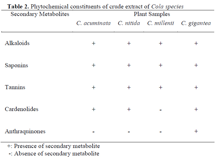
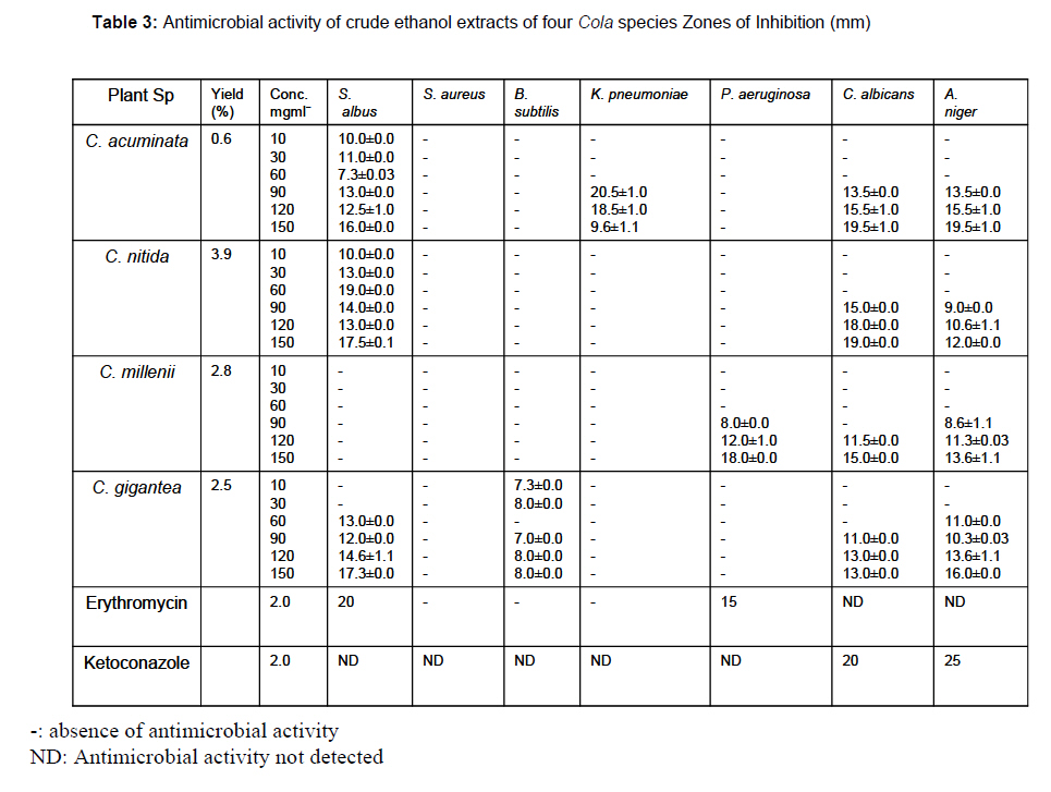
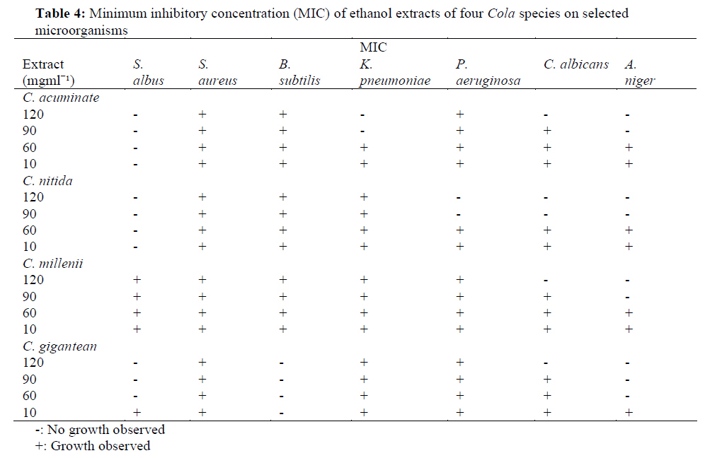
African Journal of Traditional, Complementary and Alternative Medicines, Vol. 6, No. 4, 2009, pp. 518-525 Research Paper PHYTOCHEMICAL AND ANTIMICROBIAL STUDIES OF FOUR SPECIES OF Cola SCHOTT& ENDL. (STERCULIACEAE) Mubo Adeola Sonibare1 , Micheal O. Soladoye2, Oyedokun O. Esan 2, Oluwadayo O. Sonibare3 1Department of Pharmacognosy, Faculty of Pharmacy, University of Ibadan, Nigeria Code Number: tc09041 Abstract The in-vitro antimicrobial evaluation of ethanol extracts of four species of Cola Schott & Endl. was done using human isolated strains of Staphylococcus aureus, Staphylococcus albus, Bacillus subtilis, Klebsiella pneumonia, Pseudomonas aeruginosa, Candida albicans, Aspergillus niger as test organisms. The assays were carried out by agar well diffusion, erythromycin and ketoconazole served as the control drugs. The leaf ethanol extracts of the plants were found to be more effective against the tested fungi than the bacteria at high concentrations. None of the extracts was active against Staphylococcus aureus. Plant extract of C. acuminata (P. Beauv.) Schott & Endl. and C. nitida (Vent) Schott & Endl. showed activity on S. albus at concentrations ranging from 10-150 mgmlˉ¹ having comparable diameters of zone of inhibition of 7.3±0.03-16.0±0.0 for C. acuminata and 10.0±0.0-19.0±0.0 for C. nitida. Also, these two species of Cola demonstrated activities on C. albicans and C. millenii at concentrations ranging from 90-150mgmlˉ¹ with relatively close diameters of zone of inhibition. Only C. acuminata inhibited the growth of K. pneumoniae at the MIC of 90mgmlˉ¹ whereas, C. albicans was inhibited by C. acuminata, C. millenii K. Schum and C. gigantea A.Chev. at the MIC of 120mgmlˉ¹. Phytochemical screening of the four species of Cola showed the presence of alkaloids, saponins, tannins and cardenolides in all the plants which apart from showing the probable closeness of the species could also be responsible for the observed activities. The antimicrobial property shown by the plant extracts is an evidence of the ethnomedicinal uses of the plants. The similarity observed in the phytochemical constituents and antimicrobial activities demonstrated by C nitida (Vent.) Schott & Endl., C. millenii and C.gigantea A. Chev. and C. acuminata suggest a probable closeness among these species. The results obtained in this study provide preliminary evidence of the chemotaxonomic significance of secondary metabolites and antimicrobial activities in infra-generic taxonomy of species of Cola. Key words: Cola species, Sterculiaceae, phytochemical screening, antimicrobial studies, taxonomy. Introduction Cola Schott & Endl. (Sterculiaceae) is a genus of about 125 species of trees indigenous to the tropical rain-forest African region (Ratsch, 2005). Phylogenetic information reveals that the genus was formerly classified in the family Malvaceae, subfamily Sterculioideae and was later transfered into the separate family Sterculiaceae. Cola is one of the largest in the family Sterculiaceae and is related to the South American genus Theobroma. It comprises of evergreen moderately sized trees often growing to a height of 20m with glossy ovoid leaves up to 30cm long. Cola species are found mostly in the relatively dry parts of the rain forest, although Cola millenii and Cola gigantea are widely distributed in wet and dry forest environments (Kuoame and Sacande, 2006; Olorode, 1984). According to Russell (1955), the systematics of Cola species was in a state of “indescribable confusion”. In an attempt to resolve this confusion, Chevalier and Perrot (1911) created the Subgenus Eucola containing five species of edible kolanuts – Cola nitida (important for trade), Cola acuminata (important for socio-cultural values), Cola ballayi, Cola verticillata and Cola sphaerocarpa. The latter three species are not known to be cultivated The mature fruit of Cola species is a nut known as kolanut (Duke, 2001). It has a bitter flavour and high caffeine content (Blades, 2000; Benjamin et al., 1991). It is chewed in many West African cultures individually or in a group setting. It is often used ceremonially, presented to tribal chiefs or to guests. Chewing kolanut can ease hunger pangs. Kolanuts are used mainly for their stimulant and euphoriant qualities. They have effects similar to other xanthine containing herbs like cocoa, tea etc. However, the effects are distinctively different, producing a stronger state of euphoria and well being (Benjamin et al., 1991). They have stimulant effects on the central nervous system and heart. Kolanuts are used as a source of alkaloids in pharmaceutical preparations (Newall et al., 1996; Bradley, 1992; Opeke, 1992). Various medicinal and pharmacological values have been observed in species of Cola (Daels- Rakotoarison et al., 2003; Steinegger and Hansel, 1992). Kolanuts are often used to treat whooping cough and asthma. The caffeine present acts as a bronchodilator, expanding the bronchial air passages (Jayeola, 2001; Kim, 2001). Kolanuts are also employed in the treatment of malaria and fever (Odugbemi, 2006). Experiments using animals indicate that kolanuts have analeptic and lipolytic properties and stimulate the secretion of gastric juices (GRIN, 2007). Odugbemi (2006) reported that the leaves of Cola millenii are used in the treatment of ringworm, scabies, gonorrhoea, dysentery and opthalmia. Traditionally, the leaves, twigs, flowers, fruit follicles and the bark of Cola nitida and Cola acuminata are used to prepare a tonic as a remedy for dysentery, coughs, diarrhoea, vomiting and chest complaints (Burkill, 1995; Irvine, 1961). This paper reports the phytochemical and antimicrobial activities of four species of Cola with a possible evaluation of their chemotaxonomic significance. Materials and methods Plant materials Fresh leaves of Cola acuminata was collected in Ibadan, Oyo State, Nigeria. Cola nitida, Cola millenii and Cola gigantea were collected at different location in Ago-Iwoye, Ogun State, Nigeria in June 2007. The plants were identified and authenticated by Mr. T.K Odewo at the Forest Herbarium Ibadan (FHI) where the voucher specimens were also deposited under the following numbers: Cola acuminata (P. Beauv.) Schott& Endl. FHI 107892, Cola milleni K. Schum. FHI 107893, Cola nitida (Vent) Schott & Endl. FHI 107894, Cola gigantea A. Chev. var. gigantea Bull. FHI 107895. The voucher information which includes locality of collection and herbarium numbers of the Cola species are presented in Table 1. Extraction of Plant Material The air-dried and powdered leaves were extracted by maceration of 200g of the dried, pulverized leaves at room temperature for 48 hours in 2.5 litres of 96% ethanol. The mixture was filtered using Whatman No. 1 filter paper and the filtrate solution was evaporated in a water bath at 70ºC to obtain a paste. The extracts gave yields of 3.9%, 0.6%, 2.8% and 2.5%, respectively, for C. nitida, C. acuminata, C. millenii and C. gigantea. Phytochemical screening The dried, pulverized leaves were subjected to phytochemical analysis to screen for the presence of secondary metabolites such as alkaloids, saponins, anthraquinones, cardenolides and tannins. The phytochemical screening was carried out using standard procedure (Ajaiyeoba et al., 2003; Trease and Evans, 1989). Brief description is as follows: Alkaloids: 70ml of 10% HCl was added to 4g of each sample in appropriately labeled conical flasks and boiled for 10 mins. Each boiled sample was filtered and allowed to cool. The filtrates were poured into four labeled test tubes. Few drops of Dragendoff’s, Mayer’s, Wagner’s reagents were added to each test tube separately. Alkaloids were recorded as present in the sample if turbidity or a brownish precipitate was observed. Saponins: 4g of each sample was dissolved in distilled water and heated for 2-5 mins. The mixtures were filtered, allowed to cool and shaken continuously for 2 mins to induce the production of froth. They were then left to stand for 15 mins. The observation of frothing was indicative of presence of saponin. Test for Tannins: 1g of each sample was heated with 20ml of water for 5 mins in appropriately labeled testtubes. Each solution was allowed to cool and then filtered. 1m of each filtrate was diluted with 5ml distilled water in a test tube; few drops of 0.1% ferric chloride solution were added. A characteristic blue, blue-black, green or blue-green colour and precipitate indicate the presence of tannin. Anthraquinones: 1g of each sample was shaken with 10ml of ferric chloride solution mixed with 5ml of HCL. Each mixture was heated in a water bath for 10-15 mins, filtered and allowed to cool. The filtrate was extracted with chloroform and shaken gently. The clear layers at the base were pipette into test tubes and 2ml each of ammonia solution was added. An observation of a delicate pink rose colour indicated the presence of anthraquinones. Cardenolides: 4g of each sample were extracted in test tubes with 80% ethanol, all appropriately labeled. They were then divided into two portions for Kedde’s test and Keller-Killiani’s test. For Kedde’s Test, few drops of 10% lead acetate were added to each of the tubes, followed by few drops of distilled water and chloroform. The contents were then evaporated to dryness in a water bath. 5% NaOH was added to each residue and then 2% of 3.5 dinitrobenezene acid. For Keller – Killiani’s Test, few drops of 10% lead acetate, water and chloroform were added to each test sample. The mixtures were also evaporated to dryness in a water bath and subsequently, few drops of concentrated sulphuric acid were added. For Keller-Killiani’s test, a brown ring indicated the presence of cardenolides while for the Kedde’s test, a brown to purple colour was indicative of presence of cardenolides. Antimicrobial screening Microorganisms: Four human pathogenic bacteria made up of two Gram-positive (Staphylococcus aureus, Staphylococcus albus: ATCC: 27856 (Wilson and Stuart, 1965; Stich, 1932), and two Gram-negative (Klebsiella pneumoniae, Pseudomonas aeruginosa: ATCC: 27856) and Bacillus subtilis (ATCC: 1457), were used for the antibacterial assay. One yeast (Candida albicans (MTCC: 227) and one mold (Aspergillus niger (MTCC: 227) were used for the antifungal assay. All the organisms were local isolates from the Laboratory bacterial stock of the Department of Plant Science and Applied Zoology, Olabisi Onabanjo University, Ogun State, Nigeria. Three to five identical colonies from stored slopes of microorganisms (bacteria and fungi) were lifted with a sterile wire loop and transferred into a 5ml single strength nutrient broth (Biochemica, Spain) contained in well labeled screw cap bottles for each bacterium and fungus respectively. The bottles were well shaken and incubated at room temperature for 18-24h for bacteria and 72h for fungi. The agar well diffusion method was used to test the plant extracts for antimicrobial activity. Briefly, 15ml of melted and cooled nutrient agar (Himedia Laboratories, India) and potato dextrose agar (Himedia Laboratories, India) were added to 0.2ml of 1 in 100 dilutions of bacteria and fungal cultures respectively in sterile Petri dishes. The contents were mixed. After the agar in each plate solidified, 6 wells of 5mm each were bored in each plate using an aseptic cork borer. 0.1ml of plant extracts at varying concentrations (10mgmlˉ¹, 30mgmlˉ¹, 90mgmlˉ¹, 120mgmlˉ¹, 150mgmlˉ¹) as well as the standard antibiotic solution was loaded into the wells. Control experiments were set up using erythromycin and ketoconazole (2mgmlˉ¹) for the bacterial and fungal assays respectively. The plates were incubated at 370C for 24h for bacteria and 48h for fungi. All inoculation procedures were undertaken under aseptic conditions. According to pharmacological and biometric specifications, the antimicrobial studies were done in triplicates. With the aid of a transparent ruler the diameters of zones of inhibition around the wells were measured in mm for all the three replicates and the average of the three measurements was calculated as an indication of activity. The minimum inhibitory concentration (MIC) of plant extracts was determined using the broth dilution method as described by Sahm and Washington (1990). Briefly, 1ml of the extract solution at the concentration of 120mgmlˉ¹ was added to 1ml of nutrient broth and subsequently transferred to make solutions of varying concentrations (120, 90, 60, 10 mgmlˉ¹) in different test tubes. Then 1ml of bacterial and fungal suspensions and 0.1ml of plant extracts at the different concentrations was added to each test tube and incubated at 370C for 24h for bacteria and 48h for fungi. The test tube with the concentration of plant extract at which no detectable growth was observed was considered as the MIC. Results The result of the phytochemical screening of the leaf samples is presented in Table 2. The secondary metabolites tested for were alkaloids, saponins, tannins, cardenolides and anthraquinones. The result shows that alkaloids, saponins and tannins are present in all the four species. Anthraquinones are absent in all species except C. gigantea. Cardenolides are present in C. acuminata, C. nitida and C. gigantea but absent in C. millenii. The results of the antimicrobial screening of the ethanol crude plant extracts of the species are presented in Table 3 while Table 4 shows the minimum inhibitory concentration (MIC) of each extract. The plant extracts were found to be more effective on the tested fungi than they were on bacteria. All the extracts showed important inhibition of fungal growth at the concentrations of 90, 120 and 150mgmlˉ¹. Only a few bacteria were susceptible to the extracts at high concentration. No antibacterial activity was observed against Staphylococcus aureus, a Gram-positive bacterium. Only the crude extract of C. acuminata was active against Klebsiella pneumoniae, (a Gram-negative bacterium) with the diameter of the zone of inhibition of 9.6mm and the MIC of 90mgmlˉ¹. Likewise, the extract of C. millenii was the only extract that inhibited the growth of Pseudomonas aeruginosa (a Gram negative bacterium) at a minimum diameter of zone of inhibition of 8.3mm and an MIC of 90mgmlˉ¹. Bacillus subtilis was resistant to all the extracts, except to that of C. gigantea, which was active against the organism at an MIC of 10mgmlˉ¹. Extracts of C. acuminata and C. nitida were active against the two fungi Aspergillus niger and Candida albicans, both showed activity at MIC of 90mgmlˉ¹. C. millenii extract inhibited the growth of Aspergillus niger at an MIC 90mgmlˉ¹ but was only able to inhibit the growth of Candida albicans at higher concentrations of 120mgmlˉ¹ and 150mgmlˉ¹. The extract of C. gigantea was active against Aspergillus niger and Candida albicans at MICs of 60mgmlˉ¹ and 90mgmlˉ¹ respectively. Discussion. The presence of the secondary metabolites (alkaloids, saponins and tannins) in Cola acuminata, Cola nitida , Cola millenii and Cola gigantea partly enhances the chemotaxonomic characterization of the four Cola species. Although the presence of similar secondary metabolites may not necessarily justify the generic closeness of these species, it is a noteworthy observation that requires further studies in attempting to resolve the relationship among the three species. Owing to the presence of these three secondary metabolites and the similarity of their occurrence, it can be proposed that the four Cola species are appropriately classified into the same genus. This suggests that the four plants are closely related and could be assumed to have a common origin, a claim supported by the previous work (Adegoke et al., 1968; Keay, 1989; Oliver-Bever, 1986; Purseglove, 1968; Oliver, 1960). This claim could be strengthened with a further evaluation of the active principles responsible for the antimicrobial activities observed in these species. The environment is known to potentially influence the morphology and expression of compounds in plants (Folkers et al., 2008; Shen et al., 2008; Tsukaya et al., 2007; Braga et al., 2006; Cybulskill et al., 2000). Plant physiologists have reported that a particular compound may be produced only at certain times or under certain conditions. This may be the case with Cola gigantea , the only plant that showed a rare presence of anthraquinones. C. gigantea is mainly found in forests while the other three species are more domesticated in their habitat. Therefore, the presence of anthraquinones in C. gigantea only could be due to environmental conditions. The absence of cardenolides in Cola millenii alone is an unusual occurrence; more research is needed to make a meaningful taxonomic deduction of this condition. Antimicrobial activities shown by the four Cola species are in line with previous antimicrobial works on the species of Cola (Reid et al., 2005; Adeniyi et al., 2004; Ebana et al., 1991) where Cola extracts were found to exhibit important inhibitory activities against the growth of certain bacteria and fungi. The crude ethanolic extract of C. acuminata, C. nitida and C. gigantea showed important activity against Staphyloccus albus. The diameters of the zones of inhibition of these extracts were found to be remarkably close to that of the control drug: erythromycin. The MICs were 10mgmlˉ¹, 10mgmlˉ¹ and 60mgmlˉ¹ respectively. However, the leaf ethanol extract of C. millenii was inactive against this organism. C. acuminata showed the most important activity against Staphylococcus albus, Klebsiella pnuemoniae, Aspergillus niger and Candida albicans. This is probably due to the strong presence of alkaloids in C. acuminata as reported by Adegoke et al., (1968). C. gigantea also had a high antimicrobial activity against Staphylococcus albus, Bacillus subtilis, and on Aspergillus niger and Candida albicans whereas, C. nitida and C. millenii had weak inhibitory effects on the growth of all the microorganisms. There is a need for further study to ascertain if the yield in these species would be increased by using stronger fractionating solvents such as ethyl acetone or methyl acetone. These solvents have been reported to be more vigorous than other solvents used in crude extraction of plants (Ajayeioba and Fadare, 2006). An important occurrence is that none of the extracts was effective against Staphylococcus aureus. This is in contrast to the observations in some other studies where the tested plants were more active against Grampositive bacteria (Aladesanmi et al., 2007; Ajaiyeoba and Fadare, 2006; Isu, 2005; Onocha et al., 2003). Generally, the antifungal activities of the extracts as reported in the results were stronger and more pronounced than the antibacterial activities. C. acuminata and C. nitida showed high inhibitory activity against the two fungi Aspergillus niger and Candida albicans. The presence of different secondary metabolites in the species of Cola is probably offering the therapeutic basis for the antimicrobial activities observed in these species which is in agreement with other work linking antimicrobial activities with the presence of secondary metabolites (Kisangau, et al., 2007; Kubmamwa et al., 2007; Ajaiyeoba and Sama, 2006; Reid at al., 2005). This claim is further strengthened by the work of Tiew et al. (2003) where the antifungal properties of other members of the Sterculiaceae family were reported. These plants could be a source of new antibiotic compounds. Further work is needed to isolate the secondary metabolites from the extracts studied in order to test specific antimicrobial activity. In addition, the minimun inhibitory concentration (MIC) of the plants yielded promising results that are worthy of note. C. acuminata, C. nitida and C. gigantea had low MICs of 10mgmlˉ¹, 10mgmlˉ¹ and 60mgmlˉ¹ respectively for Staphylococcus albus. This suggests that they can be gainfully employed in the production of antibiotics, as low MICs mean that only a small quantity of the extract will be required to impair bacterial growth. The average minimum MIC of the plants on Pseudomonas aeruginosa, Klebsiella pnuemoniae, Bacillus subtilis, Aspergillus niger and Candida albicans was 90mgmlˉ¹, a value which is still low enough to be of great antimicrobial advantage. The closeness observed in the antimicrobial activities demonstrated by C. nitida and C. millenii as revealed by values obtained for the MIC could also indicate a close relationship between the two species. In conclusion, the occurrence of a similar pattern of secondary metabolites in three out of the four species of Cola studied is suggestive of an important trend in the species. Further studies on structural elucidation of the compounds may offer some information which may become very useful in the knowledge of the natural relationship of the four plants. The ethanol crude extract of C. acuminata had the most important effect against both bacteria and fungi. Ethanol crude extracts of the other three species also had important effects on some of the microorganisms. Therefore, the plants are justified in their ethnomedicinal uses in the treatment of certain diseases, especially fungal diseases. The comparison of data obtained suggests a close relationship between C. gigantea and C. acuminata and also between C. nitida and C. millenii. Based on the presence of the typical secondary metabolites in the ethanol leaf extracts of the four species together with the antimicrobial activities demonstrated against various organisms, our study has highlighted the possible usefulness of phytochemical and antimicrobial studies as taxonomic tools in evaluating closeness among four species of the genus Cola. This provides evidence for further research in the chemical profile of the genus. It is important to mention that C. acuminata and C. gigantea gave the best all-round results. Acknowledgements We are grateful to Mr T. K. Odewo of the Forest Herbarium, Ibadan for helping with the identification and authentication of the plants and also to the anonymous reviewer for helpful comments on the earlier version of the manuscript. References
© Copyright 2009 - African. Journal. Traditional, Complementary and Alternative Medicines The following images related to this document are available:Photo images[tc09041t3.jpg] [tc09041t1.jpg] [tc09041t2.jpg] [tc09041t4.jpg] |
| |||||||||
