 |
| About Bioline | All Journals | Testimonials | Membership | News |
|
||||||
|
||||||
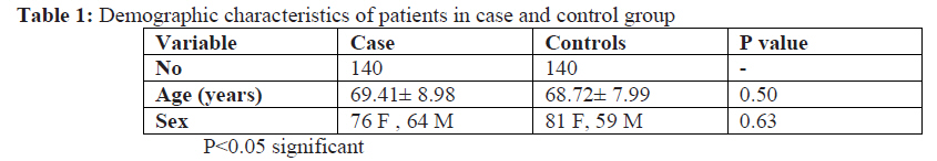
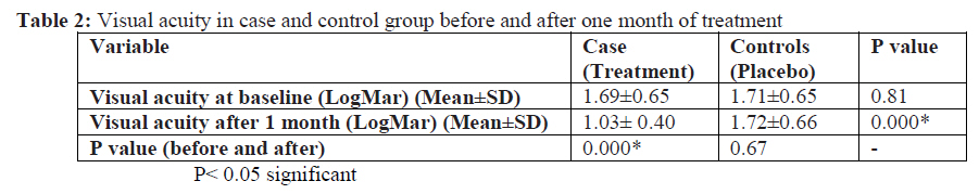
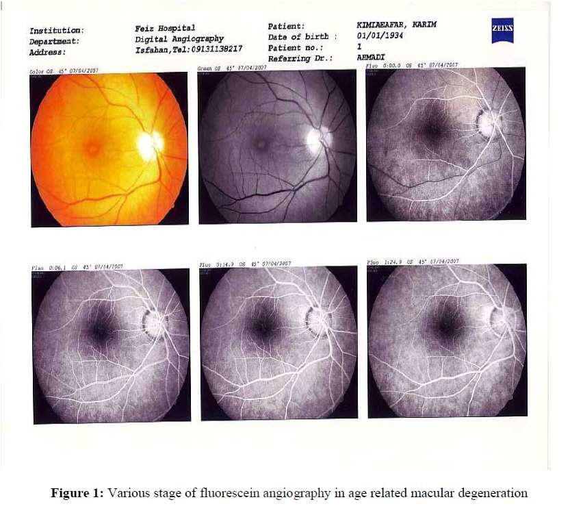
African Journal of Traditional, Complementary and Alternative Medicines, Vol. 6, No. 4, 2009, pp. 549-553 Research Paper THE EFFECT OF HESA-A (NATURAL DRUG) ON VISUAL ACUITY IN AGE RELATED MACULAR DEGENERATION: A RANDOMIZED DOUBLE BLIND CONTROLLED CLINICAL TRIAL Amrollah Ahmadi1 ; Heshmatollah Ghanbari2; Masoud Soheilian3; Mohsen Naseri4 1Institute of Cancer Research Center, Medical Sciences/University of Tehran, P.O.Box: 13185-1678,
Tehran, Iran Code Number: rc09046 Abstract We investigated the clinical efficacy and safety of HESA-A (a drug of herbal-marine origin) in the treatment of age related macular degeneration (AMD). In a randomized double blind clinical trial 280 eyes of 280 (157 F, 123 M) patients with wet and dry AMD were randomly assigned in treatment or placebo groups. Patients in treatment group received HESA-A tablet 25 mg/Kg twice a day orally and controls received placebo with the same method for 4 weeks. Visual acuity at baseline and after one month of treatment was measured and compared between two groups. All patients were followed up for 5 months after treatment. Mean patients’ age was 69.06±8.49 years. At the end of study visual acuity improved significantly from 1.69±0.65 LogMar to 1.03± 0.40 LogMar in treatment group but not in controls (P: 0.000 and P: 0.67 in treatment and control groups respectively). No drug reaction or recurrence was reported during the study and 5-month post treatment follow up period in HESA-A treated group. This study showed significant efficacy and safety of HESA-A in improvement of visual acuity in AMD patients in short term. Key words: age related macular degeneration, HESA-A, Introduction Age-related macular degeneration (AMD) is the most common cause of severe visual loss and blindness in people over 50 years of age (Klein et al., 1992; Imrie and Bailey, 2007). The age-adjusted prevalence of AMD is about 3.1% which rapidly increases with age (Klein et al., 1992; Mitchell et al., 1995; Vingerling et al.,1995; Nirmalan et al., 2004). Approximately 50-60% of patients with advanced AMD develop severe visual loss during five years. Considering this visual loss, AMD and its following consequences role decrease quality of life in aged patients and any improvement in visual acuity by treatment could improve quality of life of these patients (Kyo et al., 2007). However by increasing the life expectancy and proportion of the aged population (doubling of the proportion of individuals over 65 years of age by 2030), AMD is becoming a growing socio-medical problem all over the world. On the basis of clinical appearance, AMD is classified as dry (non-neovascular) or wet (neovascular). There is no specific treatment for dry AMD. Wet AMD stand for only 10% of the overall disease prevalence, but is responsible of 90% of severe visual loss (Ferris et al., 1984) The main purpose of treatment is to reduce visual loss and its associated physical and emotional impairment and to optimize vision related quality of life (Schmidt-Erfurth et al., 2007). During the past years many different treatments such as phototherapy, photocoagulation, antagonists of vascular endothelial growth factor (VEGF) and steroids have been used to manage AMD but they couldn’t achieve desirable results (Giuliari and Ciardella, 2007). The results of visual outcomes after treatment with these new therapeutic modalities not only are not satisfactory but also have been associated with high rates of reactivation and recurrence which needs frequent retreatments (Blumenkranz, et al., 2002; Gillies, et al., 2003; Gragoudas, et al., 2004; Spaide, 2006). HESA-A is a recently developed natural drug in Iran which contains elements such as CaO (43.787%), P2O5 (6.63%), Na2O (3.689), MgO (2.897%), SO3 (2.193%), K2O (1.988%), SiO2 (1.09%), Fe2O3 (0.375%), Al2O3 (0.354%) as well as Tm, Zn, Cu, Ag, As, Mn, Ti, Sr, Br, Ca, Se, Te, Cd, Cs, Er, Lu and other trace elements at very low quantities (Ahmadi et al., 2001a; Ahmadi and Sadeghi-Aliabadi, 2003). The anti-tumor properties of some of these elements have been demonstrated (Sadeghi Aliabadi and Ahmadi 2001). HESA-A is currently approved for use in humans for cancer treatment and its anticancer effects has been studied in vivo and in vitro (Sadeghi Aliabadi and Ahmadi, 2001; Ahmadi et al., 2001b; Ahmadi and Sadeghi-Aliabadi, 2003), also hepatoprotective effect of HESA-A against hepatic damage was approved in animals in our previous study (Ahmadi et al., 2005). Considering the physiological properties of components in HESA-A, the present study was conducted to investigate the clinical efficacy and safety of this natural drug in the treatment of AMD. Materials and Methods This randomized double blind clinical trial was conducted on 280 patients (157 F, 123 M) with wet and dry AMD. Patients with a clinical diagnosis of dry or wet AMD confirmed by Fluorescein angiography were entered the study and randomly assigned to treatment (140 patients) or control group (140 patients). Patients were selected from outpatient hospital clinic and private ophthalmology clinics in Tehran and Isfahan between December 2006 and April 2007. The study protocol was approved by Institute of Cancer Research, Medical Sciences/ University of Tehran, Shahid Beheshti University of Medical Sciences and Isfahan University of Medical Sciences. The study was performed in accordance with the ethical standards of the 1964 Declaration of Helsinki. All patients gave informed consent before enrollment to the study. Patients in treatment group received HESA-A tablet 25 mg/Kg twice a day orally and controls received placebo with the same method for 4 weeks. Patient and physician were blind about the drug or placebo group. Patients with diagnosis of cataract, glaucoma, corneal lesions and other macular pathologies were excluded from the study. All examinations and assessments were performed by a single physician. All patients were followed for 6 months after treatment and were reassessed monthly for any drug reaction or disease progress or reactivation. Best-corrected visual acuity (BCVA) was measured at baseline and in each visit (monthly) using early treatment diabetic retinopathy study (ETDRS) chart. Visual acuity was converted to logMAR score (Minimum Angle of Resolution) before analysis. Data analysis was performed using SPSS software version 11.5 for Windows. The data were analyzed with descriptive statistics. Paired and independent sample t-test were applied for analysis of continues data and P <0.05 was considered to be significant. Results In this clinical trial, 280 patients (56.1% female, 43.9% male) with wet and dry AMD were included. Mean age of patients was 69.06±8.49 years (49-90 years), which was not different between cases and controls (p>0.05, independent t-test). Demographic characteristic of the patients have been shown in Table 1. The mean BCVA was not different between 2 groups before treatment. At the end of study BCVA improved significantly from 20/400 (1/400- 120/400) to 59/400 (5/400- 280/400) in treatment group but not in controls (P: 0.000 and P: 0.67 in treatment and control groups, respectively) (Table 2). Visual acuity improved in all patients in treatment group (100%) following 4 weeks of treatment and 5 months follow up. The same effect was not observed in control group. There was a significant correlation between visual acuity before and after the treatment (r: 0.8. P: 0.000) and patients who had better visual acuity before treatment achieved better visual acuity after treatment. No adverse drug reaction or drug incompliance was reported during the 4 week treatment and 5 months follow up study. Discussion This study showed significant efficacy of HESA-A in improvement of visual acuity in AMD patients without any side effect in short term. The recently new options offered for treatment of AMD are antiangiogenic drugs, of them anti-VEGFs are the most recent ones and are subjected to investigation. The main disadvantages of anti-VEGF therapies (pegaptanib and ranibizumab) are the need for repeated intravitreal injections (with a 0.1% risk of endophthalmitis in each injection), high cost and the need for long term (2 or more years) treatment (Korotkin et al., 2006). Although, anti-angiogenic treatments provide vision maintenance in over 90% and considerable improvement in 25-40% of patients, but yet the recurrence rate is high and visual improvement is not satisfactory (Ruiz-Moreno et al. 2006). With photodynamic therapy alone for treatment of AMD, the need for retreatment is 90.8% after 3 months and slightly less after 6 months (Schmidt-Erfurth and Pruente, 2007) which is associated with high cost and is time consuming. However with present treatments using either phototherapy or one of the anti- VEGF drugs, the need for several retreatments and the lack of significant improvement in vision are major concerns. HESA-A is a recently developed natural drug in Iran with herbal-marine origin. The safety and efficacy of this drug in cancer patients has been confirmed previously (Ahmadi et al., 2001a; Ahmadi et al., 2001b; Sadeghi Aliabadi and Ahmadi, 2001; Ahmadi and Sadeghi-Aliabadi, 2003; Ahmadi et al., 2005). In the present study after 4 weeks of treatment with HESA-A there was no need for retreatment in our patients up to 5 months follow up. HESAA in comparison to present treatments of AMD seems to be superior due to its efficacy, simple oral usage and short treatment course. The exact mechanism of drug action for this natural drug in present study is not known, but its antiinflammatory and antioxidant effects which have been confirmed in previous studies (unpublished data). As it is recently developed natural drug its pharmacological effect, long term side effect and optimal therapeutic dosage should be determined in future studies. Conclusion In this study treatment of HESA-A for 4 weeks to AMD patients improved visual acuity. The effect was obvious up to 5 months post treatment. Acknowledgement The authors would like to thank Dr Fatemeh Bandarian for critical review of the manuscript and Farzan Institute for Research and Technology for technical assistance. References
© Copyright 2009 - African. Journal. Traditional, Complementary and Alternative Medicines The following images related to this document are available:Photo images[tc09046f1.jpg] [tc09046t1.jpg] [tc09046t2.jpg] |
| |||||||||
