 |
| About Bioline | All Journals | Testimonials | Membership | News |
|
||||||
|
||||||
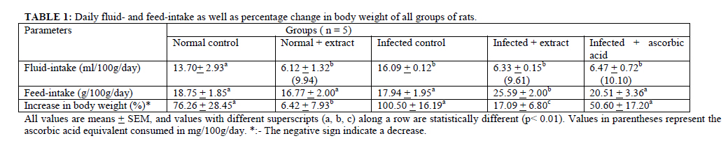
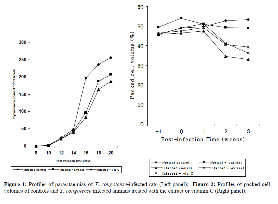
African Journal of Traditional, Complementary and Alternative Medicines, Vol. 6, No. 4, 2009, pp. 585-591 Research Paper THE EFFECT OF AQUEOUS EXTRACTS OF Hibiscus sabdariffa (SORREL) CALYCES ON HEAMATOLOGICAL PROFILE AND ORGAN PATHOLOGICAL CHANGES IN TRYPANASOMA CONGOLENSE – INFECTED RATS Ismaila A. Umar a *, Nelson G. Maryomsb, Emmanuel Daikwob, Abubakar Gidadob, Lawan B. Burataib, Ikechukwu O. Igbokwec and Mohammed A. Ibrahima aDepartment of Biochemistry, Faculty of sciences, Ahmadu Bello University, Zaria, Nigeria. Code Number: tc09051 Abstract The effects of aqueous extract of Hibiscus sabdariffa calyces on haematology and pathological changes in some selected organs during experimental Trypanosoma congolense infection of rats were investigated. Three groups of rats were intraperitoneally infected with T. congolense (Karu stock). One group was administered with the aqueous extract and another given a solution of vitamin C in drinking water; the remaining infected group was left untreated. Data from these groups were compared with those of two groups of healthy rats, one of which was similarly treated with the aqueous extract. The experiment was terminated three weeks, post-infection (pi). The uninfected and infected rats administered the extract consumed the equivalent of 9.94 mg – and 9.61 mg ascorbic acid / 100g / day during the experiment. Consumption of the extract significantly (p<0.01) retarded the rate of weight gain in both healthy and infected rats; even though the feed-intake was not significantly affected. After two weeks of infection the extract and vitamin C kept the parasitaemia significantly (p<0.01) lower than the untreated infected group. The anaemia in the untreated infected group was significantly (p<0.01) more severe than that of the corresponding extract- or vitamin-treated groups. Trypanosoma congolense infection caused significant (p<0.01) decreases in serum total proteins and albumin; serum and organ ascorbic acid as well as significant (p<0.01) elevation of serum alanine amino transferase levels in untreated rats. Consumption of the extract or vitamin C, however, prevented these disease–induced anomalies in the treated infected rats. Serum creatinine and urea levels were not affected by infection but the extract elevated these parameters significantly (p<0.01) above infection levels. It was concluded that consumption of the extract ameliorated the pathological changes in blood and organs of T. congolense-infected rats. Key words: Trypanosoma congolense, Anaemia, organ pathology, Hibiscus sabdariffa. Introduction Animal trypanosomosis is still a major factor retarding the growth of the livestock industry in Africa. Economic loss due to this disease runs into hundreds of millions of Naira in Nigeria; and perhaps even higher in other parts of Africa (Doko et al., 1991, Gaturaga et al., 1991). The important trypanosome species that cause the disease in livestock include; Trypanosoma brucei, T. simiea, T. evansi, T. vivax and T. congolense. The disease is characterized by tissue and organ degenerative changes. One factor implicated in the pathogenesis of the disease is oxidative stress (Vray et al., 1991, Igbokwe, 1994, Igbokwe et al., 1998) imposed by trypanosome and macrophageal activities. Oxidative stress has been alleviated in experimental infections with various species of trypanosomes (Umar et al., 1999a, 1999b, 2000, 2001) by administration of exogenous antioxidants, such as ascorbic acid (vitamin C) and/or vitamin E, to infected rats and rabbits. This vitamin therapy considerably reduced the degree and rate of degeneration of the tissues and organs; and in some instances significantly reduced the parasitaemia and anaemia (Umar et al., 1999a) in the trypanosome–infected animals. The use of these, or other antioxidants (along with a trypanocidal agent) would, no doubt, improve the efficacy and efficiency of chemotherapy. However, these vitamins are relatively expensive and majority of animal husbands may not be able to afford them. This necessitates the search for and use of an easily accessible and cheap local source of any of these vitamins. The aqueous extracts of Hibiscus sabdariffa (sorrel) leaves and/ or calyx is commonly used as a beverage and have been reported to be beneficial in the management of cough, asthma, bronchitis and wound as well as hypertension (Owolabi et al., 1994), obesity and diabetes (Ajabonna and Adegunloye, 1999). More relevant to the present investigation however, is its high content of ascorbic acid (Ado, 1983). This experiment was designed to determine the effect of the aqueous extract of H. sabdariffa calyces on the pathogenesis of trypanosomiasis in rats. Materials and Methods Animals Twenty-five adult male Wister strain rats were obtained from the Nigerian Institute for Trypanosomiasis and Onchorcerciasis Research (NITOR), Vom, Nigeria. The animals were acclimatized to the laboratory conditions and a commercial pelleted poultry Grower’s mash (ECWA, Feeds, Jos) diet for two weeks before commencement of experiment. A standard protocol was drawn up in accordance with the good laboratory practice (GLP) regulations of World Health Organisation (WHO). The principles of animal care were also duely followed in this study. The animals were then divided into five groups of five rats each and treated as follows: Uninfected controls: The five rats in this group were neither infected nor treated with the extract and/or ascorbic acid. Extract control: This group consisted of five uninfected rats that were given, ad libitum, H. sabdariffa aqueous extract that was estimated to contain 156mg/100ml ascorbic acid as drinking water. Infected controls: Five rats were each intraperitoneally infected with about 106 Trypanosoma congolense (Karu strain) in 0.5 ml of cold-saline diluted tail–blood from a donor rat. No further treatment was administered to these animals. Infected and Extract-treated group: The rats in this group were similarly infected with the parasites and maintained, ad libitum, on H. sabdariffa aqueous extract that was estimated to contain 156mg/100ml ascorbic acid as drinking water. Infected and ascorbic acid treated group: The rats in this group were also infected as earlier described and given a solution of ascorbic acid (156mg/100ml) ad libitum as drinking water. All animals in groups given the extract or ascorbic acid solution were acclimatized to the extract or the vitamin respectively, for two weeks before infection of rats with T. congolense and tap water was supplied to the remaining groups of rats throughout the duration of the experiment. Feed– and fluid–intake were monitored by presenting measured quantities to the rats and measuring the amount remaining at time of change of feed or drink. The infection was monitored for three weeks, at the end of which the rats were humanely decapitated and samples collected. Collection and preparation of samples: During the course of the experiment tail-blood was collected from each rat daily for estimation of parasitaemia and packed cell volume (PCV). The rats were sacrificed three weeks post infection (pi), blood collected in plain containers and serum prepared. The serum was stored at -15° C until required. The liver and kidneys of the rats were quickly extracted, blotted and weighed. One gram each of liver or kidney was placed in a 50ml tube and cold physiological saline added to the 20ml mark, this was then homogenized using a tissue homogenizer. The homogenate was centrifuged at 3000x g for 20 mins; the supernatant was used for estimation of liver and kidney ascorbic acid. Parasiteamia was estimated by the wet-mount method of Herbert and Lumsden (1976) while PCV was determined by the standard microheamatocrit method. Serum creatinine, urea and albumin were determined by methods described by Tietz (1982). Serum alanine amino transferase activity was estimated by the method of Reitman and Frankel (1957) using a commercial reagent kit (Randox Laboratories, Ireland) and total proteins in the serum was assayed by the method of Reinhold (1953). Ascorbic acid in the serum, aqueous extracts of liver and kidney as well as the aqueous extract of H. sabdariffa was estimated by the method of Roe (1973). The strain of Trypanosoma congolense (Karu stock) was obtained from NITOR, Vom, and passaged into a rat. At peak parasitaemia, tail-blood was collected from the donor rat and appropriately diluted with cold physiological saline. The parasites in the diluted blood were counted and a volume (0.5 ml) containing approximately 106 trypanosomes was injected intraperitoneally to experimental rats. Preparation of H. sabdariffa Extract: Fresh calyces of H. sabdariffa (voucher No. 1056 ) were collected from farms around Maiduguri metropolis during the rainy season. They were dried under shade and then pounded to a fine powder. Hot distilled water (70-80oC) was added to 40g of the powdered calyces in a measuring cylinder to the 100ml mark. The suspension was allowed to stand for 15 mins, after which it was filtered through cheesecloth. The filtrate was further filtered through a Whatman’s No. 1 filter paper and allowed to cool to room temperature. The ascorbic acid concentration of this filtrate was then estimated. This extract was presented to the appropriate groups of rats in a graduated bottle with a standard nipple. The extract given to the rats was always prepared fresh and changed twice daily. Statistical Analyses The data are presented as means ± SEM of five replicate values. The data were analyzed by one-way ANOVA and Students’t-test. Results The mean concentration of ascorbic acid in the aqueous extract of H. sabdariffa prepared was 156.98 ± 2.08mg/100ml. The two groups given the extract recorded significantly (p<0.01) lower fluid-intake (Table 1) than their respective control groups. The normal rats given the extract, consumed an equivalent of 9.94 mg ascorbic acid/100 g bd.wt./day, and the infected rats given the extract consumed the equivalent of 9.61 mg ascorbic acid/100g bd.wt./day, while the infected rat given solution of ascorbic acid consumed 10.10 mg ascorbic acid/100g bd. wt./day; as calculated from the daily fluid in-take of the various groups. Feed-intake (Table 1) was not significantly affected by either infection or extract. Percentage increase in body weight (Table 1) was not significantly affected by Trypanosoma congolense infection; but consumption of the extract by normal rats caused a significant drop in body weight while it considerably reduced the percentage increase in body weight of infected rats given the extract. Consumption of ascorbic acid solution by infected rats had no effect on both feed-intake and percentage increase in body weight, but caused a significant (p<0.01) decrease in fluid-intake. The parasitaemic profiles (Figure 1) indicate that parasitaemia was first detected, in both infected groups, on day 8, pi, after which it consistently increased. After the second week of infection the extract–treated and vitamin C-treated infected rats recorded significantly (p<0.01) lower parasitaemia values than the infected untreated rats (infected controls). The profiles of PCV (Figure 2) indicate that there was no fall in PCV for the two healthy groups of rats. The PCV of the three T. congolense infected groups recorded sharp decreases in the second week of infection. The profiles for the extract- and vitamin-treated infected rats were displaced above that of the infected controls. The percentage decrease in PCV; terminal compared to pre-infection values (Table 2) was significantly (p<0.01) higher in the extract–free infected group than in the corresponding groups that consumed the extract or vitamin C. Trypanosoma congolense infection had no significant effect on serum creatinine and urea levels (Table 2) of the rats. The consumption of the extract by infected rats significantly (p<0.01) elevated the levels of these two parameters, but had no similar effect in normal rats. Consumption of vitamin C had no effect on the levels of these parameters in infected rats. Hypoproteinaemia, hypoalbuminaemia and significantly (p<0.01) elevated levels of serum alanine aminotransferase were recorded for untreated infected animals. These disease-induced anomalies in the infected rats were reversed by consumption of the extract and vitamin C. Table 3 presents the ascorbic acid concentrations in serum, livers and kidneys of the rats. Trypanosoma congolense infection significantly (p<0.01) depleted the ascorbic acid reserves in the serum, livers and kidneys of infected rats. Consumption of the extract or vitamin C by infected rats prevented this disease-induced generalized drop in levels of ascorbic acid. Discussion The observed effect of the extract on body weight gain had been reported earlier (Ajabonna and Adegunloye 1999). Consumption of the extract or vitamin C solution significantly lowered the parasitaemia three weeks, pi; a pattern observed in an earlier experiment (Umar et al., 2007) in which T. congolense-infected rabbits were given daily intraperitoneal doses of vitamin C (100 mg/kg). The infected rats in the present experiment consumed approximately the equivalent of 9.61mg ascorbic acid/100g/day. While not discounting the possible role(s) of other components in the extract, it is reasonable to suggest that the ascorbic acid content played a significant role in the observed effect on parasitaemia since vitamin C has been reported (Passmore and Eastwood, 1989) to enhance cell-mediated immunity in supplemented humans. Anaemia is a consistent feature in the pathology of trypanosomosis (Anosa, 1988); however, the anaemia in the extract- or vitamin C-treated infected rats was less severe than the one recorded for the untreated infected rats. This may be partly due to the relatively lower parasitaemia in the former group, since the degree of anaemia in trypanosomosis has been positively correlated with the onset and level of parasitaemia (Dargie et al., 1979). Hypoproteinaemia, hypoalbuminaemia and elevated serum alanine aminotransferase activity have been reported in trypanosome infections (Kalu et al., 1989, Adah et al., 1992) and were, respectively, attributed to proteinuria (Bruinjn et al., 1987) and hepatocellular damage (Anosa and Isoun, 1983). These disease-associated conditions were, however, restored by consumption of the extract or vitamin C. Serum creatinine and urea were not affected by the T. congolense infection, which agreed with earlier reports in which cattle (Wellde et al., 1974) were infected with T. congolense. Consumption of the extract by infected rats caused a significant elevation of the creatinine and urea in the serum; even though, it did not have a similar effect in healthy rats. Consumption of vitamin C by infected rats had no significant effect on these parameters. Consumption of the extract or vitamin C appeared to have ameliorated the pathological processes in the blood, liver and kidney of T. congolense–infected rats; in much the same way vitamins C and/or E did in earlier experiments. These two vitamins spare endogenous antioxidant reserves and aid in “quenching” the large amount of oxidative species produced during the course of the disease. These oxidative species in unsupplemented trypanosome-infected animals presumably cause damage to certain membrane components, such as polyunsaturated fatty acids and proteins (Slater, 1984), and deplete systemic antioxidants reserves, such as reduced glutathione (Igbokwe et al., 1998) and ascorbic acid (Nyden, 1949). In the present investigation, the ascorbic acid component, and perhaps other antioxidants, of the extract presumably kept the free radical load in infected rats low as well as preventing the disease-associated depletion in systemic antioxidants, such as blood and organ ascorbic acid. This would provide greater protection for cell membrane components as well as other susceptible cellular components, hence significantly retarding tissue and organ damage. It is, of course, possible that components other than ascorbic acid, in the extract may have played a role. We thus conclude that consumption of the aqueous extract of H. sabdariffa calyces by T. congolense– infected rats prevented fall in systemic antioxidant reserves and alleviated disease–induced damage to red blood cells, as well as hepatic and renal structures. Acknowledgement The authors are grateful to NITOR, Vom, Nigeria, for supplying the trypanosomes. References
© Copyright 2009 - African. Journal. Traditional, Complementary and Alternative Medicines The following images related to this document are available:Photo images[tc09051f1.jpg] [tc09051t1.jpg] [tc09051t2.jpg] [tc09051t3.jpg] |
| |||||||||
