 |
| About Bioline | All Journals | Testimonials | Membership | News |
|
||||||
|
||||||
Research Paper NEW ANTITRYPANOSOMAL TETRANOTRITERPENOIDS FROM AZADIRACHTA INDICA*Mercy Githua1, Ahmed Hassanali2, Joseph Keriko1, Grace Murilla3, Mary Ndungu1 and Gathu Nyagah1 1Department of Chemistry, Jomo Kenyatta University of Agriculture
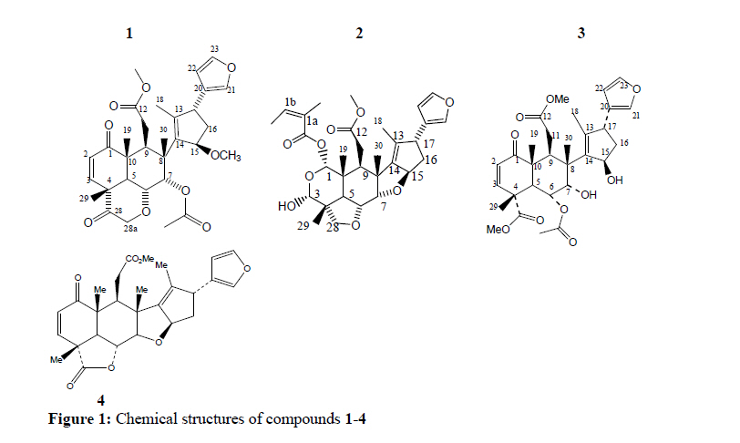
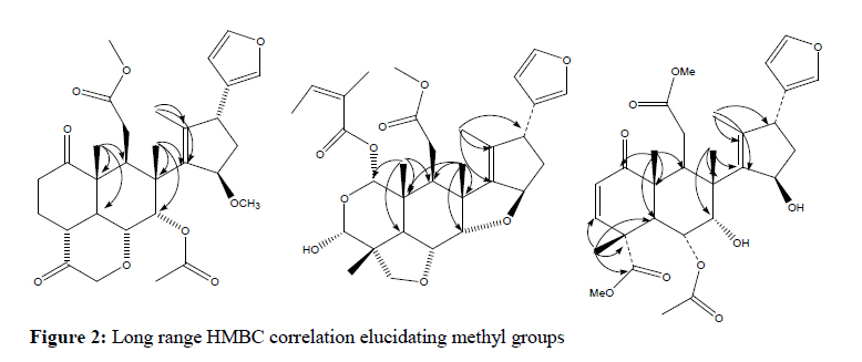
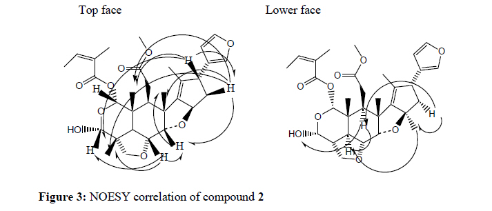
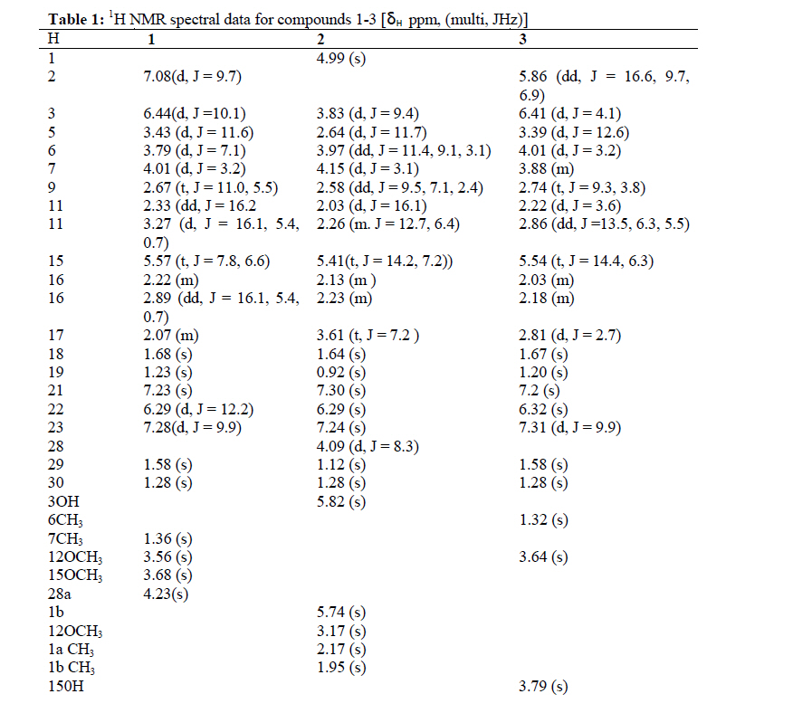
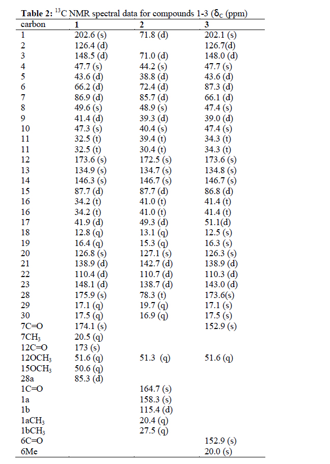
and Technology (JKUAT), P.O Box 62000-00200 Nairobi, Kenya. Code Number: tc10029 Abstract Organic extracts of the leaves of Azadirachta indica A. Juss. yielded ten antitrypanosomal terpenoids. Three of these (1 – 3), are novel and are derivatives of nimbolide and nimbin. They were extracted from chloroform fraction of methanol extract. These compounds were found to exhibit strong antitrypanosomal activities against Trypanosoma brucei rhodesiense with MIC values ranging of 6.9, 15.6 and 7.8 µg/ml respectively and were more active than Cymerlarsan ( a standard drug), which had an MIC value of 187.5 µg/ml when tested against T. b. rhodesiense The structures were elucidated by spectroscopic methods including; NMR, MS, UV and IR. Key words: Meliaceae, limonoids, Trypanosoma brucei rhodesiense, Azadirachta indica, antitrypanosomal activity. IntroductionAzadirachta indica (Meliaceae) is a large tree growing in tropical/subtropical regions. The leaves, bark, wood, roots and fruits are intensely bitter. Extracts from different parts of A. indica have shown activity against several diseases and their vectors that affect human and animals (Nagui, 1987; Mackinnon et al., 1997). According to Ayurveda (Hindu System of Medicine) the different parts of this tree possess different medicinal properties. A tea prepared from the leaves and bark is used to treat fever (Kokwaro, 1993); a decoction of fresh leaves is used as a favorite wash to cure old ulcers of long standing and the aqueous extract of the leaves in particular is used as remedy for malaria, similar to the practice in Nigeria. The methanol extract of A. indica exerts a pronounced antiinflammatory (rat paw oedema) and a fairly good antipyretic effect (pyrogen induced hyperpyrexia) in rabbits, and thus may justify its use in the treatment of fever resulting from malaria (Okpanyi and Ezeukwu, 1981). The biological activities of A. indica have been widely attributed to the presence of limonoids; modified triterpenes with or derived from a precursor with a 4, 4,8-trimethyl-17-furanylsteroid skeleton. Crude extracts of some of the limonoids constituents of the A. indica have been shown to exhibit antimalarial activities against chloroquine resistant K1 strain of Plasmodium falciparum (Bray et al., 1990). We now report the structures of three new limonoids that were isolated from the leaves of A. indica and their activities against T. b. rhodesiense. Materials and Methods Plant materials The leaves of A. indica were collected from Shimba Hills National Park, Kwale, South coast of Kenya and a voucher specimen No MG/1/2002 has been deposited in the herbarium of the Botany department of University of Nairobi (Kenya). Extraction and isolation of compounds from A. indicaAir dried and grounded powder of A. Indica leaves (6 Kg), was soaked in methanol (10 liters) for 48 hours and then filtered. . The residue was soaked for another 48 hours in methanol (10liters) and then filtered. The combined filtrates were concentrated to dryness under reduced pressure at 45оC to yield a viscous oil (636 g). The extract was partitioned between water (100 ml) and chloroform (250 ml) x 3. The combined organic layer was evaporated to dryness under reduced pressure. Thirty grams of the chloroform layer was column chromatographed on silica gel (column size, 81 x 4.5 cm; 230 -400 mesh) using hexane/ethyl acetate gradient (90 -10% hexane). For each solvent system, 1 liter of solvent was used to elute and 50ml fractions collected. Separation was monitored using thin layer chromatography (TLC). The plates were developed in hexane: ethylacetate (2:1), then sprayed with Ehrlich’s reagent (2% 4-dimethylaminobenzaldehyde) and developed in hydrogen chloride gas chamber. Typical purplish spots were obtained for limonoids rich fractions, which eluted at 55 -100% ethylacetate (Maier and Edward, 1970). A limonoid-bearing fraction (12 mg) was analyzed by semi-preparative HPLC on Beckmann Ultrasphere ODS reverse phase column (250 x 10 mm i.d) on Varian 5000 liquid chromatograph using 1:1 water/acetonitrile isocratic system at a flow rate of 2.5 ml per min and detected at 215 nm. Three new limonoids 1(6.9 mg), 2 (15.6 mg) and 3 (7.4 mg), were obtained and tested for antitrypanosomal activity. NMR experiments on the compounds were recorded on Brucker Avance (500 MHz) Spectrophotometer. The 1H and 13C spectra were recorded in CDCl3 solvent. Chemical shifts (δ) were reported in parts per million (ppm) relative to tetramethylsilane (TMS) as the internal standard and coupling constants (J), in hertz (Hz). Peaks on 1H NMR are reported as singlets (s), doublets (d), doublet of doublet (dd), triplets (t) and multlplets (m). The 13C multiplicity was determined by DEPT experiments, which gave chemical shifts values in ppm for assignments. The IR spectra of the compounds were recorded on a Fourier Transform infrared spectrometer (FT-IR8101, DR-8001). Electron Impact Mass Spectra (EIMS) were obtained on a fission VG Platform II GC/LC-MS spectrometer at 70 eV by solid probe. Melting points of the compounds were determined on Sanyo Gallenkamp electronic melting point apparatus (Cal No. MPD350. BM 3.5 Serial No. SG97/12/425 UK and are uncorrected. Propagation of typanosomes culturesTrypanosomiasis brucei rhodesiense (KETRI 3438) procyclic forms were obtained from Kenya Trypanosomiasis Research Institute (KETRI) trypanosomiasis bank and stored in liquid nitrogen. They were resuscitated and cultured until correct parastaemia for in vitro screening was obtained. The strain was isolated on 7th July 1997 from a human patient and passaged through mice. The pre-patent period was six days while the prefreezing period was twenty three days. Antitrypanosomal bioassayThe procedure developed by Burri et al. (1993) to determine the levels of melarsoprol in serum samples of animals after drug treatment was employed. The bioassay was carried out in 96 flat-bottomed well plates with a lid (Corning Glass Works, Corning, New York) arranged in a matrix of 8 rows A -H and 12 columns 1 -12 (Figure 1). The tests were carried out in columns 11 -2 and rows B -G. The wells in columns 1 and 12 and rows A and H were not used due to high rate of evaporation. The stock solutions were prepared in sterilized distilled water (1 mg/ml) and diluted to appropriate concentrations. For every extract, three different concentrations were prepared, that is, 500, 250 and 125 µg/ml. Trypanosomes in logarithmic growth phase below 106/ml one day after sub-passaging were diluted to 2 x 10³ cells/ml in medium. Hundred µl of this trypanosome suspension was placed into each well. 100 µl extract solution of each concentration was added to two adjacent wells of column 11. Serial dilutions were carried out by transferring 100 µl from wells of column 11 to wells of column 10 by multi-channel pipettes, then from wells of column 10 to 9, from 9 to 8 and so on up to wells of column 4. For every test, the highest concentration was in column 11 and in twofold dilution to the lowest in column 4 in 128 fold range, so that, for every extract, the highest concentration was 250 µg/ml and the lowest was 0.488 µg/ml. Columns 3 and 2 were the control, that is, no extract was added. Plates were incubated for 72 hrs at 27оC in 5% CO2. Determination of minimum inhibition concentrationThe test was observed under inverted microscope at 100-fold magnification. In every row the highest dilution of either standard or samples with less than 5 motile trypanosomes of normal shape was determined. The concentration in this well is defined as the minimum inhibitory concentration (MIC). MIC*D*2n = CX MIC = Minimum Inhibitory Concentration D = Dilution of extract by medium in wells of column 11 (maximum concentration) n = Steps of dilution to the first well with no living trypanosomes Cx = Concentration of extract solution * = Multiplication Results and DiscussionThe HREIMS of compound 1gave a molecular ion at m/z 554.2254 corresponding to C31H38O9. 1H and 13C NMR data (Tables 1 and 2) showed resonances for four distinctive tertiary methyl groups δH 1.23 (H-19), 1.68 (H18), 1.58 (H-29), 1.28 (H-30) on carbons at δC 16.4 (q), δC 12.8 (q), δC 17.1 (q), δC 17.5 (q), respectively. The positions of these methyl groups were assigned by analysis of HMBC spectrum. Correlations of H-18 with the carbon resonance at δC 134.9 (C-13) and δC 146.3 (C-14); H-19 with the carbon resonance at δC 202.6(C-1), δC 43.6 (C-5), δC 41.4(C-9) and δC 47.3(C-10); H-29 with the carbon resonance at δC 148.5(C-3), δC 47.7(C-4), δC 43.6(C5) and δC 175.9(C-28) and H-30 with the carbon resonance at δC 86.9(C-7), δC 49.6(C-8) and δC 146.3(C-14) (Figure 2). 1H NMR spectrum indicated presence of a β-substituted furan moiety at δH 7.23, 6.29 and 7.28. Examination of COSY spectra revealed that methine protons at δH 6.29 coupled with methine protons at δH 7.28, which allowed the assignment of the hydrogen atoms at δH 6.29 to C-22, 7.28 to C-23 and 7.23 to C-21. The carbonyl group on C-1 was deduced from the absence of H-1 resonance and was confirmed from HMBC correlation of H-2 δH 7.08 (d, J = 9.7Hz), H-3 δH 6.44 (d, J = 10.1Hz), H-5 δH 3.43( d, J = 11.6Hz), H-9 δH 2.67 ( t, J = 11.0, 5.5Hz) and methyl protons at C-19 δH 1.23( s) with the carbon resonance at δC 202.6. Cross peaks between methyl protons at δH 1.58(H-29), to carbons at δC 148.5(C-3), δC 47.7(C-4), δC 43.6(C-5) and δC 175.9(C-28) confirmed the position of H-29 and also confirmed C-28 as a ketone. The position of methoxy groups was deduced from HMBC correlations of the methoxy protons on C-12 with the carbon resonance at δC 51.6 (OCH3-12) and of the methoxy protons on C-15 with the carbon resonance at δC 50.6 (OCH3-15) . The structure is proposed to be 7αacetyl-15β-methoxy-29 methylene 7,15-deoxo nimbolide. HREIMS of compound 2 gave a molecular ion at m/z 556.1342 corresponding to C31H40O9. IR spectrum indicated the presence of a hydroxyl (3446 cm-1), CH aliphatic stretch (2997 cm-1), C=O (1729 cm-1), olefinic double bond (1652 cm-1) and C-O (1232-1150 cm-1). NMR spectral data showed a basic structure similar to that of nimbolide (4) a known compound isolated from ethanoic extract of A. indica (Sudaratana et al., 1985). 1H and 13C NMR spectral data (Tables 1 and 2), showed presence of six tertiary methyl groups δH 0.925 (s, H-19), 1.64 (s, H18), 1.12 (s, H-29), 1.28 (s, H-30), 2.17 (s, H-1a) and 1.95 (s, H-1b). The positions of these methyl groups were assigned by analysis of HMQC and HMBC correlations (Figure 2). Examination of COSY spectrum revealed that methine protons at δH 2.58 (dd, J = 9.5, 7.1, 2.4 Hz, 1H) coupled with methylene protons at δH 2.03 (d, J = 16.1Hz, 2H). HMBC correlation of the proton at δH 2.03 with carbons at resonance δC 172.5 (carbonyl carbon) and 39.3 allowed the assignment of protons at δH 2.03 to C-11 and 2.58 to C-9. From HMBC correlations, cross peaks between methyl protons at δH 1.95 (s), δH 2.17 (s) with carbons at δC 158.3 and δC 115.4 and cross peaks between protons at δH 5.74 (s) with carbons at δC 164.7, 20.4 and 27.5 suggests the side chain to be tigloyl. HMBC correlation of methine proton at δH 3.61 (t, 7.2Hz, 1H) with furan carbons allowed the assignment of that proton to C-17 (δC 49.3). The HMBC cross peaks between methyl protons at δH (1.64, s, 3H) with carbons at δC 134.7, 146.7 and 49.3 permitted the assignment of quartenary carbons C-13 and C-14. There is a double bond between them as they have no HMQC correlation and no COSY coupling. From 1H and 13C NMR there is one methoxy group whose position was revealed by the HMBC correlation. The assignment of relative stereochemistry was based on the NOESY experiments. The H-9 resonance showed a NOESY correlation with OH-3, H-5, H-15 and H-16 confirming the orientation of H’s on C-5, C-15 and hydroxyl group on C-3. H-16 resonance showed a NOESY correlation with 3H-30, H-7, 3H-19 and 3H-29 whereas H-17 resonance showed a NOESY correlation with 3H19, 3H-29 and H-16. NOESY correlation of H-6 with H-7, H-3, 3H-29 and 3H-30 confirmed the -orientation of H’s on C-3, C-6, C-7 and C-17 (Figure 3). The compound is proposed to be 2-oxo-3-deacetyl salannin. Compound 3 has a molecular formula C30H38O10 (m/z 558). The IR spectrum indicated presence of a hydroxyl group (3427 cm-1), CH aliphatic stretch (2924 cm-1), C=O (1732 cm-1), olefinic double bonds (1680 cm-1) and C-O (1234 cm-1). 1H and 13C NMR spectral data (Tables 1 and 2 indicated the presence of a β-substituted furan moiety at δH 7.2 (H-21, s), 6.32 (H-22, s) and 7.31 (H-23, d, 9.9 Hz) corresponding to carbons at δC 138.9 (d), 110.3 (d) and 143.0 (d), respectively. The signals at δC 148.0 (d), 126.7 (d) and 202.1 (s) corresponding to protons at δH 6.41 (1H, d, J = 4.1 Hz) and 5.8 (1H, dd, J = 16.6, 9.7, 6.9 Hz) are typical of α, β-unsaturated ketone moiety in the A-ring. HMBC correlation of protons at δH (5.86, dd, J = 16.6, 9.7, 6.9 Hz), 6.41 (d, J =4.1 Hz), 3.39 (d, J = 12.6 Hz), 2.74, (t, J = 9.3, 3.8 Hz) and methyl protons at 1.20(s) with the carbon resonance at δC 202.1(s) allowed the assignment of δH 6.41 to C-3 and proton at δH 5.86 to C-2 and carbonyl to C-1. This was confirmed by the examination of COSY spectra which revealed that methine protons at δH 6.41 (1H, d, J = 4.1 Hz) coupled with methine protons at δH 5.86 (1H, dd, J = 16.6, 9.7, 6.9 Hz). 1H and 13C NMR indicate the presence of 2 methoxy groups attached to keto carbons. The compound is proposed to be 7α-hydroxy-15β-hydroxy-7,15-deoxo nimbin Antitrypanosomal Activity Crude methanolic extracts and chloroform fractions of A. indica leaves exhibited significant antitrypanosomal in vitro activities against T. b. rhodesiense procyclic forms (KETRI 3438) whose MIC values are 51.2±0 and 4.4 ± 0 µg/ml, respectively and exhibited higher activities than cymerlarsan (drug in the market) which had an MIC value of 187.5 µg/ml. Chloroform extracts were more potent than the methanol extracts, indicating that the active compounds are of medium polarity. Repeated chromatographic separations of the chloroform fraction yielded three new tetranotriterpenoids (13) that were tested against T. b. rhodesiense procyclic forms (KETRI 3438). The MIC values were 6.9 ± 0, 15.6 ± 0 and 7.8 ± 0 µg/ml, respectively. All the three compounds had lower activities than that of the chloroform fraction suggesting that there are other antitrypanosomal compounds present or possible synergistic blend effects. Based on their spectroscopic data and comparison with published data, the structures of the compounds were readily elucidated and were found to have the nimbolide (4) basic structure (C-seco limonoids). Compound 2 which has an epoxy group between C7 and C15 has the lowest activity whereas compounds 1 and 3 with no epoxy group and with more methoxy groups exhibited higher activities. This suggests that antitrypanosomal activity can be enhanced by chemical modifications of the basic structure (nimbolide) and its derivatives to generate new effective antitrypanocidal drugs with novel modes of action. AcknowledgmentsWe are grateful to Mr. S. G. Mathenge and Mr. Mutiso both of University of Nairobi, Botany Department and Mr. D. Gitau for their assistance in collection and identification of plant materials. We acknowledge Dr. Antony Hooper of IACR-Rothamsted for performing the Nuclear Magnetic Resonance (NMR) experiments and his valuable advice and guidance on the interpretation of the data. The staff of tissue culture section of Tyrpanosomiasis Research Center (TRC) are acknowledged for their assistance in bioassays. References
Copyright 2010 - Afr. J. Trad. CAM The following images related to this document are available:Photo images[tc10029t2.jpg] [tc10029t1.jpg] [tc10029f3.jpg] [tc10029f1.jpg] [tc10029f2.jpg] |
| |||||||||
