 |
| About Bioline | All Journals | Testimonials | Membership | News |
|
||||||
|
||||||
African Journal of Traditional, Complementary and Alternative Medicines, Vol. 7, No. 3, 2010, pp. 258-263 Research Paper EFFECT OF VIRGIN FATTY OIL OF PISTACIA LENTISCUS ON EXPERIMENTAL BURN WOUND’S HEALING IN RABBITS Zouhir Djerrou*, Z. Maameri*, Y. Hamdi-Pacha*, M. Serakta*, F. Riachi*, H. Djaalab and A. Boukeloua*. * Department of Veterinary Sciences, Pharmacotoxicology Laboratory,
Mentouri Constantine
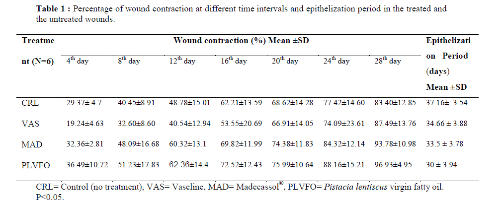
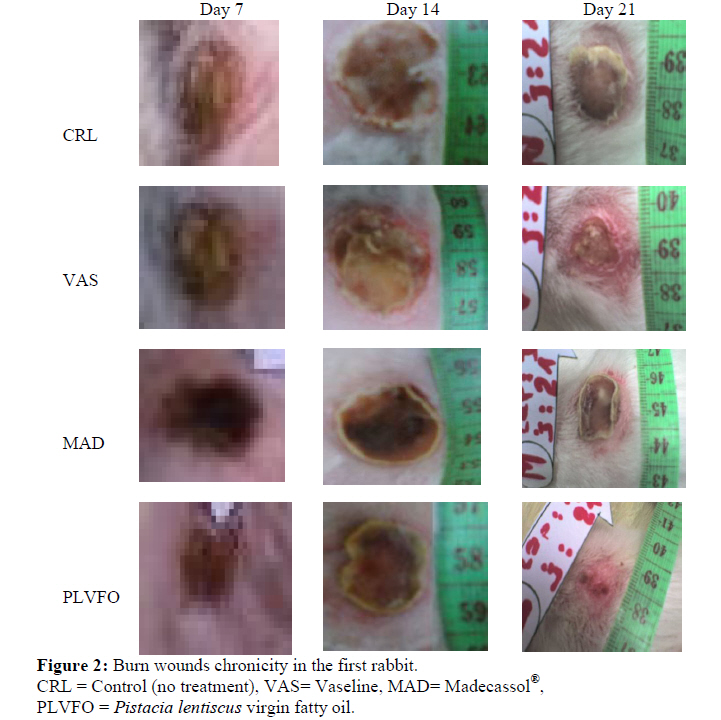
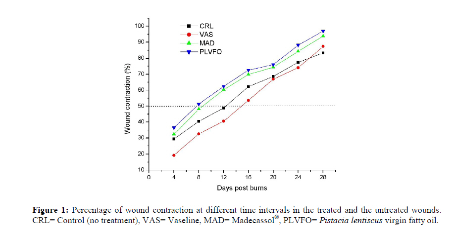
University, Algeria. Code Number: tc10036 Abstract This study aimed to assess the efficiency of the virgin fatty oil of Pistacia lentiscus (PLVFO) for burn wounds healing. It was carried out on 6 adult male New Zealand rabbits. Four burn wounds of deep third degree were made on the back of each animal. The first was not treated and served as control (CRL group); the others were covered immediately after burning procedure by 0.5g of one of the following products: Vaseline gel (VAS group), Madecassol® cream 1% (MAD group) or 1ml of PLVFO (PLVFO group). The treatments were repeated once daily until complete healing. For four days post burns, the percentage of wound contraction was assessed. Also, the different healing times were noted. The results showed that both PLVFO and Madecassol® significantly accelerated wound healing activity compared to wounds dressed with Vaseline and the untreated wounds. However, the level of wound contraction was significantly higher and the healing time was faster in PLVFO group than those of the MAD group, VAS group and CRL group. The different epithelization periods obtained in days were respectively: 30±3.94 (PLVFO group), 33.5±3.78 (MAD group), 34.66±3.88 (VAS group) and 37.16±3.54 (CRL group). We conclude that Pistacia lentiscus virgin fatty oil promotes significantly (p< 0.05) wound contraction and reduces epithelization period in rabbit model. Key words: Virgin fatty oil, Pistacia lentiscus, burn, wound healing, rabbits. Introduction Wound healing is a complex process of the replacement of the dead tissue by living one. It runs in three basic phases, inflammatory, proliferative and maturation phase (Toporcer et al., 2006). All these steps are orchestrated in controlled manner by a variety of cytokines including growth factors (Umachigi et al., 2007) such as epidermal growth factor (EGF), fibroblast growth factor (FGF) and transforming growth factor beta (TGFβ) (Ali Kokuludağ, 1999; Kwon et al., 2006; Tanaka et al., 2005; Varedi and Englander, 2006). Findings from many studies clearly indicate that appropriate treatment and wound care accelerate the healing process and prevent infection (Alizadeh et al., 2009). Different synthetic drugs are available to enhance the wound healing in modern medicine. Moreover herbal medicines are crucial in wound healing since they initiate disinfection, debridement and provide a moist environment for natural healing process (Habbu et al., 2007). In the Mediterranean area, much attention has been focused on the potential properties of Pistacia lentiscus L. (Anacardiaceae). Duru et al. (2003) collected the different virtues of Pistacia genus. Its various species are used in eczema treatment, paralysis, diarrhea, throat infections, renal stones, jaundice, asthma and stomach-ache, and as astringent, anti-inflammatory, antipyretic, antibacterial, antiviral, pectoral and stimulant. The essential oil of Pistacia lentiscus L. was obtained by hydrodistillation of leaves, fruits or from trunk exudates called mastic gum (Castola et al., 2000). This essential oil has been proven to exhibit antioxidant, antiinflammatory, antimicrobial (Benhammou et al., 2008), antifungal (Duru et al., 2003; Kordali et al., 2003) and antiatherogenic activities (Dedoussis et al., 2004). According to Tounes et al. (2008), many studies of the chemical composition of this oil have been carried out by some authors: Duru et al. (2003), Zrira et al. (2003), Uidrich et al. (2004), Benyoucef et al. (2005). Benhammou et al., (2008) reported that the fatty oil has good nutritive quality because of its content in unsaturated fatty acids (Oleic + linoleic = 73%) and saturated fatty acids (Palmitic + stearic = 25.8%). The purpose of this study was to assess clinically the effect of this oil (PLVFO) on burn wounds healing in rabbit model. Material and MethodsThe study was carried out at Department of Veterinary Sciences, University of Mentouri, Constantine, Algeria. DrugsPistacia lentiscus fruits were collected from Skikda region, Algeria in December 2008. They were air dried in the shade for 14 days, and then oil was extracted by traditional cold-pression in different steps. First, the fruits were ground using millstones into a paste, which was then mixed for 30 mins. After grinding, the paste was spread on fiber disks and pressed. Cold water was run down the sides of the disks to increase the filtration of the oil. The liquids were then separated by decantation. At the end of this phase Virgin Pistacia Lentiscus oil was produced. Vaseline gel and Madecassol® (cream 1%) are commercial products; they were obtained from the local pharmacy. Madecassol® contains: Hydrocotyle*(*Centella asiatica ; reconstituted titrated dry extract containing asiaticoside 40% and madecassic and Asiatic acids 60%). Other ingredients: Ethylene glycol (mono+diester), palmito-stearate, propylene glycol, liquid paraffin, essential oil of lavendar, essential oil of geranium, purified water. AnimalsSix healthy adult male New-Zealand rabbits, weighing between 1.58–2.88 kg, were used for the study. Animals were kept in individual standard cages in standardized environmental condition with an ambient temperature of 22 ± 2ºC and a 12 h light-dark cycle. Food and water were provided ad libitum. All the experimental procedures adopted were in accordance with the International Guidelines for Animal Care. Burn wound modelThe procedure used in this study was described by Hamdi-pacha et al. (2002). On day zero, rabbits were anesthetized by 50 mg/kg ketamine hydrochloride, intramuscularly injected, along with 5 mg/kg diazepam. The hairs on the skin of the animals' back were shaved with a sterilized razor blade. Then, four burns of identical size (3 cm in diameter) were created on the back of each animal, two cranially ( left and right) and two caudally (left and right), by a metal cylinder weighing 200 g immersed in prior in boiled water for 3 mins and maintained on animal skin 15 seconds. Each animal served as his own control. Treatment and assessment of healing processEach wound was assigned to a test substance in such a way that each drug would be applied at different wound location in each animal. This was done to prevent experimental bias consecutive to animal body movement (Bae et al., 2005). Specific treatments were as follows: Madecassol® cream 1% at the dose of 0.5g (MAD group), Pistacia Lentiscus virgin fatty oil 1ml (PLVFO group), Vaseline gel at the dose of 0.5g (VAS group) and no treatment representing the control group (CRL group). All the drugs were applied topically slowly using sterilized cotton swabs immediately after burning procedure and they were repeated once daily until complete epithelization had taken place. The wound size was traced on a transparent paper every 4 days, and the wound surface area was evaluated. This later parameter was then employed to calculate the percentage of wound contraction, taking the initial size of the wound, 706.5 mm2, as 100 %, by using the following equation: Percentage of wound contraction= [(Initial wound size – specific day wound size) / Initial wound size] × 100 (Srivastava and Durgaprasad, 2008). Statistical analysisThe results obtained were statistically analyzed using Students’ t-test. All values were expressed as Mean ± Standard deviation. The level of significance was set at p < 0.05. ResultsDuring the experimentation period, no mortality occurred in the animals. Morphological parameter was used to evaluate efficacy of PLVFO for burn wounds healing compared to Madecassol®, Vaseline and the untreated wounds. The different percentages presented in Table1 represented the means calculated for six wounds treated with the same drug in the different rabbits. Generally, there was a progressive reduction in the wound surface area with time in the different wounds (Figure 2 and Table 1). Higher mean percentage of wound contraction was obtained in wounds treated with the oil followed by Madecassol®. Contraction in Vaseline treated wounds was less than untreated wounds from day 4 to day 24. After this day and until healing process had taken place, contraction in Vaseline was higher than control (Table 1 and Figure 1). The epithelization period was significantly shortened (p<0.05) in the oil (30 ± 3.94 days) compared to the control groups. Also, wounds treated with Madecassol® healed significantly faster (p<0.05) than wounds treated with Vaseline (33.5 ± 3.78 and 34.66 ± 3.88 days respectively). The Vaseline group, however, showed better healing time compared to the untreated wounds (37.16 ± 3.54 days) (Table 1). DiscussionSeveral drugs for the management of wound healing take their origin from plants. Habbu et al. (2007) presented a detailed review of literature on natural prohealers, phytoconstituants, polyherbal formulations and various nutraceuticals responsible for wound healing activity. They reported 81 plants which were studied by different authors. However, other plants used in our folk medicine with good reputation in healing process, such as Pistacia lentiscus L., needed to be studied in this field of research. Wound assessment has several dimensions such as: clinical, physical, physiological, biochemical, histological and genetic. The study adopted morphological parameter including wound contraction percentage and epithelization period. The results showed that both Madecassol® and PLVFOl significantly (p<0.05) promoted wound contraction and shortened epithelization period. However, the PLVFO seemed to be more effective. In the case of Vaseline, the percentage wound contraction obtained was less than that of untreated wound until day 24. Pu et al. (1999) reported that, in the early period of healing process Vaseline was capable of inhibiting wound evaporation. So, it is known that a moist physiological environment should be formed in the wound for skin regeneration repair of burn wounds. However, Vaseline therapy may evoke suffocated and macerated tissues (Xu and Xiao, 2003) as seen in some wounds from the current study. Madecassol® is one of the local commonly used products for burn management. Shetty et al. (2008) reported that the principal ingredients of titrated extract of Centella asiatica (Asiaticoside, asiatic acid and madecassic) were effective on systemic scleroderma, abnormal scar formation and keloids and also significantly shorten the wound healing time. Their most beneficial effect appears to be the stimulation of maturation of scar by the production of type I collagen and resulting decrease in the inflammatory reaction and myofibroblast production. In the case of our oil, its fatty composition revealed that the three dominant fatty acids found are: Palmitic 16.3 %, oleic 55.3 % and linoleic 17.6 %. The oil contains an appreciable amount of unsaturated fatty acids 78.8 % (Charef et al., 2008). The unsaponifiable fraction contains tocopherols, sterols and phenolic components. However, no study has been published which concern this fraction. It is important to mention that Mattähaus and Özcan (2006) quantified fatty acids, tocopherols and sterols in Pistacia terebinthus Chia. Generally, fatty acids and triglycerides are able to reduce trans epidermal water loss and so increase skin hydration (Dweck, 2002). Oleic and Linoleic acids are known for their anti-inflammatory properties. Linoleic and alpha linoleic acid provide lipids necessary for cell membrane repair and cellular respiration (Loden and Andersson, 1996). Phenolic components have been identified as having antibacterial and antioxidant properties (Siger et al., 2007; Waterman et al., 2007). It has been found that plants that have cicatrizing and vulnerary properties often have a high level of plant sterols (Dweck, 2002). Anecdotal reports claim that vitamin E speeds wound healing and improves the cosmetic outcome of burns and other wounds (Baumann and Spencer, 1999). Martin (1996) and Palmeri et al. (1995) reported that alpha tocopherol has been proven to have powerful antioxidant effect or free scavenging properties and acts as humectants. Different antioxidants reduce freeradicals damage, there by preventing impairment at the cellular level. They inhibit inflammation, which leads to collagen depletion, and they offer protection against photo damage and skin cancer. Finally, perhaps the different previous properties of these fatty oil components, particularly unsaponifiable fraction may explain its promising healing effect. The current study concludes that the virgin fatty oil of Pistacia lentiscus fruits is effective in burns healing process. It promotes wound contraction and shortens epithelization period. References
Copyright 2010 - Afr. J. Trad. CAM The following images related to this document are available:Photo images[tc10036f1.jpg] [tc10036t1.jpg] [tc10036f2.jpg] |
| |||||||||
