 |
| About Bioline | All Journals | Testimonials | Membership | News |
|
||||||
|
||||||
African Journal of Traditional, Complementary and Alternative Medicines, Vol. 7, No. 4, 2010, pp. 350-369 Evaluation Of The Toxicity And Reversibility Profile Of The Aqueous Seed Extract Of Hunteria Umbellata (K. Schum.) Hallier F. In Rodents Adeneye A.A.1, 2 , *, Adeyemi O.O.2 , Agbaje E.O.2 , Banjo A.A.F3 1Department of Pharmacology, Faculty of Basic Medical Sciences, Lagos State University College of
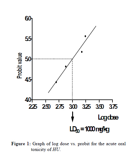
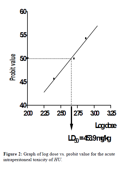
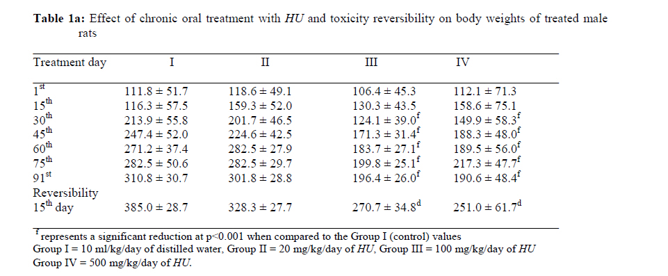
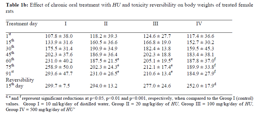
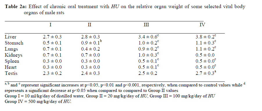
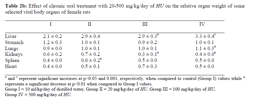
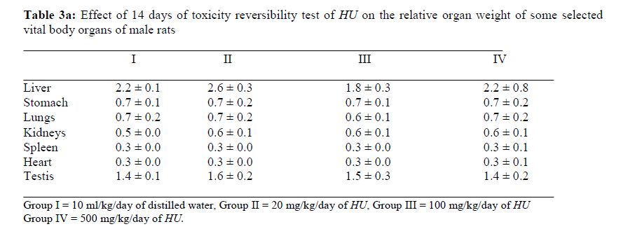
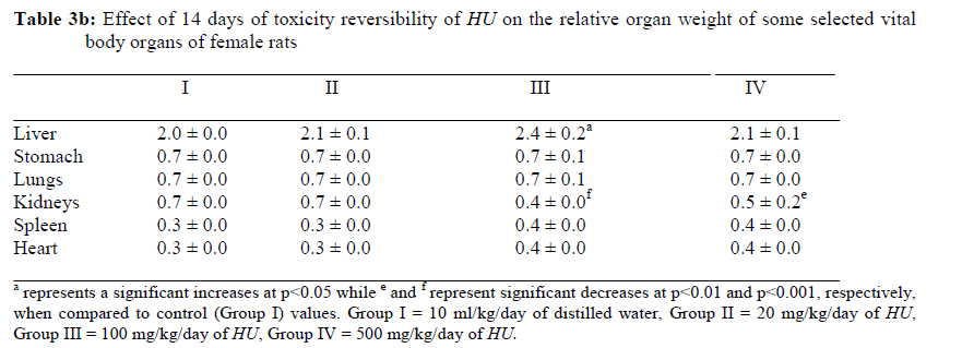
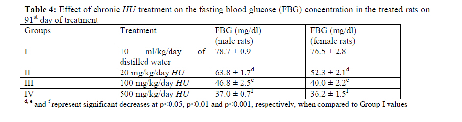
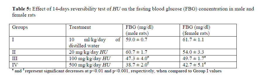
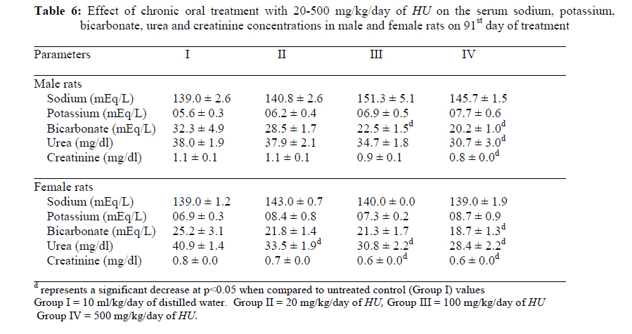
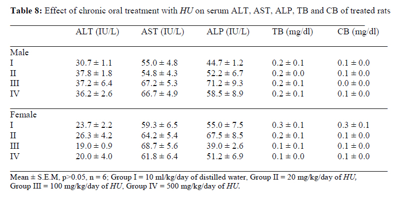
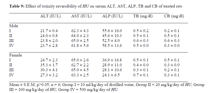
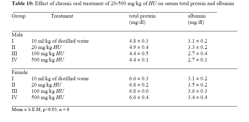
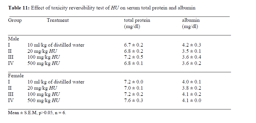
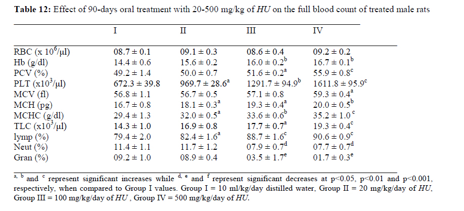
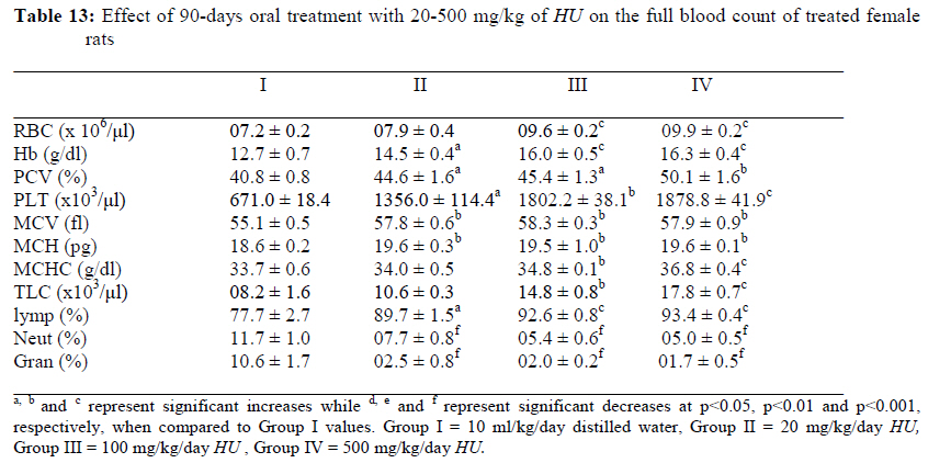
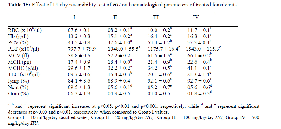
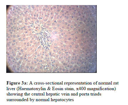
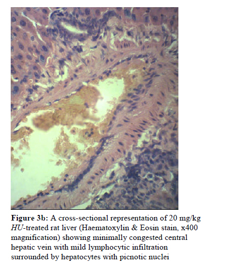
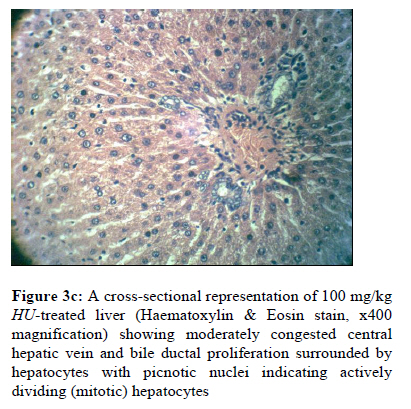
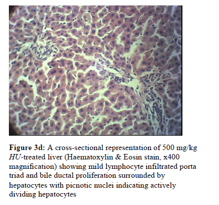
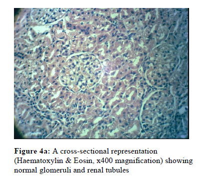
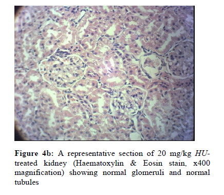
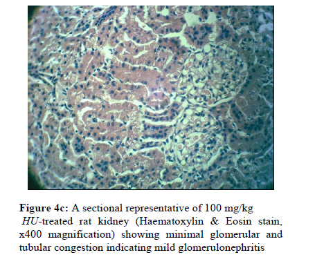
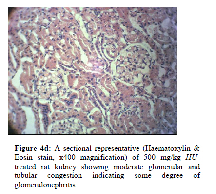
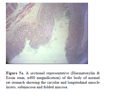
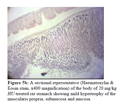
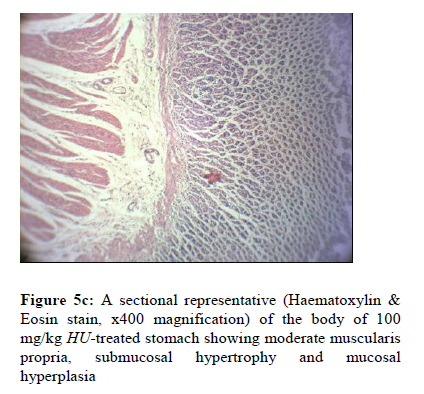
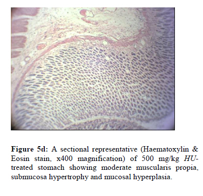
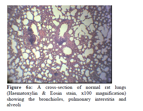
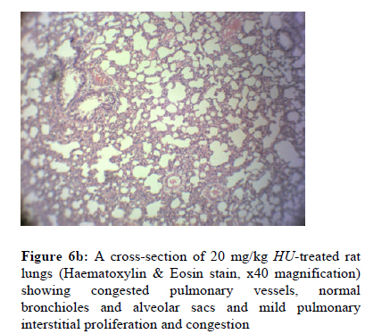
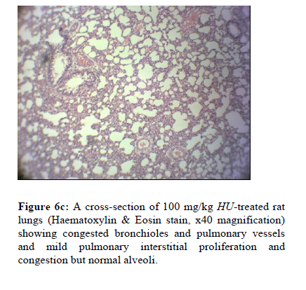
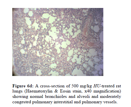
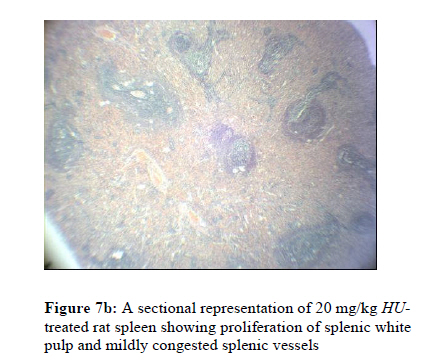
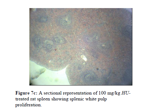
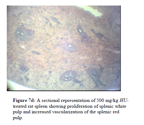
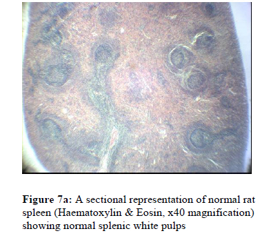
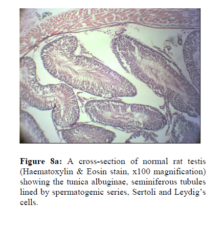
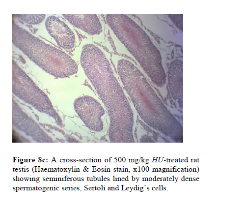
Medicine, Ikeja, Lagos State, Nigeria, Code Number: tc10049 Abstract Hunteria umbellata (K. Schum.) Hallier f. (family: Apocynaceae) is reputed for the folkloric management of labour, pain and swellings, stomach ulcers, diabetes, obesity, and anaemia, with no scientific report of its toxicity and reversibility profile. The present study was, therefore, aimed at investigating the in vivo toxicity and reversibility profile of the aqueous seed extract of Hunteria umbellata (HU). The acute oral and intraperitoneal toxicity studies of HU were determined in Swiss albino mice while its 90-day oral toxicity and toxicity reversibility profile on anthropometric, biochemical, haematological and histopathological parameters were also assessed using standard procedures. Results showed that the LD50 values for the acute oral and intraperitoneal toxicity studies for HU were estimated to be 1000 mg/kg and 459.3 mg/kg, respectively. Visible signs of immediate and delayed toxicities including starry hair coat, respiratory distress, and dyskinesia were observed. For the chronic oral toxicity study, HU administered for 90 days produced significant (p<0.001) reductions in the weight gain pattern and significant (p<0.001) and dose related increases in the relative weights of liver, stomach, spleen, testis, lungs and heart, at the 100 and 500 mg/kg of HU. Chronic HU treatment also produced significant (p<0.05, p<0.001) dose related reductions in the serum levels of fasting blood glucose, bicarbonate, urea and creatinine while causing non-significant (p>0.05) alterations in the serum levels of sodium, potassium, alaninine transaminase, aspartate transaminase, alkaline phosphatase, total and conjugated bilirubin, total protein and albumin. Also, chronic oral treatment with HU produced significant (p<0.05, p<0.01, p<0.001) and dose-related increases in the red cell count, packed cell volume, haemoglobin concentration, platelet count, total leucocyte count and lymphocyte differential while producing significant (p<0.05) reductions in neutrophil and granulocyte differentials. HU also produced histological features of proliferations of the stomach epithelia, lung tissues, splenic white and red pulps, and testicular spermatogenic series. Following 14 days of oral toxicity reversibility test, there was no significant (p>0.05) reversal in the serum levels of the biochemical and haematological parameters investigated, including the HU-induced histological lesions. Overall, results of this study showed that HU has a relatively low oral toxicity profile but its prolonged use, particularly, at high doses should be with great caution. Key words: Hunteria umbellata, Toxicity and reversibility profile, Haematology, Liver and Renal function tests; Histopathology, Rodents. IntroductionHunteria umbellata (K. Schum.) Hallier f. (Apocynaceae) is a medicinal plant with a long standing use in the treatment of various ailments in Nigeria and Ghana (Adegoke and Alo, 1986). Also, the use of the plant in herbal medicines has long been reported (Bevan et al., 1967). Among the Yoruba and Binis (Southwest Nigeria), it is locally known as “Abeere”. In African folk medicine, various extracts prepared from different parts of the plant Hunteria umbellata (K. Schum.) Hallier f. are employed in the treatment of various human diseases such as sexually transmitted infections including yaws, stomach ulcers, pains and swellings, diabetes mellitus, dysmenorrhoea and to induce or augment labour (Adegoke and Alo, 1986; Falodun et al., 2006). Water decoction made from the dried seeds of Hunteria umbellata (K. Schum.) Hallier f. is highly valued in the local management of diabetes mellitus, obesity, stomach ache, pains and swellings, hypertension and as immune booster (Boone, 2006; Adeneye and Adeyemi, 2009a). Recently, Falodun et al. (2006) and Igbe et al. (2009) reported the oxytocic effect of the leaf aqueous extract and antipyretic and analgesic effect of the fresh fruit pulp of Hunteria umbellata, respectively. The oral hypoglycaemic effect of the aqueous seed extract of Hunteria umbellata (K. Schum.) Hallier f. (HU) in various in vivo models of experimental diabetes was also recently reported (Adeneye and Adeyemi, 2009a; 2009b). In addition, the anti-obesity and hyperlipidaemic activities of HU have also been reported to be mediated via inhibitions of intestinal lipid absorption and de novo cholesterol and triglyceride syntheses (Adeneye et al, 2010). Despite its wide application in human health, the folkloric therapeutic efficacy and the safety profile of the seed extract are yet to be scientifically validated. Therefore, the current study was designed to evaluate both the toxicity and reversibility profile of HU in rodents, which is strongly in line with the World Health Organization set goals on determining the safety profile of any medicinal plants before it can become acceptable for human use. Materials and methods Collection of plant materials Plant collection, identification and authentication were made as previously described by Adeneye and Adeyemi (2009a). Cold aqueous extraction In the preparation of the cold aqueous extract of the seeds of Hunteria umbellata (K. Schum.) Hallier f., 100 g of the dry seeds was pulverized to white-to-light brown fine powder using domestic blender. Thirty grams of the fine powdered sample was dissolved in 500 ml of distilled water in a 1 litre Pyrex beaker and was left to stand in the refrigerator at -4 ºC for 72 h. After 72 h, the homogenate was then rigorously shaken intermittently for 6 hours and was rapidly filtered through a piece of clean white cloth. The filtrate was then transferred to an aerated oven preset at 40 ºC and completely dried until a deep brown, aromatic solid residue was obtained. The weight of the solid residue left behind was 23 g, giving a yield of 76.67% (w/w). This procedure was repeated for three more times. The residues, thus obtained, were pooled, and stored in air- and moisture-tight container which was kept in a refrigerator maintained at -4 ºC. From this, a fresh stock was reconstituted in distilled water at a concentration of 100 mg/ml (pH = 4.96), whenever needed. Experimental animals and their care Young adult white albino rats and Swiss albino mice (aged 8-14 weeks old) that were used in this study were obtained from the Animal House of the Lagos State University College of Medicine, Ikeja, Lagos State, Nigeria, after ethical approval was obtained. The rats were handled in accordance with international principles guiding the Use and Handling of experimental animals (United States National Institutes for Health, 1985). The rats were maintained on standard rat feed (Ladokun Feeds, Ibadan, Nigeria) and water which were made available ad libitum. The rats were maintained at an ambient temperature between 28-30 ºC, humidity of 55± 5%, and standard (natural) photoperiod of approximately 12 hour of light (06:30 h – 18:30 h) alternating with approximately 12 hour of darkness (18:30 h - 06:30 h). Acute oral toxicity study of HU using Miller and Tainter method Overnight fasted Swiss albino mice were randomly divided into eight groups with six mice in each group such that the differences within and between groups do not exceed ±20% of the average weight of the sample size. Group I mice served as the untreated control and were orally administered with 10 ml/kg of distilled water while Groups II-VIII mice were orally gavaged 125, 250, 500, 750, 1500, 1750 and 2000 mg/kg of the aqueous seed extract of Hunteria umbellata, respectively. The animals were closely monitored for behavioural and general signs of toxicity such as feeding and drinking pattern, restlessness, and mortality, etc. within the first 24 hrs. The median lethal dose (LD50) was estimated by log-dose probit analysis of Miller and Tainter (1944) and as adopted by Agbaje et al. (2009). Surviving mice were further observed for 14 days for delayed toxicities or death. Acute intraperitoneal toxicity studies of HU Using the method of Miller and Tainter (1944) described for the acute oral toxicity, overnight fasted mice were randomly divided into five treatment groups (Groups II-VI) and given 62.5, 125, 250, 500, 750 and 1500 mg/kg of the HU intraperitoneally. In addition, mice in the untreated control (Group I) were given 1 ml/kg of distilled water intraperitoneally. Chronic oral toxicity of HU A total of 96 white albino Wistar rats, 6-8 weeks old and of either sex were randomly allotted to 4 groups of 12 rats of per sex per group and such that the difference within and between groups do not exceed± 20% of the average weight of the sample size. In either sex of rats, Group I rats served as the untreated control and were orally administered with 10 ml/kg of distilled water while Groups II-IV rats were orally treated with single, daily doses of 20 mg/kg body weight (a-fifth of the pharmacologically active dose), 100 mg/kg (pharmacologically active dose), and 500 mg/kg (5 folds the pharmacologically active dose) (Tanira et al., 1988; Thanarbon et al., 2006) of HU, respectively, for 90 days. The rats were closely observed for the general and behavioural signs of toxicity, body weight changes and mortality. At the end of the 90-day treatment period, 6 rats from each group of treated rats of either sex were randomly anaesthetized with inhaled diethyl ether and blood samples were withdrawn directly from the heart chamber with 21 G needle mounted on a 5 ml syringe plunger (Unique Pharmaceuticals, Sango-Otta, Ogun State, Nigeria). After the samples were collected into the sample bottles, the animals were sacrificed humanely and selected internal organs such as the liver, heart, kidneys, spleen, stomach, and testes were collected. The blood and selected organs were processed for biochemical, haematological and histopathological studies. Oral toxicity reversibility test of HU After the initial sacrifice, the remain six rats per sex of the experimental animal from each treatment group were left untreated with the extract but given drinking potable water and allowed free access to feed for additional 2 weeks before they were also sacrificed humanely using same procedure described earlier. Effect of HU on body weight in rats In the course of the 90-day oral treatment, body weights of rats were regularly taken at 2 weeks interval with electronic Mettler weighing balance (Mettler Toledo Type BD6000, Mettler-Toledo GmbH, Greifensee, Switzerland). Absolute and percentage (%) weight changes were calculated in respect of the initial body weight on day 1. Effect of HU on relative weights of selected vital internal organs Rat internal organs including heart, lungs, liver, spleen, kidneys, stomach, and testis were carefully dissected out and freed from adjoining supporting connective tissues. The organs were gently rinsed in normal saline, blotted with filter paper (Whatmann’s No. 1 filter paper) and weighed. Each weighed organ was grossly observed for visible lesions and thereafter standardized for 100 g body weight for the corresponding animal weight (Yemitan and Adeyemi, 2004). Haematological assessment Blood samples were collected directly from the heart chamber from anaesthetized rats with 12 G needle mounted on a 5 ml syringe plunger (Unique Pharmaceuticals, Sango-Otta, Ogun State, Nigeria). Blood collection into EDTA-coated sample bottles (BD Vacutainer®, BD-Plymouth, Plymouth, U.K.) was for determination of red cell count (RBC), packed cell volume (PCV), haemoglobin concentration (Hb), platelet count (PLT), erythrocyte indices, total white blood cell counts and its differentials using Automated Haematology System (Sysmex Haematology-Coagulation Systems®, Model KX-21N, Sysmex Incorporation, Kobe, Japan). Biochemical assays The blood samples drawn directly from the heart chamber were collected into non-heparinised and allowed to clot and then centrifuged at 5000 rpm to separate clear sera from the clotted blood samples. The clear samples were obtained for assays of the following biochemical parameters: alanine aminotransferase (ALT), aspartate aminotransferase (AST), alkaline phosphatase (ALP), fasting blood glucose (FBG), urea, creatinine, total protein, albumin, triglyceride, total cholesterol and cholesterol fractions [high density lipoprotein cholesterol (HDL-c), low density lipoprotein cholesterol (LDL-c), and very low density lipoprotein cholesterol (VLDL-c)], total (TB) and conjugated bilirubin (CB). Serum ALT, AST and ALP were measured using the enzyme kinetic method of Reitman and Frankel (1957). The readings were done at 546 nm for ALT and AST and 590 nm for ALP. One unit of ALT and AST activities were defined as the amount of protein that liberated one μmole pyruvate/ml per min and one μmole oxaloacetate/ml per min, respectively, under experimental condition Urea was measured using modified diacetylmonoamine method, read at 546 nm (Marsh, 1965). Other biochemical determinations include triglyceride, total cholesterol and cholesterol fractions using method of Fossati and Principe (1982), while glucose was estimated using modified oxidase method of Trinder (1969). Estimation of creatinine was done by the Jaffe’s reaction method (Biod and Sirota, 1948; Owen et al., 1954; Chawla, 1999). The total protein was estimated by Biuret method (Treitz, 1970) while that of albumin was determined by bromocresol green (Lowry et al., 1957). The total bilirubin and the conjugated bilirubin were determined by Jendrassik–Grof method (Spencer and Price, 1977). In addition, serum concentrations of sodium, potassium, and bicarbonate were estimated using standard procedures. Histopathological studies After the animals were sacrificed, postmortem examination was performed on the six randomly selected rats from each treatment and control groups with their vital organs identified and carefully dissected out en bloc for histopathological examinations. After rinsing in normal saline, the organs were preserved in 10% formosaline before they were completely dehydrated in absolute (100%) ethanol. The organs were then embedded in routine paraffin blocks. From the embedded paraffin blocks, 4-5 μm thick sections of each tissue was prepared and stained with haematoxylin-eosin. These were examined under a photomicroscope (Model N-400ME, CELTECH Diagnostics, Hamburg, Germany) connected with a host computer. Sections were illuminated with white light from a 12V halogen lamp (100 W) after filtering with a 520nm monochromatic filter. The slides were examined for associated histopathological lesions (Thanarbon et al., 2006). Statistical Analysis Results were presented as mean ± S.D. for body weights, organ weights and relative organ weights while data for haematological and, biochemical indices were expressed as mean ± S.E.M. of six observations. Statistical analysis was done using two-way analysis of variance followed by post-hoc test, Student-Newman- Keuls test on SYSTAT 10.6. Statistical significance were considered at p<0.05, p<0.01, and p<0.001. Results Acute toxicity studies The results of the acute oral and intraperitoneal toxicity tests of HU using the Miller and Tainter (1944) are shown in Figures 1 and 2, respectively. Treatment with HU did not cause mortality for up to 125 mg/kg body weight but increasing mortality was recorded with increasing doses of 250, 500, 750, 1500, 1750 and 2000 mg/kg body weight orally. However, at 2000 mg/kg body weight orally and 1500 mg/kg body weight intraperitoneally, 100% mortality was recorded. Death in each case was preceded by peri-oral tremor, followed by decreased locomotor activity and generalized tonic-clonic contraction and asphyxia. Similar pattern of behavioural toxicity was recorded in the acute intraperitoneal toxicity determination in rats given the test doses of 62.5, 125, 250, 500, 750 and 1500 mg/kg body weight. Chronic oral toxicity studies Effect of 90-days oral treatment with 20-500 mg/kg/day of HU on body weight of treated rats Chronic treatment with 20-500 mg/kg/day caused significant (p<0.001) reductions in the pattern of weight gain in the 100 and 500 mg/kg of HU-treated rats when compared to the control values. Reductions in the weight gain pattern became noticeable from the 30th day to the 91st day of treatment in the male rats (Table 1a) while it became noticeable only from 60th day to the 91st day in the female rats (Table 1b). However, there were no significant (p>0.05) differences between the 20 mg/kg/day of HU-treated and the control rats (Tables 1a and 1b) Effect of 14-day toxicity reversibility of HU on body weight of treated rats On withdrawing HU treatment, there was reversal in the pattern of weight gain in the rats such that pattern of weight gain in the 20 mg/kg of HU-treated rats was not significantly (p>0.05) different from those of the control group (Tables 1a and 1b). However, the pattern of weight gain in 100 and 500 mg/kg of HU-treated rats was still significantly (p<0.05) lower than those of control and 20 mg/kg of HU-treated rats (Tables 1a and 1b). Effect of 90-days oral treatment with 20-500 mg/kg/day of HU and its reversibility test on the relative organ weights of treated rats Chronic oral treatment with HU caused a significant (p<0.001) and dose related increases in the relative weights of liver, stomach, spleen, testis, lungs and heart in the male rats (Tables 2a). At the oral dose of 500 mg/kg/day of HU, there was a significant (p<0.05) reduction in the relative kidney weight when compared to the 100 mg/kg/day of HU-treated values in the male rats (Table 2a). In the female rats, HU treatment caused significant (p<0.05, p<0.001) and dose-related increases in the relative weights of liver and lungs, a significant (p<0.01) decrease in the relative kidney weight while causing non-significant (p>0.05) decreases in the relative weights of stomach and heart (Table 2b). However, following withdrawal of HU treatment for 14 days, there was no significant (p>0.05) difference in the relative organ weight among the control and treated groups of rats (Tables 3a and 3b) except in the 100 mg/kg of HU-treated rats that still had significant (p<0.05) increase in the relative liver weight and significant (p<0.001) decreases in the relative kidney weight (Table 3b). Similarly, there was still a significant (p<0.01) decrease in the relative kidney weight of 500 mg/kg of HU-treated female rats after the treatment withdrawal (Table 3b). Effect of 90-days oral treatment with 20-500 mg/kg/day of HU and reversibility test on the fasting blood glucose of treated rats Prolonged oral treatment with HU caused significant and dose-related (p<0.05, p<0.01 and p<0.001) reductions in the fasting glucose levels in both sexes of treated rats, with the most significant hypoglycaemic effect recorded in rats treated with 500 mg/kg/day of HU by reducing the glucose level from 78.7 ± 0.9 mg/dl to 37.0 ± 0.7 mg/dl and 76.5 ± 2.8 mg/dl to 36.2 ± 1.5 mg/dl in the male and female rats, respectively (Table 4). Despite the withdrawal of oral treatment with HU for 14 days, there was persistence in the significant (p<0.01, p<0.001) dose-related hypoglycaemic effect at 100 mg/kg and 500 mg/kg of the extract when compared to those of control and 20 mg/kg of HU-treated groups (Table 5). Effect of 90-day oral treatment with 20-500 mg/kg/day and toxicity reversibility of HU on the serum sodium, potassium, bicarbonate, urea and creatinine concentrations of treated rats Tables 6 and 7 show the effect of chronic oral treatment with HU and its reversibility on the serum concentration of sodium, potassium, chloride, bicarbonates, urea and creatinine in the treated rats. HU treatment produced significant (p<0.05) dose-related reductions in the serum levels of bicarbonate, urea and creatinine while causing no significant (p>0.05) alterations in the serum levels of sodium and potassium (Table 6). Upon HU treatment withdrawal, the significant (p<0.05 and p<0.01) dose-dependent reductions in the serum level of urea still persisted in the male rats while there was no significant (p>0.05) alteration in the level of urea in the female rats (Table 7). In a similar pattern, HU withdrawal did not significantly (p>0.05) affect the serum levels of sodium and potassium (Table 7). However, withdrawal of HU resulted in a reversal to about that of the control values in the serum level of bicarbonates in the female rats while there was a significant (p<0.05) increase in the serum level of bicarbonates in the male (Table 7). Effect of 90-day oral treatment with 20-500 mg/kg/day of HU and toxicity reversibility on the serum alanine transaminase, aspartate transaminase, alkaline phosphatase , total and conjugated bilirubin concentrations of rats Chronic treatment with 20-500 mg/kg HU did not significantly (p>0.05) affect the serum concentrations of ALT, AST, ALP in both sexes of treated rats (Table 8). Similar effect was recorded in the toxicity reversibility tested rats (Table 9). Effect of 90-day oral treatment with 20-500 mg/kg/day of HU and its toxicity reversibility test on the serum total protein and albumin concentrations of treated rats Chronic oral treatment with 20-500 mg/kg HU did not significantly (p>0.05) alter the serum concentrations of TP and ALB in both sexes of treated rats (Table 10). Similar effect was recorded in the toxicity reversibility tested rats (Table 11). Effect of 90-day oral treatment with 20-500 mg/kg/day of HU on the full blood counts of treated rats Treatment with HU significantly and dose-dependently increased the haemoglobin concentration (Hb), packed cell volume (PCV), platelet counts (PLT), mean corpuscular volume (MCV), mean corpuscular haemoglobin (MCH), mean corpuscular haemoglobin concentration (MCHC), total leucocyte count (TLC), and lymphocyte while causing significant and dose-related (p<0.05 and p<0.01) reductions in neutrophils and granulocyte (Tables 12 and 13). These results suggest that HU could have neutropaenic and granulopaenic effect despites its significant haematopoietic effect. Effect of 14-days reversibility test of HU on the full blood counts of treated rats Haematological data in Tables 15 and 16 indicated that despite the withdrawal of HU treatment for 14 days, the significant (p<0.001) elevations in the measured haematological parameters induced by HU treatment still persisted in both sexes of treated rats. Also, the significant reductions in % Neut and % Gran remained sustained after 14 days of the extract withdrawal (Tables 14 and 15). Effect of 90-days oral treatment with 20-500 mg/kg/day of HU on the histopathology of selected vital organs of treated rats Histopathological effect of chronic HU treatment on liver Figures 3a, 3b, 3c, 3d are representative sections of normal rat liver, 20 mg/kg HU-treated, 100 mg/kg HUtreated and 500 mg/kg HU-treated rat livers, respectively. Prolonged oral HU treatment produced mild-tomoderate central hepatic congestion with varying degree of lymphocytic infiltration and hepatocytes with picnotic nuclei, indicating varying degree of hyperplasia in the hepatocytes of HU-treated livers (Figures 3b, 3c, 3d). Histopathological effect of chronic HU treatment on kidneys of rats Figures 4a, 4b, 4c, 4d are representative sections of normal rat kidney, 20 mg/kg HU-treated, 100 mg/kg HUtreated and 500 mg/kg HU-treated rat kidneys, respectively. Prolonged HU treatment at 20 mg/kg produced no obvious histological lesion (Figure 4b). At the oral doses of 100 mg/kg and 500 mg/kg, HU produced mild and moderate glomerular and tubular congestions indicating glomerulonephritis (Figures 4c-4d). Histopathological effect of chronic HU treatment on stomach of rats Figures 5a, 5b, 5c, 5d are representative sections of normal rat stomach, 20 mg/kg HU-treated, 100 mg/kg HUtreated and 500 mg/kg HU-treated rat stomach, respectively. Prolonged HU treatment at 20 mg/kg produced mild hypertrophy of the muscularis propria (Figure 5b). At the oral doses of 100 mg/kg and 500 mg/kg, HU produced moderate muscularis propria, submucosal and mucosal hypertrophy ( Figures 5c-5d) when compared to that of normal rat stomach (Figure 5a). Histopathological effect of chronic HU treatment on lungs of rats Figures 6a, 6b, 6c, 6d are representative sections of normal rat lungs, 20 mg/kg HU-treated, 100 mg/kg HUtreated and 500 mg/kg HU-treated rat lungs, respectively. Prolonged HU treatment at 20 mg/kg produced mild interstitial proliferation (Figure 6b). At the oral doses of 100 mg/kg and 500 mg/kg, HU produced mild-tomoderate interstitial proliferation (Figures 6c-6d) when compared to that of normal lungs (Figure 6a). Histopathological effect of chronic HU treatment on rat spleen Prolonged HU treatment caused splenic white and red pulp proliferations in the spleens of treated rats in a dose-related fashion (Figures 7b, 7c, 7d) when compared to normal spleen (Figure 7a). Histopathological effect of chronic HU treatment on rat testes Prolonged HU treatment at 20 mg/kg produced mild interstitial proliferation (Figure 8b). At the oral doses of 100 mg/kg and 500 mg/kg, HU produced mild-to-moderate interstitial proliferation (Figures 8c-8d) when compared to that of normal lungs (Figure 8a). Histopathological effect of HU treatment on rat heart muscle Chronic oral treatment with 20-500 mg/kg of HU did not produce any significant histological lesions on the heart muscles when compared to normal heart muscles. Effect of 14-days toxicity reversibility on the histopathology of selected vital organs of rats Histological examination of selected vital organs after 14 days of reversibility test showed no reversal in the earlier documented histological lesions which were associated with chronic HU treatment. Discussion Acute oral toxicity studies showed that HU was orally tolerated for up to 1000 mg/kg body weight. According to the American Society for Testing and Materials (1987), any chemical substance with LD50 estimate less than 2000 mg/kg/oral route but greater than 1000 mg/kg/oral could be considered to be slightly toxic, although Clarke and Clarke (1977) consider any compound with an estimated LD50 equal to or greater than 1000 mg/kg/oral to be safe. Based on the latter recommendation, HU can, thus, be considered relatively safe on acute exposure. Similarly, the intraperitoneal LD50 of HU was estimated to be 453.9 mg/kg body weight. Substances with intraperitoneal LD50 value of 500-5000 mg/kg body weight in toxicity rating are classified as being slightly toxic. Thus, HU can be considered slightly toxic on acute exposure. The acute oral toxicity of HU could be due to some of the toxic phytocomponents in HU such as tannin and saponin. Saponin has been documented to induce haemolysis and behavioural toxicity in rats treated with extracts containing high concentration of saponin (Ajagbonna et al., 1999). It is, therefore, possible that saponin and/or other phytocomponents present in HU to be responsible for the observed behavioural toxicity of the extract. In the chronic oral toxicity study, HU at the oral doses of 100 mg/kg and 500 mg/kg produced significant reductions in the pattern of body weight gain over the 90 days of treatment, indicating the inherent weight losing potential of the extract. Reductions in body weight gain and internal organ weights are simple but strong and sensitive indices of toxicity after exposure to toxic substances (Teo et al., 2002). The observed significant weight loss could have been mediated via appetite inhibiting or lipid lowering effect or other mechanisms. The presence of saponins in high concentrations has earlier been reported to induce weight loss in animal exposed to such plants due to its appetite-inhibiting effect (Ajagbonna et al., 1999). Thus, the presence of this phytocomponent in HU could account for the weight losing effect of HU. In respect of relative organ weight, HU produced variable effect on the selected organs. In the male and female rats, HU produced increases in the relative organ weight of all the selected organs except in the relative kidney weight of the female rat where HU produced significant reduction. The increases in the relative organ weight are strong indications of either organ hyperplasia/hypertrophy (a physiological enlargement of an organ) or organomegally (a diseased enlargement of an organ). However, the presence of the histological features of rapid cellular proliferation within the organs particularly the liver, lungs, stomach and testis, strongly strengthen the former assertion. Blood is an important index of physiological and pathological status in man and animals and the parameters usually measured are total red blood counts and its indices, haemoglobin, packed cell volume, total white blood cell count and its differential counts, platelets count and biochemical parameters such as liver and renal function tests (Raza et al., 2002; Oduola et al., 2007). The normal range of these parameters can be altered by the ingestion of some toxic plants (Abatan and Arowolo, 1989; Ajagbonna et al., 1999; Adedapo et al., 2004). In the present study, HU treatment for 90 days produced significant elevations in the measured haematological parameters indicating the haematopoietic effect of HU. Thus, the significant elevations in RBC, PCV and Hb strongly suggest that HU could be useful in the management of anaemia. However, the significant thrombocytosis produced by HU treatment suggests that while its haemopoietic effect could be beneficial in the management of anaemia, the resultant thrombocytosis could invoke predisposition to thrombotic stroke and ishaemic heart disease. The relative lymphocytosis and the splenic lymphoid proliferation induced by HU indicate its lymphopoetic effect and possible immunostimulatory potential. This result may be due to the immune response of the rats to the extract, which led to the mobilization of immune-competent cells. The implication of this finding is that HU could be immunogenic and this finding appears to be in agreement with that previously reported in rabbits (Ibeh et al., 2007). On the other hand, the HU-induced neutropaenia and granulocytopaenia are indicative of its cytotoxic effect on neutrophil and granulocyte lineages. The neutrophils are the first line of defence in any microbial infection and are often significantly elevated in acute inflammatory conditions (Guyton and Hall, 2000) while lymphocytes on the other hand produce antibodies that bind to pathogens to enable their destruction and are more involved in defence against intracellular microbes and tumour cells (Ganong, 2001). The neutropaenic and granulocytopaenic effects of HU could suggest that the extract could have damaging effect on this first line of body defence while its lymphocyte-forming effect suggest that HU could have immune boosting effect. The non reversal in the haematopoietic effect of HU despite the extract withdrawal indicates that the haematopoietic effect of the extract is long lasting. The liver is one of the most important organs in the body and is responsible for breaking down all ingested xenobiotics. Liver function tests conducted through blood assays provide in-depth information about the state of the liver, describing its functionality (e.g. albumin, total proteins), cellular integrity (e.g. aminotransaminases) and its link with the biliary tract (e.g. gamma-glutamyl transferase and alkaline phosphatase) (Boyde and Latner, 1961; Adeoye and Oyedapo, 2004). ALT is the enzyme produced within the cells of the liver, recording increases in conditions where liver cells have been inflammed or undergone cell death (Boyde and Latner, 1961; Adeoye and Oyedapo, 2004). As the cells are damaged, the ALT leaks into the bloodstream leading to a rise in its serum concentrations (Adedapo et al., 2004). However, of these hepatic enzymes, ALT is the most sensitive and reliable marker of hepatocellular injury since AST is known to be present in abundance in the cardiac muscles, skeletal muscles, kidneys and testes, and ALP abundant in the growing bone (Friedman et al., 1996). As a result, any disease state affecting any of these extrahepatic tissues significantly elevates the serum levels of these enzymes (Friedman, 1996). From the results of chronic oral toxicity study of HU, serum analyses of the treated rats showed that HU treatment caused no significant alterations in the serum ALT and AST levels despite elevation in the serum ALP concentration indicating that HU has no deleterious effect on liver functions. This is a strong indication of the oral safety of HU on liver function. Similarly, the effect of HU on serum electrolytes, urea and creatinine which are reliable markers of renal function is also a strong indication of the extract’s safety on the renal function and the possible inherent nephroprotective potential of the extract despite the minimal and reversible histological lesions associated with chronic HU use. However, reduction in the serum bicarbonate level indicates the tendency of chronic HU use to be associated with metabolic acidosis. The presence of flavonoids and alkaloids in HU could account for these protective effects since these phytocomponents have been documented to confer protection on the liver and kidneys through prevention of tissue lipid peroxidation which is mediated their anti-oxidant and free-radical scavenging activities (Fraga et al., 1987; Laughton et al., 1989; Sanz et al., 1994). Apart from liver enzymes, serum proteins and albumin assays are also used as reliable and sensitive indicators of liver function status since they are synthesized and metabolized in the liver (Ganong, 2001). Reductions in their levels are reflective of the hepatocellular damage, particularly in chronic liver disease, while their levels are elevated during the anabolic phase of protein metabolism, ingestion of high protein diets or in certain cancerous states like multiple myeloma (Mayne, 1996; Guyton and Hall, 2000). The profound increase in the serum levels of these proteins couple with the histological lesion of hepatic hyperplasia may indicate the anabolic effect of HU. The profound increase in the relative testicular weight and the histological evidence of enhanced spermatogenesis in the testes of HU-treated rat not only strengthen the anabolic theory of HU but also indicate the fertility-enhancing of the extract in although further studies will be required in this area. The proliferative effect of HU on the body organs particularly the stomach, liver, lungs, testes and the spleen is indicative of the potential tumorogenesis of HU, particularly when used on long-term basis. Thus, the chronic toxicity results indicate that HU should be used with great caution, particularly, when used on long-term basis. Results of the oral toxicity reversibility test suggest that despite the withdrawal of HU treatment for 14 days, most of the toxic biological effects and histological lesions induced by HU remain irreversible strongly indicating that the extract may contain toxic phytocomponents with prolonged half-lives. In the same vein, the results of the oral toxicity reversibility results indicate that HU should be used with great caution, particularly, when used on long-term basis as the toxicities emanating from the chronic use of HU may not readily become reversible on short-term withdrawal. In conclusion, HU can be considered relatively safe on acute and chronic exposures, although its prolonged use should be with a great caution as it could have tumour-promoting tendency. Acknowledgements This study was partly sponsored by the Thesis Support Grant which was awarded to the corresponding author by the Lagos State University College of Medicine, Ikeja, Lagos State, Nigeria. References
Copyright 2010 - Afr. J. Trad. CAM The following images related to this document are available:Photo images[tc10049f4d.jpg] [tc10049f8a.jpg] [tc10049f8b.jpg] [tc10049f2.jpg] [tc10049t5.jpg] [tc10049t2a.jpg] [tc10049f4b.jpg] [tc10049t10.jpg] [tc10049f3c.jpg] [tc10049t3b.jpg] [tc10049f7b.jpg] [tc10049f6d.jpg] [tc10049t13.jpg] [tc10049t1b.jpg] [tc10049f6b.jpg] [tc10049f6a.jpg] [tc10049f4a.jpg] [tc10049f8c.jpg] [tc10049f5d.jpg] [tc10049t15.jpg] [tc10049f3d.jpg] [tc10049t4.jpg] [tc10049t3a.jpg] [tc10049f3b.jpg] [tc10049t11.jpg] [tc10049t9.jpg] [tc10049f5a.jpg] [tc10049f6c.jpg] [tc10049f7a.jpg] [tc10049t2b.jpg] [tc10049f5c.jpg] [tc10049t6.jpg] [tc10049t14.jpg] [tc10049f4c.jpg] [tc10049t12.jpg] [tc10049f1.jpg] [tc10049t1a.jpg] [tc10049t7.jpg] [tc10049f3a.jpg] [tc10049f7c.jpg] [tc10049t8.jpg] [tc10049f5b.jpg] [tc10049f7d.jpg] |
| |||||||||
