 |
| About Bioline | All Journals | Testimonials | Membership | News |
|
||||||
|
||||||
African Journal of Traditional, Complementary and Alternative Medicines, Vol. 8, No. 2, 2011, pp. 98-103 INVESTIGATION OF WOUND HEALING ACTIVITY OF METHANOLIC EXTRACT OF STEM BARK OF MIMUSOPS ELENGI LINN Gupta, N. and Jain, U. K*. Bhopal Institute of Technology & Science-Pharmacy
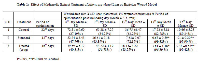
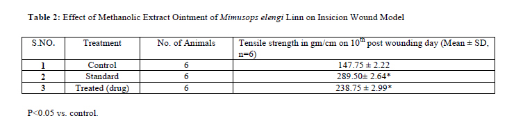
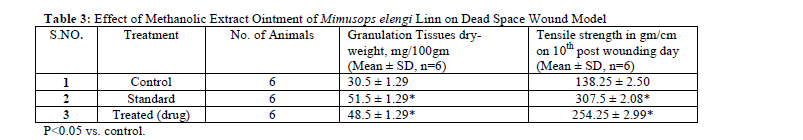
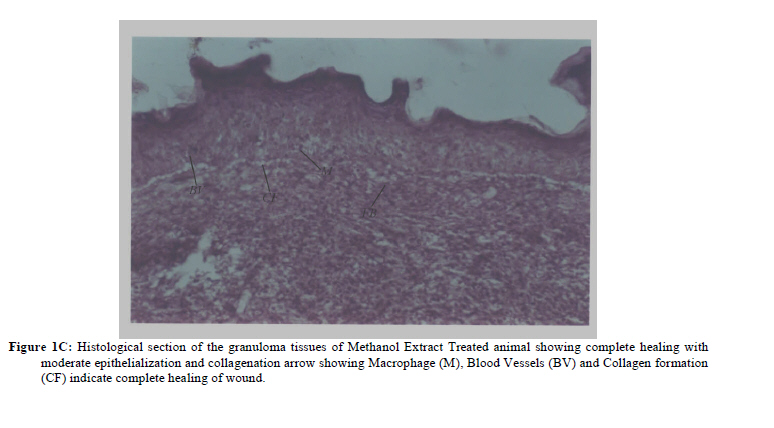
Bhojpur Road, Bangrasia, Bhopal (M.P.), 462045 Code Number: tc11016 Abstract The present study was aimed to evaluate the wound healing activity of extract of bark part of Mimusops elengi. It is well-known plant in Indian traditional medicines. On the basis of traditional use and literature references, this plant was selected for wound healing potential. A methanolic extract of bark parts of Mimusops elengi was examined for wound healing activity in the form of ointment in three types of wound models on mice: the excision, the incision and dead space wound model. The extract ointments showed considerable response in all the above said wound models as comparable to those of a standard drug Betadine ointment in terms of wound contracting ability, wound closure time, tensile strength and dry granuloma weight. Histological analysis was also consistent with the proposal that Mimusops elengi bark extract exhibits significant wound healing Key words: Mimusops elengi, Wound healing, Betadine, methanolic extract IntroductionWounds are visible results of individual cell death or damage. It is a disruption of tissue integrity that is typically associated with a loss of substance. Therefore wound is a loss of cellular and anatomic or functional continuity of living tissues (Charde et al., 2005). More than 80% of the world population still depends upon traditional medicines for various skin diseases (Shamuga et al., 2002). Herbal medicines are crucial in wound healing since they initiate disinfection, debridement and providing a moist environment to encourage the establishment of the suitable environment for natural healing process (Purna and Babu, 2000). Mimusops elengi Linn (Sapotaceae) commonly known as Bakul, is a small to large evergreen tree found all over the different parts of India. It is cultivated in gardens as an ornamental tree. It has been used in the indigenous system of medicine for the treatment of various ailments. Several therapeutic uses as cardiotonic, alexipharmic, stomachic, anthelmintic and astringent have been ascribed to the bark of Mimusops elengi (Kirtikar, 1935). It has been reported as dantarogahara (treats and prevent tooth decay and tooth disease) in Ayurveda (Satyavati and Gupta, 1987). A decoction of the bark is used as a gargle in salivation in weak and spongy gums, pyorrhea, stomatitis and ulcerated throat (Gogte et al., 2001). Compound powder made of the bark is recommended as toothpowder in cases of spongy gums (Raghunathan and Mitra 1982). Phytochemical review shows the presence of taraxerol, taraxerone, ursolic acid, betulinic acid, V-spinosterol, W-sitosterol, lupeol, alkaloid isoretronecyl tiglate and mixture of triterpenoid saponins in the bark of Mimusops elengi (Misra and Mitra.,1967,1968; Hart et al., 1968; Varsheny and Badhawar, 1972). Literature search revealed that there is no report available regarding wound healing activity of Mimusops elengi bark. The present study was, therefore undertaken to evaluate the wound healing activity of methanolic extract of Mimusops elengi bark. Material and Methods Plant material The stem bark of Mimusops elengi was collected from mature trees and its botanical identification was confirmed from National Botanical Research Institute Lucknow. A voucher specimen NBRI/CIF/Re./08/2008/32 was deposited in the herbarium of NBRI Lucknow. The plant material was dried in shade, powdered and sieved through 40-mesh size and material was stored in well-closed container. Extraction and preparation of formulationBark of Mimusops elengi Linn was finely pulverized (100 g) and extracted in Soxhlet apparatus for 24 hr with methanol then concentrated and dried under reduced pressure (British Pharmacopoeia, 1953). The extract was weighed and the yield was 28.22% (w/w). The semisolid mass (dark brown colour) was obtained and used as ingredient for 5% ointment Animals Healthy Albino mice of either sex (35–45 g) with no prior drug treatment were used for all the present in-vivo studies. The animals were fed on a commercial pellet diet (Hindustan Lever, Bangalore, India), and water ad libitum. The animals were acclimatized to laboratory hygienic conditions for 10 days before starting the experiment. Animal study was performed in Division of pharmacology, Bhopal Institute of Technology & Science-Pharmacy Bhojpur Road, Bangrasia, Bhopal (M.P) with due permission from institutional animal ethical committee. Wound healing activity Grouping of animals Screening for wound healing activity was performed. For incision, excision and dead space wound model, 54 animals of either sex weighing between 35 and 40 g were divided into three groups in each model consisting of six animals as follows: group I – simple ointment base; group II – 5% methanolic extract ointment of Mimusops elengi and group III – 5% Betadine (Win-Medicare, India) ointment was used. The hairs on the skin of white surface of animals were removed by using a suitable depilatory (Anne-French hair removing cream). Excision wound modelCircular wounds of approximately 10mm diameter were inflicted on the cleared skin by cutting under mild Xylocain 4% topical anesthesia. The areas of the wounds were measured (sq. mm) immediately by using vernier calipers. This was taken as the initial wound area reading. Group-I animals served as negative control, which received ointment I.P. Group-II served as positive control to which Betadine (5% w/w in ointment I.P.) was applied topically. Group-III animals were treated with the extract (5% w/w) in a similar manner. All the test samples were applied once daily. The wound area of each animal was measured on 1st, 4th, 7th, 10th, 13th and 16th post wounding day (Table 1). The wound closure was measured at regular intervals of time to see the percentage of wound closure and epithelialization time that indicates the formation of new epithelial tissue to cover the wound. The number of days required for falling of the scar without any residual of the raw wound gave the period of epithelialization. Incision wound modelAll animals were anaesthetized before wound creation and paravertebral long incisions were made through the skin at the distance of about 1.5 cm from midline on the depilated back of mice. No local or systemic antimicrobials were used throughout the experiment. All groups were treated same as in excision model, the both edges were kept together and stitched with black silk surgical thread (no. 000) and a curved needle (no. 11) was used for stitching. The continuous threads on wound edges were tightened for good closure of the wound. After stitching, wound was left undressed then simple ointment base, extract ointment and standard ointment were applied daily up to 9 days; when wounds were cured thoroughly the sutures were removed on the 10th day and tensile strength (Hemalata et al., 2001) of cured wound skin was measured using tensiometer (Table 2). The skin breaking strength is expressed as the minimum weight (in grams) of water necessary to bring about the gapping of the wound. Dead space wound modelThis model was used for the study of granuloma tissue. Animals were anaesthetized by Xylocain 4% topical anesthesia and wound was made by implantation of cotton pellets (10 mg), (2.0×0.5), one on side, in the lumber region on the dorsal surface in each animal. On the tenth post-wounding day, granuloma tissue formed on implanted cotton pellets was dissected out carefully. Granuloma tissue from one part was dried (600C) and weight granulomas, while the other part of granuloma tissue (Shirwaikar et al., 2003; Patil et al., 2001) was used for determination of tensile strength (Table 3). Histopathological Studies Wound tissue specimens from control, test and standard groups were taken after complete healing of excision wound. After usual processing, 6-mm thick sections were cut and 10% of nautral formalin solution was used to fix the granulation tissues for 24 hr and dehydrated with a sequence of ethanol-xylene series of solutions. The inflicted materials were embedded with paraffin at 40-60 0c. Microtome sections were taken at10μ thicknesses. The processed sections were stained with haematoxylin eosin (Varshney et al., 1997) and observed under microscope. An excision wound margin was traced after wound creation by using transparent paper and area measured by vernier calipers. Wound contraction was measured in each 3 days interval, until complete wound healing and expressed in percentage of healed wound area. The epitheliazation time (Rashed et al., 2003) was measured from initial day. Measurement of tensile strength Tensile strength is the resistance to breaking under tension. It indicates how much the repaired tissue resists to breaking under tension and may indicate in part the quality of repaired tissue. Sutures were removed on the 9th day after wound creation and the tensile strength was measured. For this purpose, the newly formed tissue including scar was excised and tensile strength was measured with the help of tensiometer, which is based on method of Kuwano (Kuwano et al., 1994). In this method wound breaking strength was measured as the weight of water at the time of wound breaking per area of the specimen. Dry granulation weightFrom the collected tissue 10th day skin sample of dead space wound (granulomas) was excised from cotton pellets and granuloma mass was dried and weighed, while the other part of granuloma tissue was used for determination of tensile strength. Histopathological studiesThe healing tissues obtained on the 16th day from all three groups of animals of the excision wound model were processed for histological study. Sections were qualitatively assessed under the light microscope and observed in respect of fibroblast proliferation, collagen formation, epithellization and blood vessels. Statistical analysisTreated group was compared with the control group. The results were analyzed statistically using Student’s t-test to identify the differences between the treated and control. The data were considered significant at p<0.05. Results and Discussion The percentage wound contraction (Srivastava et al., 2008) was determined using the following formula:
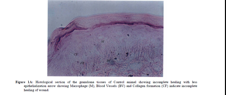
Wound area was measured by tracing the wound margin using vernier calipers in each 3 day-interval and healed area was calculated by subtracting from the original wound area. On day 7, the wound contraction of standard and extract ointment treated groups were found to be significant (P<0.05) in comparison to simple ointment base treated group. On day 13, standard ointment treated wound was completely healed while extract ointment treated wound was also almost at complete healing stage. On day 16, extract ointment treated group healed 99.42% and simple ointment base treated group showed 89.34% healing. It was also observed that epitheliazation period of treated and standard group were less in comparison to simple ointment base treated group (Table 1). The time required for complete epitheliazation of the excision wound is an important parameter to assess the wound healing process. The studies on excision wound healing model revealed that all the three groups showed day to day decrease in wound area. However, on 16th post wounding day, control animals group-I showed 10.66 ± 5.23 of wound area (which may be due to self immunity of the animals) whereas group-II Betadine treated animal, showed 0.1 ± 0.20 wound area and the treated group-III exhibited 0.58 ± 0.68 wound area. When compared with the control, the activity of extract was found to be highly significant (P<0.05). The promotion of wound healing activity is also well gazed by its tensile strength of the incision wound. Generally wound-healing agents have the properties to enhance the deposition of collagen content, which provides strength to the tissues and forms cross-linkages between collagen fibers. The tensile strength of the extract treated group was found to be (238.75± 2.99) which was higher than that of control treated group (147.75 ± 2.22) of animals and slightly lesser than that of standard treated group (289.50± 2.64) of animals on 10th post wounding day. The effect of topical administration of the extract ointment treated group and control treated group on dead space wound model was assessed by increase in the weight of granulation tissue and increase tensile strength. The data is depicted in table-3. This indicates enhanced collagen maturation by increased cross-linking of collagen fibers. The increased weight of the granulation tissue also indicated the presence of higher protein content. Among these treated animals the response was shown to be the best in extract ointment treated animals. Histology of the wound tissue of the control animals showed the presence of acute inflammatory cells, fibroblastic connective tissue and very little number of blood vessels (Figure 1A). The lesser epithelialization and lesser collagen formation indicated incomplete healing of the wound in control animals. Where as, in the sections of Betadine treated animals (Figure 1B) increased collagen deposition was observed. The sections of the granuloma tissue of the animals treated with methanolic extract showed moderate epithelialization, fibrosis, collagen formation and increased number of blood vessels (Figure 1C) Wound healing process consists of different phases such as contraction, epithelization, granulation, collagenation, collagen maturation and scar maturation which are concurrent but independent to each other. Hence in this study three different models were used to assess the effect of herbal ointment on various phases. The result showed that methanolic extract ointment possesses a definite prohealing action. This was demonstrated by a significant increase in the rate of wound contraction and by enhanced epithelialization. Significant increase (P<0.05) in tensile strength, collagen levels and weight of granulomas were observed, which were further supported histopathological studies and gain in granuloma breaking strength. This indicated improved collagen maturation by increased cross-linking while an increase in dry granuloma weight indicated accumulation of higher protein content. The study has showed that the methanolic extract ointment of Mimusops elengi effectively stimulated wound contraction; increase tensile strength of incision and dead space wounds as compared to control group. These finding could justify the pronounced wound healing activity of this plant. AcknowledgementThe authors are thankful to Bhopal Institute of Technology & Science-Pharmacy, Bhojpur Road, Bangrasia, Bhopal for providing facilities to carry out the research work. References
The following images related to this document are available:Photo images[tc11016t2.jpg] [tc11016f1a.jpg] [tc11016t3.jpg] [tc11016f1c.jpg] [tc11016t1.jpg] [tc11016f1b.jpg] |
| |||||||||
