 |
| About Bioline | All Journals | Testimonials | Membership | News |
|
||||||
|
||||||
African Journal of Traditional, Complementary and Alternative Medicines, Vol. 8, No. 2, 2011, pp. 165-169 USE OF ETHNOVETERINARY REMEDIES IN THE MANAGEMENT OF FOOT AND MOUTH DISEASE LESIONS IN A DIARY HERD Gakuya, D. W. 1* Mulei, C.M.1 and Wekesa, S. B.2 1Department of Clinical Studies, Faculty of Veterinary Medicine, University of Nairobi, P.O.Box
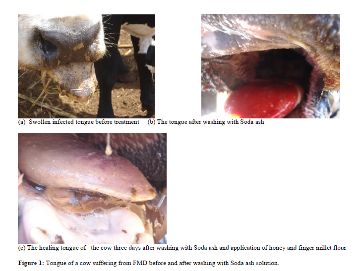
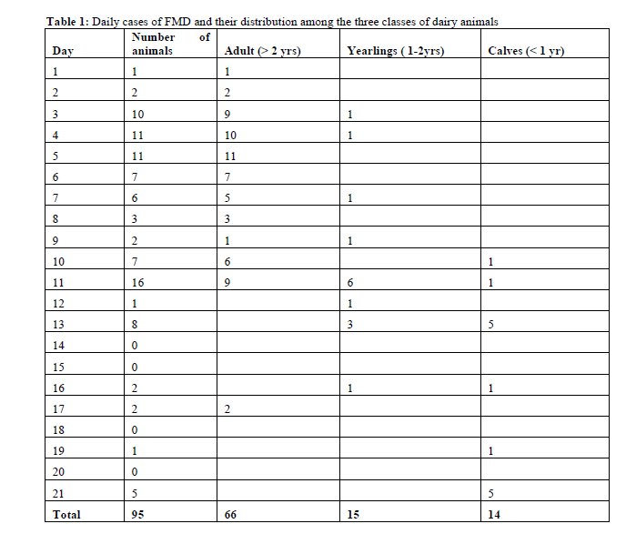
29053,00625 Kangemi, Nairobi, Code Number: tc11023 Abstract An outbreak of Foot and Mouth Disease (FMD) affecting 95 (57.2%) out of 166 cattle occurred in a medium-scale dairy farm in Kikuyu district, Kenya. Ethnoveterinary remedies of natural Soda ash solution (97% sodium bicarbonate), honey and finger millet flour were used to manage the FMD lesions. The lesions were washed with soda ash solution to remove the necrotic tissue after which raw honey and finger millet flour were applied to the cleaned lesions. The lesions were examined daily and those with necrotic material washed again with the Soda ash solution. Honey and finger millet flour were applied daily for three days. There was rapid healing of the lesions with the animals resuming feeding after three days. The fast healing of the lesions vindicates the use of these cheap, locally available and easy to apply products in the management of FMD lesions. However, more studies are needed to evaluate further their potencies. Keywords: Foot and Mouth Disease lesions, Ethnoveterinary remedies, Soda ash, Honey, Finger millet Introduction Foot and mouth-disease (FMD) is a highly infectious viral disease of cattle, pigs, sheep, goats and artiodactyls wildlife species, caused by FMD virus which has 7 major serotypes: A,O,C,Asia1 and SAT 1,2,3, (Kahn et al. 2005). The disease is characterized by fever, salivation and vesicles in the mouth, muzzle, dental pad, tongue, teats and feet. The rupture of the vesicles results in marked painful swelling of the coronary band, depression, innapetence, lameness, recumbency, loss of body condition, severe mastitis and abortions (Radostitis et al. 2000). The serotypes have a large number of strains that have a spectrum of antigenic characteristics requiring more than one vaccine strain for each serotype (Kahn et al. 2005). Five of the seven serotypes of FMD virus (O, A, C, SAT1 and SAT2) have been reported in Kenya (Ngichabe, 1984; Vosloo et al. 2002). There is no cross immunity between serotypes and this presents difficulties to vaccination programs (Radostitis et al. 2000). The inability to detect all the serotypes in a multiple serotype infection may also result in vaccination and disease control failure as the vaccine being used may be deficient of all relevant serotype combination (Sangula et al. 2005). The mode of transmission from infected animal to susceptible one is by inhalation, where exhaled air containing the virus infects the other animal via the respiratory or oral route. All body excretions and secretions from infected animal contain the virus (Kahn et al. 2005). Transmission to calves is via infected milk and fodder which has come into contact with infected animal. Other sources of infection include; milk tankers, mechanical animal vectors like horses, cat, dogs, avian species and humans. Pigs fed on infected feed derived from infected animals become infected and may spread the disease via aerosol to the cattle (Kahn et al. 2005). Factors encouraging the rapid spread of the disease within the herds include; the speed and direction of the wind, ambient temperature and humidity, time of the day and the extent of human and animal movement (Terpstra, 1972; Donaldson and Ferris, 1980). There is no specific treatment for FMD. The conventional method of treating infected animals mainly involves the use of antibiotics, flunixin meglumine and mild disinfectants (Radostitis et al. 2000). Ethnoveterinary practices in the treatment and control of livestock diseases has widely been documented in Kenya (Ohta, 1984, Illes, 1990, Wanyama, 1997, 2000, Miaron, 2003). FMD has been managed traditionally by use of natural soda ash solution for washing of the lesions and other communities have applied honey and even finger millet flour to the lesions (personal communication). These traditional remedies have been reported elsewhere in the management of wounds and ulcers (Hedge, 2005; Molan, 1992, 2001; Nuzov, 1990a and b). This paper reports on the use of ethnoveterinary remedies in the management of FMD lesions following FMD outbreak in a medium scale dairy farm in Kikuyu District, Kenya. Case historyThe dairy cattle in the affected farm are vaccinated against FMD three times in a year. The animals were vaccinated with a quadrivalent vaccine containing serotypes A.K(5/80), O.K(77/88), SAT1.T(155/71) and SAT2.K(52/84) before the outbreak of the FMD. Four days after the vaccination, one adult cow was reported with signs of respiratory distress, hypersalivation and smacking of the jaws, which on clinical examination had vesicles in the mouth and a body temperature of 41.8o C. From the clinical signs, a tentative diagnosis of FMD was made. On the second day two more cows developed the same clinical symptoms. For confirmation of the disease, two samples (K114/09 and K115/09) of the vesicular fluid and epithelium in the mouth were collected and submitted to the Foot and Mouth disease laboratories of the Ministry of Livestock Development (Embakasi, Kenya) for confirmation and further serotyping. The samples were later sent to WRL Pirbright, UK for further tests to ascertain the serotypes and strains involved in the outbreak. Disease management After notification of the relevant authorities, the farm was placed under quarantine and disinfection with natural soda ash (MAGADI SODA® (97% sodium bicarbonate), (Magadi soda company limited, Magadi, Kenya) applied on the two entry points of the farm. Vehicles entering and leaving the farm were sprayed with Soda ash solution and foot bath for all personnel either entering or leaving the farm were created. The Soda ash solution was prepared by dissolving 1kg of Soda ash into 20 litres of water. All new cases of infection were isolated from the rest of the herd and their ear tag number recorded. Workers dealing with the infected animals were separated from the others dealing with healthy ones. The management of the lesions in the mouth and the foot involved thorough cleaning of the lesions with Soda ash solution to remove the necrotic tissue (Figure 1b) followed by application of raw honey and finger millet flour to the cleaned lesions. The lesions were reviewed daily and only those with necrotic material were washed with Soda ash solution. In all cases honey and millet flour were applied daily for three days. Severe cases involving the tongue and the feet were given long acting oxytetracyline (ADACYCLINE (R) , Assia pharmaceutical Ltd, Nairobi, Kenya) at 20mg/kg bodyweight. Teat lesions were treated with milking salve and honey and mastitis cases were managed with Ampicillin and Cloxacillin (MASTACLOX(R)Assia pharmaceuticals Ltd, Nairobi, Kenya) intramammary tubes. The were no new cases after day 21 (Table 1) and all the animals had fully recovered by day 28. ResultsA total of 95 (57.2%) out of 166 animals were affected within three weeks (Table 1). The clinical manifestations of the disease included mouth lesions, salivation, respiratory distress, blisters, ulceration of the gums and muzzle and fever (39.7 41.8). Following treatment the lesions started healing and subsequently the animals were able to eat on the third day posttreatment. The results of the ELISA test conducted to the two samples (K114/09 and K115/09) that were submitted to FMD laboratories in Embakasi, showed that only K115/09 was positive for SAT1 with traces of serotype O. The results from World Reference Laboratory (WRL) in Pirbright (UK) also showed that sample K115/09 was positive for SAT1 while K114/09 was negative. Phylogenetic analysis showed that the strain (K115/09) that caused this outbreak had 10.56% nucleotide difference from the vaccine strain used (SAT1/T155/71). Discussion After washing of the lesions with Soda ash solution (97% Sodium bicarbonate) and daily application of honey and finger millet flour, the lesions in the mouth of the cattle healed after 3 days. The animals started eating slowly on the second day. This fast healing may be attributed to the use of Soda ash solution, honey and finger millet flour. Soda ash kills the virus within a few minutes (Radostitis et al. 2000) and therefore, the washing of the mouth with the Soda ash solution could have killed the virus thus reducing the viral load in the discharges and the desquamated necrotised epithelium. The cleaning of the FMD lesions could also have prevented any secondary bacterial infection hence the rapid healing of the wounds. Honey has been in use for treatment of infected wound as long as 2,000 years ago, even before bacteria were discovered. Recently honey has been reported to have inhibitory effect to around 60 species of bacteria (Molan, 1992). Honey has antibacterial properties due to production of hydrogen peroxide which is formed and released slowly by glucose enzymes when the honey is diluted (Vaux, 2009). Although the level of hydrogen peroxide in honey is very low, it is still effective as an antimicrobial agent (Molan, 2001). Some types of honey have also been reported to have some non identified phytochemical components which are antibacterial apart from hydrogen peroxide production (Allen et al. 1991). The clearing of infection seen when honey is applied to a wound may reflect more than just antibacterial properties (Molan, 2001). Abuharfeil et al. (1999) reported that there is proliferation of peripheral blood B-lymphocytes and T-lymphocytes stimulated by honey at concentrations as low as 0.1% in cell cultures and phagocytes are activated by honey at concentrations as low as 0.1%. Tonks et al. (2001) also reported that honey at a concentration of 1% also stimulated monocytes in cell culture to release cytokines, tumour necrosis factor (TNF)-alpha, interleukin (IL)-1 and IL-6, which activated the immune response to infection. Therefore, the mobilization of blood cells which are key in the immune response to infection together with the production of hydrogen peroxide which inhibit microbes could have contributed to the fast healing of the lesions in this case. However, there is a need to do further studies on the antibacterial properties of the honey and evaluate its efficacy in both in vivo and in vitro tests. The current use of honey in management of these cases as is often done traditionally is justified by the success of the treatment. Millet is one of the oldest foods known to humans and possibly the first cereal grain for domestic purposes. Millet is tasty, with a mildly sweet, nut-like flavor and contains a myriad of beneficial nutrients. It is nearly 11 % protein, contains high amounts of fiber, B-complex vitamins including niacin, thiamin, and riboflavin, the essential amino acid methionine, lecithin, and some vitamin E (FAO, 1995). It is particularly high in the minerals: iron, magnesium, phosphorous, and potassium. The seeds are also rich in phytochemicals, including phytic acid, believed to lower cholesterol, and phytate, which is associated with reduced cancer. The mucilaginous substance in millet when cooked has some healing action in case of gastrointestinal inflammation and ulceration (Morgan, 2004). The millet oil promotes healthy skin, has a protective function against skin damage through skin irritants and promotes regeneration of epidermis after previous skin damage (Henning, 2005). The influence of whole grain flour of finger millet (Eleusine coracana) on dermal wound healing in rats has been evaluated (Hedge et al. 2005). Finger miller flour as an aqueous paste was applied daily for 16 days to an excision wound made at the shaven back of rats (Hedge et al. 2005). There was significant increase in protein and collagen contents in the granulation tissue in the treated rats than the untreated. The rate of contraction of the wound was also higher in the treated 88-90% compared to untreated (75%). The number of days for complete closure of wounds was lower for finger millet (13 days) when compared to untreated (16 days) rats and therefore the authors concluded that finger millet accelerated the process of wound healing. This may justify the rapid healing of the lesions when millet flour was used to manage the FMD lesions in these cases. Niazov (1990a) studied the efficacy of miliaceum (millet) oil which is obtained from waste products of millet processing in local application for the treatment of purulent wounds. Using 55 rabbits with purulent wounds, the preparation caused a marked anti-inflammatory effect, promoted rapid cleansing of the wounds from the pyonecrotic contents, and significantly activated the reparative processes. Similarly, the effect of Oleum miliacei obtained from millet scraps on the regenerative processes of trophic ulcers was evaluated using 74 rabbits (Niazov, 1990b). The experiment confirmed that Oleum Miliacei had anti-inflammatory action and stimulated the processes of regeneration. This justifies the traditional usage of millet flour for wound management and in the current use for management of FMD lesions. The traditional usage of Soda ash, honey and millet for the management of FMD is supported by the rapid healing of the FMD lesions observed. The usage of these ethnoveterinary remedies is therefore recommended as these products are cheap, locally available and easy to apply at farm level. However, further studies on these products are necessary to experimentally evaluate their potency. Acknowledgement We wish to acknowledge the diagnostic services availed to us by Foot and Mouth Disease laboratories, Embakasi (Kenya) and World Reference laboratories (WRLFMD), Pirbright laboratory, UK. References
Copyright 2011 - Afr. J. Trad. CAM The following images related to this document are available:Photo images[tc11023t1.jpg] [tc11023.jpg] [tc11023f1.jpg] |
| |||||||||
