 |
| About Bioline | All Journals | Testimonials | Membership | News |
|
||||||
|
||||||
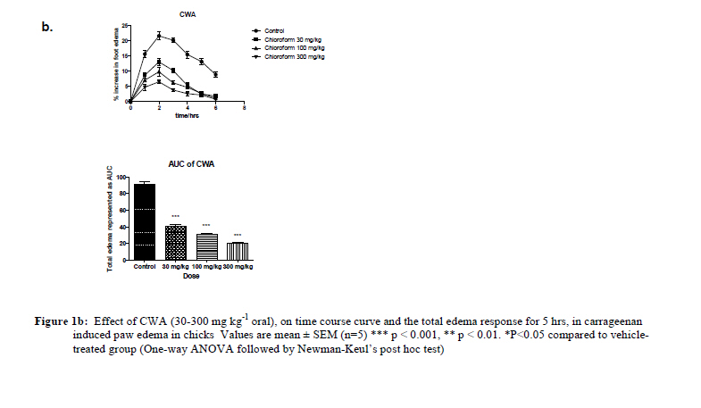
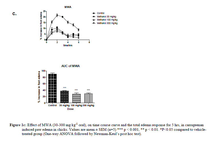
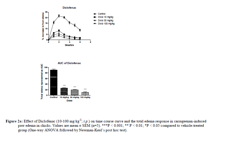
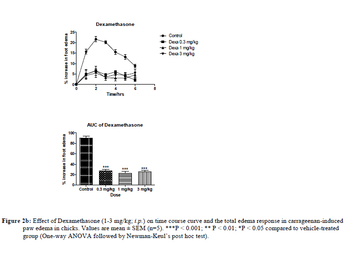
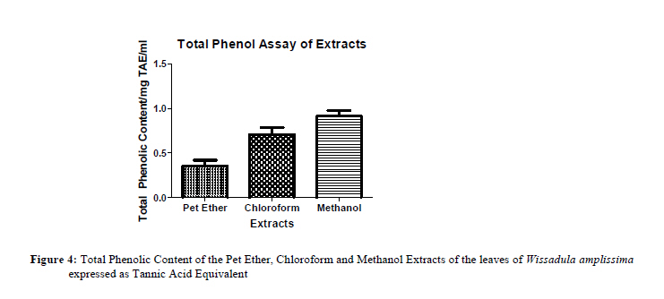
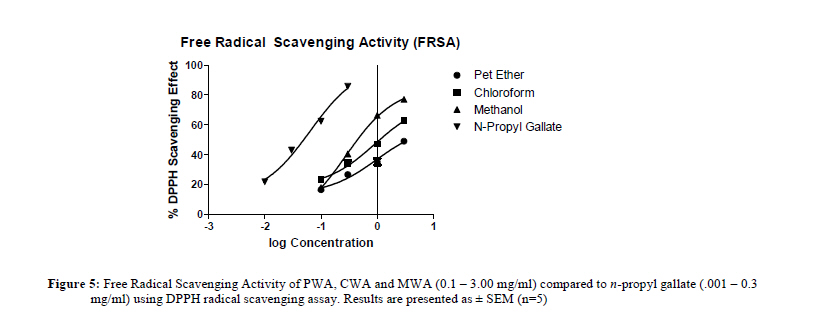
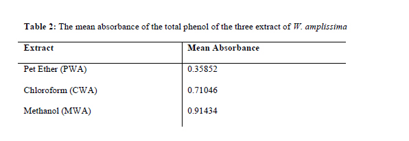
African Journal of Traditional, Complementary and Alternative Medicines, Vol. 8, No. 2, 2011, pp. 150-158 ANTI-INFLAMMATORY AND ANTIOXIDANT ACTIVITIES OF THE LEAVES OF WISSADULA AMPLISSIMA VAR ROSTRATA A.Y. Mensah*, P.O. Donkor and T.C. Fleischer Department of Pharmacognosy, Faculty of Pharmacy & Pharmaceutical Sciences, College of Health Sciences, Kwame Nkrumah University of Science and Technology, Kumasi, Ghana * Email: aymensah.pharm@knust.edu.gh Code Number: tc11026 Abstract The present study determined the anti-inflammatory activity of Wissadula amplissima var rostrata (Schum. & Thonn.), and calculated the total phenolic content and total antioxidant capacity of the plant in an attempt to justify the traditional uses of the plant in the Ashanti region of Ghana for the management of spider,wasps and bee stings. Powdered dried leaves of Wissadula amplissima were Soxhlet extracted with Petroleum Ether (PWA, yield: 1.46% w/w); Chloroform (CWA, yield: 1.18% w/w) and Methanol (MWA, yield: 3.39% w/w). These fractions were tested for anti-inflammatory activity using carrageenan-induced foot edema in 7 day old chicks. The effect before the induction of inflammation (pre-emptive protocol) paradigm was used for the assessment. Oral administration of PWA, CWA and MWA (30 – 300 mg/kg) dose dependently reduced edema with maximal effects of 68.25±2.03%, 77.83±0.81% and 62.21±2.61% respectively. Similarly the NSAID, Diclofenac (10 – 100 mg/Kg, i.p) and the steroidal anti-inflammatory drug dexamethasone (0.3 – 3 mg/Kg, i.p) used as positive controls, dose-dependently inhibited the edema with maximal effect of 87.96±1.11% and 67.47±3.51% respectively. The potencies exhibited by all three extracts were comparable to that shown by Diclofenac but higher than that of Dexamethasone. Phenols were detected in all three extracts with the highest concentration in the MWA. The extracts also scavenged DPPH with EC50 values of 0.9784, 0.9096 and 0.2767 for PWA, CWA, MWA respectively. The results of this study give scientific credence to the local use of Wissadula amplissima to modulate inflammation induced by stings of animals. Key words: Wissadula amplissima, anti-inflammatory, carrageenan, antioxidant capacity IntroductionEnvenomation by insects such as ants, bees, wasps, snakes and scorpions is common and frequent in the rural areas of the Ashanti region of Ghana where most of the inhabitants are cocoa and food stuff farmers. Although envenomation is mostly painful and causes inconvenience it is rarely life threatening except those caused by some snake bites and in previously sensitized individuals (Hutt and Houghton, 1998). Majority of inhabitants of these areas normally treat these conditions with plant extracts because of the difficulty in accessing orthodox medicines. Among the numerous plants used by the indigenes is Wissadula amplissima var rostrata (Schum. & Thonn.) family, Malvaceae. It is a herb or undershrub that is normally considered to be a weed in most food growing areas in the Guinea Savanna with ovate cordate leaves, acuminate apex and entire leaf margin The leaves are used in the treatment of spider bites as well as sting by venomous animals (Fernandez de la Pradilla, 1988; Mshana et al., 2000). It is prepared as a poultice and applied to the affected part. Several plants popularly used as anti-snake venoms and for the management of spider bites and wounds exhibit anti-inflammatory activities (Lewis, 1989; Ruppelt et al., 1991; Ribeiro et al., 1988; Mensah et al., 2006). These references suggest the possibility that W. amplissima may also have anti-inflammatory activity. Antioxidants are documented in several publications to mitigate the inflammatory processes and some of the antioxidant activity of plants have been ascribed to the phenolic compounds present in the plant particularly flavonoids (Pereira et al., 1994; Hutt and Houghton, 1998; Sakai et al., 1999; Ozgova et al., 2003). A search in the literature shows no evidence of a scientific attempt to justify the traditional uses of the plant as anti-inflammatory agent. The anti-inflammatory potential of W. amplissima was therefore investigated using the chick-carrageenan model described by Roach and Sufka (2003).The antioxidant activity of the extracts of W. amplissima was also evaluated using three different assay protocols: Total Phenol Content, Free Radical Scavenging Activity (FRSA) and Total Antioxidant Capacity. Materials and Methods Plant material and preparation The leaves of Wissadula amplissima were collected from the wild in a secondary forest in Antoa, a village near Kumasi in the Ashanti Region of Ghana in February 2009. The leaves were authenticated at the Department of Pharmacognosy and a voucher specimen (WA1/2009) deposited at the Pharmacognosy Department Herbarium KNUST, Kumasi. The leaves were then air-dried for 7 days and coarsely powdered. 1.2 kg of the coarsely powdered air-dried leaves was then serially extracted in a Soxhlet apparatus using 20 L each of petroleum ether, chloroform and methanol in the order of increasing polarity. The extraction was carried out over 24 hrs for each solvent. The different solvent extracts were collected and concentrated using a rotavapor. The percentage yields of the petroleum ether extract (PWA), chloroform extract (CWA) and methanol extract (MWA) were 1.46 %w/w, 1.18 %w/w and 3.39 %w/w respectively. Phytochemical Analyses Part of the dried powdered material was subjected to preliminary phytochemical screening for the major phytochemical groups by simple qualitative methods (Harborne, 1991). Animals Cockerels (Gallus gallus, strain Shaver 5790) weighing between 50-85 g were purchased 1-day post-hatch from Akropong Farms in Kumasi, Ghana and maintained in the Animal House of the Department of Pharmacology, KNUST, Kumasi as previously reported (Woode et al.,2007) . They were housed in stainless steel cages (34×57×18 cm3) at a population density of 12– 13 chicks per cage in a temperature regulated environment (26 -29 °C) with a 12 hr light-dark cycle. Food and water was available ad libitum. Daily maintenance of the cages was conducted during the first quarter of the light cycle. Chicks were tested at 7 days of age. Group sample sizes of five–six were utilized throughout the study. The chicks handling and experimental protocol were in accordance with the institutional animal care and use committee approved by the Ethical Review Committee of the College of Health Sciences, KNUST. Carrageenan-induced foot edema in chicks The anti-inflammatory activity of the extracts of Wissadula amplissima was evaluated using the carrageenan foot edema model of inflammation in the 7-day old chick with some modifications (Roach and Sufka, 2003, Woode et al., 2007)). The activities of the extracts (PWA, CWA and MWA) were compared with that of two reference standard anti-inflammatory drugs dexamethasone and diclofenac which served as positive controls. The different extracts were suspended in sterile water by adding 2% of tween 20 to enhance the suspension of the extract. Following the protocol described by Woode et al. (2007), 30, 100 and 300 mg/kg of the suspended extracts were administered p.o. to chicks (n = 5). One hour after the administration of the extracts, Carrageenan (10 µl of a 2% suspension in saline) was injected sub-plantar into the right footpads of the chicks. The foot volume was measured before injection and at hourly intervals for 5 hrs after injection using a digital Venier Caliper. The edema induced by the inflammation was quantified by measuring the difference in foot thickness before carrageenan injection and at the various time points. Dexamethasone (0.3-3.0 mg/kg, i.p) and diclofenac (10-100 mg/kg, i.p) were used as positive controls. The drug vehicle 2% tween 20 in sterile water was used as negative control. All drugs and extracts doses were administered in volumes not exceeding 100 ml/kg p.o. Total Phenol Content The Total Phenols of the petroleum ether, chloroform and methanol extracts of Wissadula amplissima were determined using the Folin-Ciocalteau reagent (Singleton et al., 1997; Woode et al., 2008). A range of concentrations (3.00, 1.00, 0.30, 0.10 mg/ml) of each extract (1 ml) were mixed with the Folin-Ciocalteau Phenol reagent (1ml; diluted 1:10 with distilled water). Na2CO3 (2% w/v, 1ml) was added to each mixture and incubated at room temperature (28oC) for 2 hours. Absorbance was taken at 760 nm using a Cecil UV/VIS spectrophotometer (Model: CE 7200, Milton, England). The samples were centrifuged at 3000 rpm for 10 mins and the supernatant taken for each measurement. Tannic acid was used as a reference standard. The total phenolics were expressed as milligrams per milliter of Tannic Acid Equivalents (TAEs). Scavenging of 2,2-Diphenyl-1-Picrylhydrazyl (DPPH) Antioxidant property of plant extracts may be tested in several ways and several in vitro tests are routinely conducted. These include the DPPH method that detects free radical scavengers and relies on the discoloration of the purple coloured methanolic solution of DPPH to yellow by free-radical scavengers (Houghton et al., 2005). 1 ml methanolic solutions of the extracts of W. amplissima (3.00, 1.00, 0.30, 0.10 mg/ml) were added to 3 ml 2% solution of DPPH in methanol in a test tube. The reaction mixtures were kept at 25oC for 1 hr. The absorbance of the residual DPPH was then determined at 517 nm using a Cecil UV/VIS Spectrophotometer (Model: CE 7200, Milton, England). The scavenging action of the extracts of W. amplissima (3.00, 1.00, 0.30,0.10 mg/ml in methanol) was compared to the standard, n-propyl gallate (0.30, 0.10, 0.03, 0.01 mg/ml in methanol). 1 ml methanol (99.8%) added to 3.00 ml DPPH solution, incubated at 25oC for 1 hr served as control and methanol (99.8%) was used as blank. Results were expressed as percentages of control, and the concentration of extracts required to cause a 50% decrease in the absorbance was calculated (EC50 ). The % DPPH scavenging effect (% of control) of the antioxidant is calculated as follows:
Total Antioxidant Capacity Following the protocol described by Prieto et al. (1999) with some modification, the total antioxidant capacity of the three extracts of W. amplissima was determined. The assay is based on the reduction of Molybdenum (Mo) (5) to Mo (4) by the extract and subsequent formation of a green Phosphate-Mo (5) complex at acid pH. Various concentrations (3.00, 1.00, 0.30, 0.10 mg/ml) of the extracts of W. amplissima were prepared. 3 ml of the reagent solution (0.6 M H2SO4, 28 mM Na2HPO4 and 4 mM Ammonium Molybdate) were added to 1 ml volumes of the various concentrations. The solutions were incubated at a temperature of 95oC for 90 mins. The solutions were then cooled to room temperature. Absorbance was measured at 695 nm against a blank. Antioxidant capacity was expressed as an Ascorbic Acid Equivalent (AAE). Statistical analysis Raw scores for right foot volumes were individually normalized as percentage of change from their values at time 0, then averaged for each treatment group. The time-course curves for foot volume were subjected to two-way (treatment × time) repeated measures analysis of variance with Neuman Keul’s post hoc test. Total foot volume for each treatment was calculated in arbitrary unit as the area under the curve (AUC) and to determine the percentage inhibition for each treatment. The following equation was used.
Differences in AUCs were analyzed by one way ANOVA followed by Student-Newman-Keuls’ post hoc test. Doses and concentrations responsible for 50% of the maximal effect (EC50) for each drug were determined using an iterative computer least square method with the following non-linear regression (three-parameter logistics) equation.
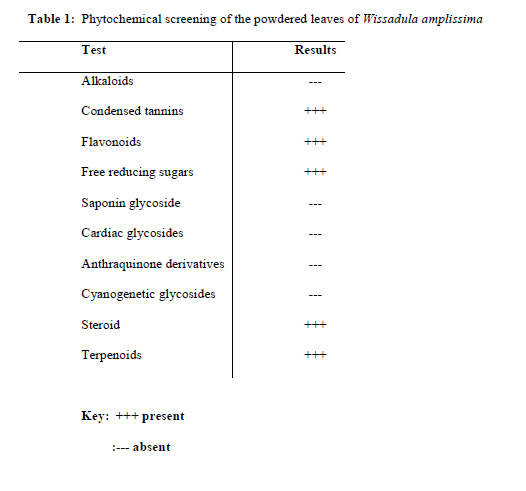
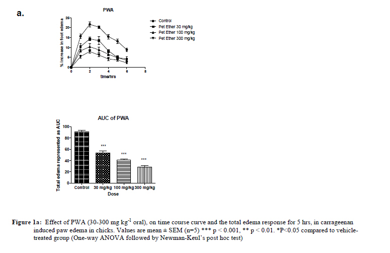
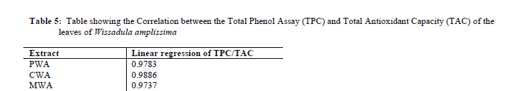
Where, X is the logarithm of dose and Y is the response. Y starts at a (the bottom) and goes to b (the top) with a sigmoid shape. The fitted midpoints (ED50s were compared statistically using F test (Miller, 2003; Motulsky and Christopoulos., 2003). Graphpad Prism for windows version 4.03 (GraphPad Software, San Diego, CA, USA) was used for all statistical analyses and ED50 determinations. P < 0.05 was considered statistically significant. Results Phytochemical screening Table 1 shows the results of the general phytochemical screening of the leaves of Wissadula amplissima. The results indicate the presence of phenolic groups particularly condensed tannins and flavonoids. The results also revealed the presence of steroids and terpenoids. The general test for reducing sugars using Fehling’s test identified the presence of free reducing sugars. However, anthraquinone glycosides, saponin, cyanogenetic glycosides and cardiac glycosides were absent. Carrageenan-induced edema The effect of the extracts in acute inflammation was assessed in the chick carrageenan-induced foot edema model using Dexamethasone and Diclofenac as positive controls. Administration of 10 µl of 2% carrageenan-induced moderate inflammation resulting in foot edema in the 7 day old chicks, peaking at 2-3 hr as described by Roach et al. (2003). Figures 1a, 1b, 1crepresents the Test Alkaloids Condensed tannins Flavonoids Free reducing sugars Saponin glycoside Cardiac glycosides Anthraquinone derivatives Cyanogenetic glycosides Steroid Terpenoids time course and total edema response for the effects of the three extracts. The results indicate that all the extracts significantly and dose dependently inhibited carrageenan-induced edema in the seven day old chicks at all doses. Figures 2a-2b indicates the dose dependent inhibition of edema by the reference drugs Diclofenac and Dexamethasone (positive controls). The NSAID Diclofenac (10-100 mg kg-1 , i.p) showed significant effect on the time course curve [P<0.001(Figures2a and 3)] and dose dependently reduced the total edema (AUC), with maximal inhibitory effect of 87.96±1.11 at 100 mg kg-1. Similarly, the steroidal anti-inflammatory agent Dexamethasone (1-3 mg Kg-1i.p) reduced the total edema with maximal inhibition of 73.12±17.65% (P<0.001). PWA, CWA and MWA all caused a significant inhibition of edema (P<0.001) and dose dependently inhibited carrageenan-induced edema with maximal inhibitions of 68.25±2.03%, 77.83±0.81% and 62.21±2.61% respectively. A comparison of the activities of the three extracts and that of Diclofenac is presented in Figure 3. The graph shows that the chloroform extract (CWA) has the highest activity followed by the petroleum ether (PWA) and methanol extract (MWA) respectively at the maximum dose of 300 mg/kg. The Diclofenac at the dose of 300 mg/kg was not tolerated by the chicks hence no value was observed on the Y-axis (% increase in foot edema) Total Phenol Content The results of the total phenol content determination of the various extracts of W. amplissima indicate that the MWA had more phenol content followed by the CWA extract and then the PWA (Figure 4) when expressed as the tannic acid equivalent (TAE) Scavenging of 2,2-Diphenyl-1-Picrylhydrazyl (DPPH) The DPPH assay is often used to measure the ability of an agent to scavenge free radicals. The W. amplissima extracts showed a concentration-dependent scavenging activity analogous to that of n-propyl gallate (Figure 5). The absorbance decreased with increasing free radical scavenging activity. The EC50 values of 0.9784, 0.9096, 0.2767 and 0.06412 obtained for the Pet Ether, Chloroform and Methanol extracts of W. amplissima and n-propyl gallate respectively suggest that W. amplissima has less ability to scavenge free radicals compared to n-propyl gallate. Comparing the scavenging activities of the three extracts (Figure 5 and Table 2), the methanol extract was more active than the chloroform and the petroleum ether extracts. DiscussionCarrageenan-induced acute foot pad edema in laboratory animals is a model widely used to screen new anti-inflammatory drugs and remains an acceptable preliminary screening test for anti-inflammatory activity (Singh et al., 2000; Roach and Sufka, 2003; Woode et al., 2007). The figures 1a, 1b, 1c depict the increases in foot volume which were carrageenan-induced effects and were prominent and significant during the first hr from the baseline and reached the maximal volume at the two hr time point. The foot volume decreased consistently with time till after the fourth to six hr time point. It is instructive that these observations have been documented in several publications confirming the reliability of the model for measuring inflammation (Roach and Sufka, 2003; Woode et al., 2007). The petroleum ether, chloroform and methanol extracts of W. amplissima demonstrated varying levels of antiinflammatory activities which was dose dependent. The antioxidant activities of the three extracts of W. amplissima were analyzed by three different assays: total phenol content, DPPH scavenging activity and total antioxidant capacity. All three assays confirmed the antioxidant properties of the extracts showing the strong correlation between the Total Phenol Assay (TPC) and Total Antioxidant Capacity (TAC) of the leaves of Wissadula amplissima as presented in Table 5. Many bioactive compounds from foods and plants are known to prevent or mitigate numerous inflammatory processes and diseases. Some of these compounds are flavonoids and are believed to function as antioxidants and possess anti-inflammatory activity (Kris-Etherton et al., 2004). The flavonoids act as antioxidants by chelating redox-reactive metals and by scavenging free radicals. They are effective scavengers of hydroxyl and peroxyl radicals as well as quenching superoxide radicals and oxygen singlet (Simic and Jovanovic, 1990; Bravo, 1998; Jovanoic and Simic, 2000). The antioxidant and free radical scavenging properties of the W. amplissima is supported by the detection of tannins and the flavonoids in the three different extracts of the plant and may explain to some extent the traditional uses of the plant for alleviating inflammatory conditions such as those induced by spider and bee stings. Conclusion The results of this study demonstrate that Wissadula amplissima possesses potential anti-inflammatory activity. The use of the plant by traditional healers in the treatment of inflammation from spider and bee stings may have some scientific justification as the plant has been shown to have anti-inflammatory activity. Bioactivity guided fractionation from the more active fractions (chloroform and methanol fractions) is currently underway. The isolation of new and effective anti-inflammatory agents is important for potential drug development as natural products provide unlimited opportunities for new drugs because of the unmatched availability of chemical diversity (Cos et al., 2006). This will eventually go a long way to validate the use of Ghanaian medicinal plants by traditional healers Acknowledgements The authors appreciate the support and assistance offered by Professor Kwame Sarpong and Dr. Eric Woode both of the Faculty of Pharmacy & Pharmaceutical Sciences, KNUST. We are also grateful to Mr. Kwadwo Kakraba of the Pharmacognosy Department KNUST and Mr. Thomas Ansah of the Pharmacology Department, KNUST for the technical support. Finally we acknowledge the sharing of the traditional knowledge of the uses of the plant by Auntie Birago of Senkyire fie, Ejisu Onwe in the Ashanti region of Ghana with us. References
The following images related to this document are available:Photo images[tc11026f3.jpg] [tc11026f5.jpg] [tc11026f1a.jpg] [tc11026f4.jpg] [tc11026t2.jpg] [tc11026f2b.jpg] [tc11026t5.jpg] [tc11026t4.jpg] [tc11026t3.jpg] [tc11026f6.jpg] [tc11026f1b.jpg] [tc11026f1c.jpg] [tc11026f2a.jpg] [tc11026t1.jpg] |
| |||||||||

