 |
| About Bioline | All Journals | Testimonials | Membership | News |
|
||||||
|
||||||
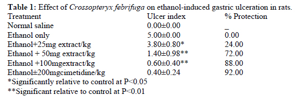
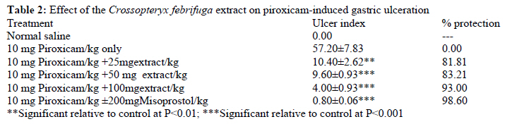
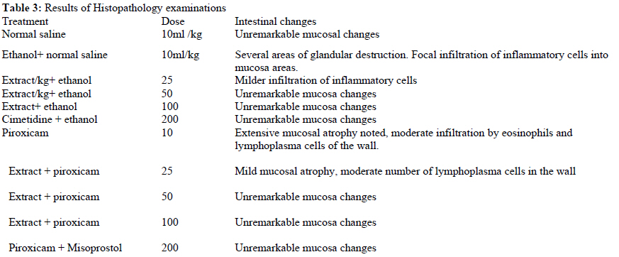
African Journal of Traditional, Complementary and Alternative Medicines, Vol. 8, No. 3, 2011, pp. 300-306 Gastro-Protective Effect Of Crossopteryx febrifuga in Wistar Rats Salawu Oluwakanyinsola Adeola1, Tijani Adeniyi Yahaya1*, Babayi Hafsatu2 , Nwaeze Angela Chinwe,1 Ezeonu Chidimma MaryJane2, Igwe Sunday3, Ndukuba Mary Adanna1
1Departments of Pharmacology and Toxicology, National Institute for Pharmaceutical Research
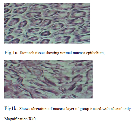
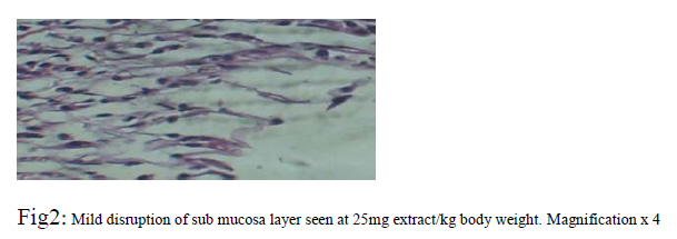
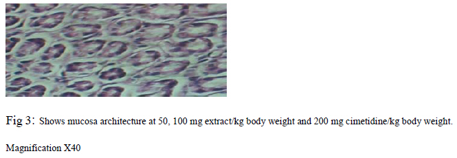
&Development, P.M.B. 21, Garki -Abuja, Nigeria., Code Number: tc11038 Abstract Preparations of Crossopteryx febrifuga (Afzel.) Benth. (Rubiaceae) are widely used in Northern Nigeria in the therapeutic management of trypanosomiasis, malaria and painful inflammatory disorders. Previous studies have shown that the methanolic stem bark extract of Crossopteryx febrifuga possesses significant analgesic and anti-inflammatory properties possibly mediated via Non-selective inhibition of cyclo-oxygenase pathways. In the present study, the methanolic stem bark extract of Crossopteryx febrifuga was evaluated against ethanol- and piroxicam-induced ulceration in rats. Histopathological studies of the rat stomach tissues were also carried out in order to determine its safety profile on the gastrointestinal tract (git). The extract (25, 50 and100 mg extract/kg body weight) significantly (P<0.05) and dose-dependently reduced ulcer index induced by ethanol (24 - 92%) and piroxicam (81.81- 98.60%). Histopathology of the rat stomach tissues from control and extract-treated groups at 25 mg/kg body weight extract showed mild inflammation characterized by infiltration of inflammatory cells, while the extract treated groups at 50 and 100mg/kg body weight and 200 mg misoprostol/kg body weight group showed no obvious lesions. These results showed that the extract had no deleterious effects and was cytoprotective on the gastrointestinal tract (git). It can thus be developed as a safe alternative to conventional non-steroidal anti-inflammatory drugs (NSAIDs) for the management of painful inflammatory disorders. Key words: Crossopteryx febrifuga, extract, analgesic, anti-inflammatory agents, gastrointestinal tract, ulcer Introduction Nonsteroidal anti-inflammatory drugs (NSAIDs), which act as non-selective cyclooxygenase (COX) inhibitors, are commonly used to treat pain and inflammation despite the risk of major upper gastrointestinal complications (Venkova et al., 2008)). The primary cause of the pathogenic effects of NSAIDs in the stomach is the deficiency of endogenous prostaglandins required to maintain the mucosal barrier to luminal acid. Oxygen free radicals and lipid peroxidation (Takeuchi et al., 1991), leukocyte adhesion and tumor necrosis factor alpha were found to play an important role in this process. However, a breakdown in the mucosal barrier against gastric acid is not the only mechanism contributing to mucosal injury during NSAIDs administration. Studies by Santo et al., (2007) have showed that rat models of NSAID treatment revealed alterations in gastroduodenal motility which resulted in delayed gastric emptying and mucosal ulceration. To avoid NSAID-induced gastrointestinal complications, patients with one or more risk factors (history of ulcer disease, high doses or use of multiple NSAIDs, advanced age etc.) receive concurrent gastro-protective therapy (Venkova et al., 2008). Recently Arthrotec® a combination of diclofenac and misoprotol was launched in Nigeria to overcome the gastrointestinal side effects of long term use of non-steroidal antiinflammatory agents in the management of arthritis. However in African countries including Nigeria, indigenous herbal medicines are widely used for the management of painful inflammatory disorders, despite an apparent lack of scientific evidence for their quality, safety and efficacy (Fennell et al., 2004). One of such therapeutically useful medicinal plant is Crossopteryx febrifuga Benth, (Family Rubiaceae), a twisted tree with conspicuous tubular flowers, which is widely distributed throughout the Savannah region of Central, East and West Africa. Preparations of the tree is used traditionally for symptomatic relief of dry cough and for treatment of septic wounds, respiratory infections, fever, dysentery and pain. In northern Nigeria, the plant has been used for treatment of pain and malaria for many years and its efficacy has been established in our laboratory (Salawu et al., 2008). Previous studies using crude methanolic extract of C. febrifuga revealed that it contains biologically active substances with potential values in the treatment of trypanosomiasis, malaria, Staph aureus infection (Hostettmann et al., 2008; Yusuf et al., 2004)). Salawu et al., (2009) have proposed that the extract produced its analgesic and anti-inflammatory effects through non selective inhibition of prostaglandin synthesis like the conventional NSAIDs. The sub- chronic toxicity of the methanolic extract revealed that it is safe below 500 mg/kg body weight when administered orally in rats (Salawu et al., 2009). Considering its widespread use in traditional medicine for the management of pain and inflammatory disorders such as arthritis, the need to investigate its safety on the gastrointestinal tract cannot be over emphasized. The present study was therefore designed to study its effect on the gastrointestinal tract when used for management of chronic pain related disorders. Materials and Methods Collection plant material Fresh stem bark of C. febrifuga was collected from Suleja, Niger State, Nigeria. It was identified and authenticated by Mallam Ibrahim Muazzam of the Department of Medicinal Plant Research and Traditional Medicine, National Institute for Pharmaceutical Research and Development (NIPRD), Abuja where herbarium specimen (voucher number 4041) was prepared and deposited. Preparation of the plant extract The stem bark was cleaned, air-dried for seven (7) days and crushed into coarse powder using a pestle and mortar. Five hundred grams of the coarse powder was cold macerated with 2.5 L of 70% v/v methanol in water for 72 hr with constant shaking using the GFL shaker (No. 3017MbH, Germany). The resultant mixture was filtered using Whatman filter paper (No.1) and the filtrate concentrated to dryness in vacuo at 40oC using Rotary Evaporator to give a yield of 20%w/w of the extract. Animals Wistar rats (180 – 200 g) of either sex maintained at the Animal Facility Centre of NIPRD, Abuja, were used. They were housed under standard conditions of temperature, (25 ± 2oC) and light, (approximately12/12h light-dark cycle), fed on standard diet and given water ad libitum. These rats were approved for use by the AFC committee after reviewing the protocol. The studies were carried out following the principles of good laboratory practice and animal handling (National Institutes of Health Guide for the Care and use for Laboratory animals; Publication No. 85-23, revised 1985) and NIPRD standard operating procedures. The study protocol was approved by the Animal Use Ethical Committee of the Department of Pharmacology and Toxicology, National Institute for Pharmaceutical Research and Development (NIPRD), Idu- Abuja, Nigeria Acute toxicity (LD50) study The oral median lethal dose (LD50) of the methanolic extract was determined in rats orally using Lorke’s (1983) method with modifications. In the first phase, nine rats randomized into 3 groups of 3 rats each and treated with the extract at doses of 10, 100 and 1000 mg/kg, p.o. in order to determine the range in which the LD50 were used. The rats were kept under the same conditions and observed for signs of toxicity which include but not limited to paw-licking, stretching, respiratory distress and mortality for the first critical 4h and thereafter daily for 7 days. In the second phase of the study, 2000, 4000 and 6000 mg extract/kg body weight orally respectively were administered to another fresh set of three groups of three rats each. These rats were also observed for signs of toxicity and mortality for the first critical 4h and thereafter daily for 7 days. The oral median lethal dose was calculated as the geometric mean of doses that caused 0 and 100% mortality respectively. The number of deaths in each group within 24 h was recorded and the final LD 50 values were calculated as the geometric mean of the highest nonlethal dose (with no deaths) and the lowest lethal dose (where deaths occurred). Ethanol-induced gastric ulceration The method described by Salawu et al., (2009) was adopted for the study. Twenty four hours fasted rats were randomized into 6 groups of 5 rats each. Group 1 rats served as the normal control (received no treatment). Group 2 rats served as the negative control and were pretreated received 10 ml normal saline/kg body weight while rats of Groups 3 to 5 received 25, 50, and 100 mg /kg body weight of methanolic stem bark extract of Crossopteryx febrifuga orally respectively and Group 6 rats which served as the positive control were each given 200 mg cimetidine/kg body weight. Thirty mins later, 1ml of absolute ethanol was administered to all the rats in groups 2-6. One hour after ethanol administration, the rats were sacrificed under diethyl ether anaesthesia. The stomachs were removed, opened along the greater curvature, rinsed with slow running water. The ulcer lesions were then observed macroscopically with a hand lens and the ulcer lesions (elongated black-red lines parallel to the long axis of the stomach), scored and fixed in 10% formal saline solution. The ulcer lesions were scored according to severity (Larach and Malagelada, 1982) as follows: 0 = No ulcer 1 = Haemorrhagic and slightly dispersed ulcers less than 2 mm length 2 = 1 ulcer, haemorrhagic and up to 5 mm length 3 = More than 1 ulcer, each up to 5 mm length 4 = 1 ulcer above 5 mm in length 5 = More than 1 ulcer above 5 mm in length. Ulcer index (UI) defined as the severity of damage caused by an ulcer inducing agent was then calculated using the formula: UI = UA/TA x 100 where UA = Ulcer area of stomach mucosa TA = Total area of stomach mucosa Preventive ratio (PR) defined as the degree of protection offered by a treatment against ulcer causing agent was calculated using the formula: PR = [({MUI control-MUI treated}/MUI control)] x 100 where MUI = Mean ulcer index. Piroxicam-induced gastric ulceration The method described by (Salawu et al., 2009) using piroxicam (10 mg/kg) dissolved in 1% tragacanth solution as the ulcerogen was adopted for this study. Twenty four hours fasted rats were randomized into 6 groups of 5 rats each. Group 1 served as the normal control and received no treatment while group 2 rats that served as the negative control were given 10ml normal saline/kg body weight orally. Groups 3 to 5 rats were treated orally with 25, 50, and 100 mg extract/kg body weight respectively while Group 6 rats were given 200 mg misoprostol/kg body weight administered as a suspension in 0.2% w/v tragacanth solution. Thirty minutes later, all the rats in groups 2-6 were treated with piroxicam. Six hours after piroxicam administration, the rats in all the groups were sacrificed under diethyl ether anesthesia. The stomachs were removed, excised along the greater curvature and the ulcer lesions observed, scored and fixed in 10% formal saline solution. Histopathological studies The stomach tissues removed from the rats were fixed in 10% formal saline for at least 48 h. These were then processed routinely and the tissues were embedded in paraffin wax. Histological sections were cut at 5 -6 μm and stained with routine haematoxylin and eosin (HE). These were then examined by a consultant histopathologist. The lesions observed were assessed for the following; mucosal atrophy, presence of inflammatory cells in the wall, eosinophils, lymphocytes and plasma cells. These were graded according to mild (+), moderate (++) or severe (+++). Photomicrographs of representative lesions were taken at various magnifications. Statistical Analysis Graph pad prism version 5.02 was used to analyse data obtained and these were expressed as mean ± standard error of mean.The differences between means of the treated and the control groups were compared using One way analysis of variance (ANOVA) followed by Dunnet’s post hoc test. P ≤0.05 were considered significant. Results Acute toxicity tests In the first phase of the oral acute toxicity study, no remarkable signs of toxicity were observed in the rats. In the second phase, paw licking, reduced activity, sedation, convulsion and death in groups given 4000 mg /kg body weight orally were observed. The oral LD50 value of the extract was calculated to be (⌡ 2000 × 4000) mg/kg = 2828.43 mg extract/kg body weight in the rats. Effects of the extract on ethanol-induced gastric ulceration Ethanol produced haemorrhagic gastric lesions mainly in the glandular segment of the stomach mucosa. The methanolic stem bark extract of Crossopteryx febrifuga at 25 mg/kg body weight significantly (P≤0.05) reduced ethanol -induced gastric ulceration. Doses of 50 and100mg extract/kg and cimetidine however produced highly significant (P≤0.01) reduction in ethanol -induced gastric ulceration in rat. There was no significant difference in protection produced by the extract at 50,100mg extract/kg body weight and 200 mg Cimetidine/kg body weight (Table 1). Effect of the extract on piroxicam-induced gastric ulceration Piroxicam produced focal haemorrhagic gastric lesions and inflammation of the stomach mucosa in the rats. Crossopteryx febrifuga stem bark extract significantly (P < 0.05) reduced gastric lesion at 25mg/kg body weight. Doses of 50 and 100 mg extract/kg and 200mg Misoprostol/kg body weight completely prtectedo the rats against gastric lesion as evident from highly significant (P < 0.01-0.001)) reduction of ulcer index and ulcer grading relative to control (Table 2). Histopathology Ethanol caused destruction of several glandular areas and focal infiltration of inflammatory cells into the sub mucosa areas. In the 25 mg extract/kg body weight group mild infiltration of inflammatory cells, destruction of glandular epithelium. No remarkable changes were observed at 50, 100mg extract/kg body weight, the cimetidine, and the normal saline only - treated groups (Table 3 and Figures 1, 2, 3). Piroxicam caused extensive mucosal atrophy, infiltration by eosinophils and lymphoplasma cells of the wall. At 25 mg extract/kg body weight, mild mucosa atrophy. In the 50 and 100 mg extract/kg body weight - treated groups, unremarkable mucosa changes were seen. At 200mg misoprostol/kg and the normal saline only - treated groups, no remarkable changes were observed (Table 3). Discussion The results obtained from the study showed that the methanolic stem bark extract of Crossopteryx febrifuga possess anti-ulcerogenic activity in rats. The data obtained in the test for acute toxicity suggest that the extract is moderately toxic (Salawu et al., 2008). The choice of models used for antiulcer evaluation is very appropriate because the protocols undertaken in the rats are those mostly used for the evaluation of antiulcer agents and are reproducible. Drugs that are effective against peptic ulcer act either by reducing the aggressive factors on the gastro-duodenal mucosa or by increasing mucosal resistance against them (Larach and Malagelada, 1982, Salawu et al., 2009). In the ethanol-induced ulcer assay, the control group treated orally with ethanol clearly produced the expected characteristic zone of necrotizing mucosal lesions. It is known that ethanol produces necrotic lesions in the gastric mucosa by its direct toxic effect, by reducing both secretion of bicarbonates and production of mucus (Marhuenda et al., 1993). The products of the 5-lipoxygenase pathway may also play a key role in the development of ulcer induced by irritant agents such as ethanol (Lange et al., 1987). The results obtained showed that the methanolic stem bark extract of Crossoptery febrifuga possesses significant antiulcer effect against ethanol- induced gastrointestinal ulceration in rats. The cytoprotective effect of the extract may have been due to its ability to promote secretion of bicarbonate and production of mucus. This observation further highlights the safety of the extract as an analgesic without gastrointestinal side effects associated with the traditional NSAIDs. Synthetic NSAIDs such as indomethacin cause mucosal damage by interfering with prostaglandin synthesis, increase acid secretion and back diffusion of H+ ions, resulting in an overproduction of leukotrienes and other 5-lipoxygenase pathway products (Rao et al., 2000) . The extract profoundly antagonized the Piroxicam- induced ulceration in rats. The anti-ulcer effect of the extract may have been produced via enhanced prostaglandin synthesis, inhibition of leukotriene biosynthesis and decreased acid secretion. Thus, illustrating the pharmacodynamic safety of the extract even in painful conditions associated with gastro-intestinal ulceration. In the stomach, prostaglandins is critical for the maintenance of gastric mucosal integrity, but the mechanism involved in the cytoprotective action of prostaglandins is still incompletely understood. Factors that may contribute to the protection of mucosa, such as mucus and bicarbonate secretion are dependent on gastric blood flow (Guth et al., 1984). A severe decrease in gastric mucosal blood flow has been reported after treatment with indomethacin (Murai et al., 1996). This observed gastroprotective effect of the methanolic stem bark extract of Crossopteryx febrifuga may have been produced by enhancing gastric mucosal defensive factors through increased gastric blood flow, increased mucus and bicarbonate secretion. Misoprostol a synthetic analogue of prostaglandin E1 completely protected the mucosal layer against piroxicam-induced lceration. Histopathological studies further confirmed the extract’s mucosal protective effect in that it inhibited both piroxicam – and ethanol induced mucosal atrophy, infilteration by eosinophils and lymphoplasma cells in the wall glandular destruction, focal infiltration of inflammatory cells into mucosa areas. The efficacy of the gastro-histoprotective effects of methanolic extract of Crossopteryx febrifuga against the piroxicam - induced gastric mucosa atrophy and ethanol-induced focal infiltration of inflammatory cells into mucosa areas, were comparable to that of misoprostol and cimetidine respectively. The phytochemical analyses carried out on methanolic stem bark extract of Crossopteryx febrifuga by Salawu et al., (2009) showed the presence of carbohydrates, monossacharides, free reducing sugar, combined reducing sugars, tannins, combined Anthraquinones, saponins, terpenes, flavonoids and Phenols. Previous studies have shown that some tannins (Salawu et al., 2008), terpenes (sesquiterpenes, diterpenes and triterpenes) or their derivatives, mostly isolated from higher plants, have in vivo anti-ulcerogenic activity (Rodriguez et al., 2003). Several plants containing high amounts of saponins have been shown to possess antiulcer activity in several experimental bioassays (Yamahara et al, 1987; Yesilada and Takaish, 1999; Morikawa et al, 2006) probably acting as an activator of mucus membrane protective factors (Saito et al., 1977). Finally, the flavonoids are the major secondary metabolites class with several descriptions of antiulcer, antioxidant and gastroprotective properties, which involves nitric oxide participation (Matsuda et al., 2003). Flavonoids have attracted the attention of many researchers because of their wide range of biological activities (Lewis and Hanson, 1991), including antiulcer properties (La Casa et al., 2000; Zayachkivska, 2005). This class of secondary compounds is able to protect the gastric mucosa against a variety of antiulcerogenic agents, particularly through scavenging properties on oxygen radicals by inhibition of nitric oxide Synthase activity (Di Carlo et al., 1999). In conclusion this study illustrated the safety of Crossopteryx febrifuga in gastrointestinal tract when used as an antiinflammatory analgesic agent that could be developed for management of painful inflammatory disorders. In addition the observed gastro-protective effect of the extract may be related to effects of several classes of active secondary compounds present in this medicinal plant Acknowledgements The authors are grateful to the management of National Institute for Pharmaceutical Research and Development for provision of enabling environment and facilities used for the study. The technical assistance of the Staff of Animal Facility center is highly appreciated. References
Copyright 2011 - African Journal of Traditional, Complementary and Alternative Medicines The following images related to this document are available:Photo images[tc11038t3.jpg] [tc11038f1.jpg] [tc11038t2.jpg] [tc11038f3.jpg] [tc11038t1.jpg] [tc11038f2.jpg] |
| |||||||||
