 |
| About Bioline | All Journals | Testimonials | Membership | News |
|
||||||
|
||||||
African Journal of Traditional, Complementary and Alternative Medicines, Vol. 8, No. 3, 2011, pp. 322-327 Detection And Extraction Of Anti-Listerial Compounds From Calligonum comosum, A Medicinal Plant From Arid Regions Of Tunisia *Hammami Riadh, Farhat Imen, Zouhir Abdelmajid and Fedhila Sinda
Unité Protéomie Fonctionnelle & Biopréservation Alimentaire. Institut Supérieur des Sciences
Biologiques Appliquées de Tunis. Université Tunis El Manar, Tunis. Tunisie
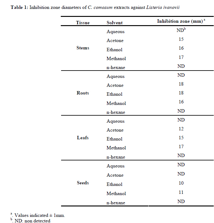
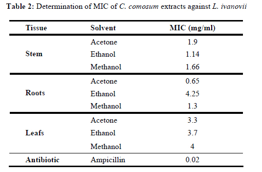
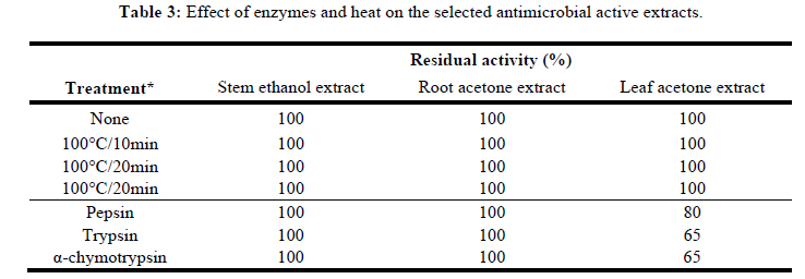
Code Number: tc11041 Abstract Calligonum comosum, a Tunisian plant from arid regions, is traditionally used in folk medicine to treat rural population microbial infections. The plant was investigated in vitro for its ability to inhibit the growth of Listeria ivanovii. Various aqueous and organic extracts were prepared from different plant tissues. Results indicated that ethanolic, methanolic and acetonic extracts from whole plant tissues except seeds, exhibited significant antibacterial activity with growth inhibition zones (9 - 18mm) as shown by the agar-well diffusion method. Minimum Inhibitory Concentration (MIC) of 0.65mg/ml was obtained in acetonic extract generated from C. comosum roots. Preliminary phytochemical analysis based on heat and protease treatments showed that bioactive extracts were stable up to 10m in heating at 100°C and that they resist protease digestion. Based on these latter results, the activity of organic extracts may be related to the presence of sterols, terpenoids, and/or phenolics. Overall, these results indicate that C. comosum organic extracts are probably useful in the control of food contamination by listerial species. Key words: Calligonum comosum; organic extracts; antimicrobial compounds; anti-listerial activity Introduction Listeriosis is a severe animal and human foodborne disease caused by gram-positive potentially pathogenic Listerial species, Listeria monocytogenes and Listeria ivanovii. Listeriosis is characterized by central nervous system infections and fetal or neonatal infections associated with a high mortality rate despite early antibiotic treatments (Vazquez-Boland et al., 2001). The foods most frequently implicated are soft cheeses and dairy products, smoked fish and in general industrially produced, refrigerated ready-to-eat products (Ryser, 1999). The high risk of contamination is mainly related to the mode of preparation of these kinds of foods in factory or at home with no thermal treatment and substantially limited use of classical microbiological barriers, such as salting and chemical additives, because of the potential risk they present for consumer's health. Besides, ubiquitous microbes such as Listeria and Bacilli species are particularly problematic in food industry. They are widely disseminated in the rural environment and, consequently, contaminate the raw materials used in the preparation of industrially processed foods (Roberts and Wiedmann, 2003). Biopreservation is an excellent alternative for food microbiological safety. This strategy is based on the use of natural antimicrobial substances as food additives. The use of plant extracts as new sources of antimicrobial agents is enjoying great popularity in the late 1990s. Plants produce a huge variety of secondary compounds (phenols, quinines, flavones, tannins, coumarins, terpenoids, alkaloids etc) as natural protection against attacks by microorganisms, insects, and herbivores (Cowan, 1999; Rios and Recio, 2005). Many of these compounds, especially those of medicinal herbs, have been used in the form of whole plants or plant extracts for food or medical applications in man (Wallace, 2004). Medicinal herbs with anti-inflammatory, antimicrobial, immunomodulatory and/or analgesic properties are used in a therapeutic way to treat acute infections and inflammatory conditions, in humans and animals. Screening of potential antimicrobial compounds from plants by clinical microbiologists is commonly performed with pure substances or crude extracts using broth dilution assay and the disc or agar well diffusion assay. Thus, these plant secondary metabolites have been demonstrated to possess a large spectrum of activity against pathogenic and non pathogenic bacteria (Shigella flexneri, diverse Staphylococcus, Streptococcus and Enterococcus species, Pseudomonas aeruginosa, Salmonella typhimurium, Mycobacterium tuberculosis, Klebsiella pneumonia, Escherichia coli etc) and fungal species as well as viruses like HIV (Saravanakumar et al., 2009) for review; (Liu, 2007; Mahady, 2005) and parasites like Trypanosoma and Plasmodium organisms (for reviews (Athanasiadou and Kyriazakis, 2004; Kokoska and Janovska, 2009). Anti-listerial properties of plant-derived compounds have been recently investigated as these may be used as natural preservatives in foods (Nair et al., 2005). Essential oils of clove, bay, cinnamon, thyme and pimento have all been found to inhibit the growth of L. monocytogenes in food, at concentrations less than 1% (Hao et al., 1998; Smith-Palmer et al., 2001; Vrinda Menon and Garg, 2001). Ethanolic extracts from the leaves of Eremophila alternifolia and Eremophila duttonii inhibited the growth of L. monocytogenes in standard laboratory media as well as in milk, salami, pâté and brie cheese (Owen and Palombo, 2007; Shah et al., 2004). It was suggested that this activity is due to organic extracts containing terpen and/or sterol antimicrobial compounds. Besides these secondary metabolites, plants produce several antimicrobial agents of protein nature designated AMPs for antimicrobial peptides. These are classified into overall seven families including Thionins, Defensins, Lipid transfer proteins (LTP), Hevein- and Knottin-like peptides, Snakins and the Cyclotides family (Hammami et al., 2009a). These peptides are ubiquitous in plants and form an essential part of the innate immunity arsenal. Demonstration of a defence role for these peptides comes from different suggestive observations: (a) antimicrobial activity in vitro against a wide range of gram-positive and gram-negative phytopathogenic bacteria; (b) gene expression, peptide distribution, and peptide concentrations in planta (before or after infection) that are congruent with a defence role; (c) correlation of the variation of expression levels (natural or genetically engineered) with the severity of symptoms; (d) correlation of the variation of the pathogen resistance to plant peptides (natural or genetically engineered) with virulence (Garcia-Olmedo et al., 1998; van Loon et al., 2006). Some of these peptides, especially the cysteine/glycine-rich small ones, have been purified from plant seeds (Garcia-Olmedo et al., 1998). Surprisingly, only few studies dealing with the effect of plant AMPs on human microbial pathogens with respect to food biopreservation have been reported in the literature. For instance, both plant antimicrobial peptides, Thionin and Snakin-1, have been recently used for in vitro inhibiting several strains of pathogenic and nonpathogenic Listeria species (Lopez-Solanilla et al., 2003). In a study conducted recently by our group, ethanol and acetone extracts of protein nature from three medicinal plants (Juniperus phoenicea (Cupressaceae), Pistacia atlantica (Anacardiaceae) and Oudneya africana (Brassicaceae)) originated from Tunisian arid regions, were found to have antimicrobial activity against L. ivanovii, Listeria innoccua, L. monocytogenes, Escherichia coli and Pseudomonas aeruginosa (Hammami et al., 2009c). Peptides weighing 1kDa were purified from O. africana and found to be active against the above bacteria (Hammami et al., 2009b). C. comosum is a pastoral plant belonging to the polygonaceae family that is frequently used as sources of medicine by rural people of south Tunisia. Indeed, anti-inflammatory, anti-ulcer and anti-cancer activities of C. comosum have been reported in rat and shrimp animal models (Badria et al., 2007; Liu X. M. et al., 2001). Moreover, tar resulting from stem combustion is used to cure dromedary scabies. Stem bark and leaf-bath serves as leather tanning and milk wineskin disinfectant. To our knowledge no studies have been reported on the effects of C. comosum antimicrobial agents against food-contaminating bacteria belonging to Listeria genus, although there are reports on its medicinal uses. In this study we report the extraction and partial phytochemical characterization of anti L. ivanovii compounds from C. comosum. Materials and Methods Plant materials C. comosum was collected from three localities in the region of Gafsa (Aguila: 34°23'47.47"N, 8°43'57.44"E; El Ksar: 34°23'24014"N, 8°47'58.55"E and Cheria: 34°22'44.29"N, 8°41'45.36"E), south Tunisia, during May 2006. Extraction protocols: For each plant, 10g of plant tissue (mature seeds, leafs, stems and roots) were ground to a powder and extracted using two extraction methods. In method one (aqueous extraction), ground tissues were homogenized in 0.02M phosphate buffer pH 7.2 containing 0.1M NaCl and then incubated overnight at room temperature. The mixture was then centrifuged at 6,000 rpm for 10min. The supernatant was finally filtrated (0.22m filter, Millipore, USA). Method 2 (organic extraction) is based on a previously described protocol (Mathabe et al., 2006) with some modifications: plant ground tissues were extracted into 150ml of 100% ethanol and incubated overnight in a shaker at 150rpm at room temperature. The homogenate was centrifuged at 10,000 rpm for 20 min and the supernatant was filtered with ethanol resistant filters (0.22m filter, Millipore, USA). The clear filtrate was then concentrated by evaporation at 37°C. This last protocol was realized again with other different organic solvents: 100% methanol and 100% acetone. All extracts were assayed for antibacterial activity as described below. Bacterial strains and growth conditions: L. ivanovii strain RBL30 was used. Bacteria were grown in tryptic soy broth (TSB; Difco Laboratories, Sparks, MD) supplemented with 0.6% (w/v) yeast extract and incubated at 30°C in aerobic conditions. Each strain was propagated at least three times in TSB before use. Assays for antimicrobial activity by agar-well diffusion method: The agar-well diffusion method used was as described previously (Perez et al., 1990). TSB agar 0.7% yeast extract medium was autoclaved and cooled to 45°C in a temperaturecontrolled water bath. An overnight culture of the bacterial strain was added at a final concentration of 1% (v/v) and 25ml of this suspension was poured into each sterile Petri plate. Plates were then stored at 4°C. Wells were dug into the set agar using the wide end of a sterile Pasteur pipette. 80l of test solutions were dispensed into each well. Before incubation, all Petri dishes were kept in the refrigerator (4°C) for 2 hr. The plates were then incubated at 30°C for at least 24hr and inhibition zone diameters were then measured. Zones with diameters greater than 6mm were considered positive. Determination of MICs: MICs (Minimum Inhibitory Concentrations) were performed with use of the critical dilution method as described previously (Eloff, 1998). This method relies on the agar-well diffusion technique with the exception that serial of twofold extract dilutions (80l) ranging from 1:2, to 1:32 were spread into TSB agar-wells. MICs were determined by visible inspection of the TSB plates. MICS were also expressed in mg proteins/ml and considered as the lowest concentration of plant extracts that completely prevented microbial growth. Protein concentrations of all extracts were determined using Bradford method. Characterization of the active compounds: To evaluate heat resistance, active samples were boiled for 10, 20, or 30min in a water bath, and then cooled before testing the residual activity. The stability of selected extracts against various enzymes was carried out using the following proteases: trypsin (10,000U/mg), α-chymotrypsin (42U/mg), pepsin (2,500U/mg) (Sigma, MO, USA). Selected samples were dissolved individually in appropriate buffers as recommended by the manufacturers and incubated with each enzyme at a final concentration of 1 mg/ml for 2 hr at 37°C. Samples containing trypsin and αchymotrypsin were incubated at 25°C. Separate aliquots with bovine serum albumin instead of enzymes were used as controls. After incubation, the samples were boiled for 3min and the residual activities were determined. Results Anti-listerial activity of the plant extract We performed aqueous and different organic extraction methods (in ethanol, methanol and acetone) from C. comosum, a medicinal plant originated from arid regions of south Tunisia. Extracts were assayed for their inhibitory effects against growth of L. ivanovii species using agar-well diffusion method. All extracts, except those isolated with the hexanesolvent, displayed antibacterial activity with variable efficiency (data not shown). Indeed, clear zones of growth inhibition ranging from 10 to 18mm, were observed for methods using acetone, ethanol or methanol during the extraction steps (Table 1), whereas, controls consisting of water or the different organic solvents used alone (data not shown), gave no inhibitory effects (complete absence of the zone of inhibition) on the tested bacterial species. Besides, activity depends on the plant tissue, as seed-extracts showed the least antibacterial activity in comparison to the other tissues. Root-organic extracts seem to be the most active against L. ivanovii. Indeed, the maximum antibacterial activity was recorded with root/acetonic or ethanolic extracts that produce zones of growth inhibition values reaching 19mm (Table 1). Analysis of the Minimum Inhibitory Concentrations of C. comosum compounds In addition to the above results, we performed MIC assays to determine the lowest concentration of extract that possess antibacterial activity. The bacterial growth of L. ivanovii was subjected to different concentrations of selected extracts namely those giving significant anti-listerial activity (stem/ethanolic, stem/methanolic and root/acetonic extracts). Results show that for all extracts tested, MICs were less than 1:32. MICs ranged from 5.9 mg/ml to 0.65 mg/ml, this latter stemming from C. comosum roots using acetone solvent-based extraction protocol (Table 2). Characterization of the antimicrobial compounds We selected active extracts from C. comosum stems, roots and leafs derived from extraction protocols that gave the lowest L. ivanovii MICs for a further characterization. The nature of active compounds was evaluated by testing their susceptibility to heat and various proteases (Pepsin, Trypsin and α-chymotrypsin). As shown in Table 3 all extracts tested retain 90-100% activity against L. ivanovii after heating at 100°C for 30 min. With regard to protease treatments, results revealed that only leaf-acetone extracts was partially inactivated by the tested enzymes. Discussion In this study, we assessed the antibacterial activity of organic extracts from C. comosum, a wild-medicinal plant from arid regions of Tunisia, against the human pathogenic specie L. ivanovii. Overall, our study clearly demonstrated that C. comosum possessed anti-infective agents active against L. ivanovii. Indeed, results revealed that L. ivanovii was susceptible to C. comosum ethanolic, methanolic and acetonic extracts since, all of them significantly inhibited growth of this bacteria with the most active ones being those obtained from leaves, stems and roots as demonstrated by agar-well diffusion method. Interestingly, MIC values differ between plant tissues and were dependent on the organic solvent used in the extraction protocol. This may be indicative of different phytochemical components producing the antibacterial activity in each extract, supporting the literature data that different active compounds are present in plant extracts (Mathabe et al., 2006). Many plants, belonging to polygonaceae family, especially to the Calligonum phylogenetically related Polygonum genera (Polygonum, amphibium, multiflorum, sachalinense, cuspidatum), have been reported to possess antimicrobial properties directed against various pathogenic and non pathogenic bacteria such as those belonging to Staphylococcus, Pseudomonas, Escherichia, Bacillus, Klebsiella, Photobacterium, Streptococcus genera (Kumagai et al., 2005; Ozbay and Alim, 2009; Song et al., 2007; Zuo et al., 2008). Nevertheless, few of them clarified the molecular basis of this antimicrobial activity. In order to further characterize the phytochemical nature of C. comosum extract components, especially those showing the highest MICs, we performed biochemical treatments consisting of subjecting extracts to heat and proteases. Data suggest that the relative majority of extracts are composed of thermostable organic molecules since their bioactivity was stable up to 10min heating at 100°C and, except the leaf-acetone extract, they were not affected by protease treatment. Interestingly, many antimicrobial agents from Polygonum and Callugonum species were found to be organic molecules. For instance, Song et al. (2007) investigations found that in vitro effects of fractions separated from Polygonum cuspidatum may be related to the presence of anthraquinones, cardiac glycosides, terpenoids, and phenolics. It has been suggested that these organic compounds are probably useful in the control of oral biofilms and subsequent dental caries development caused by Streptococci. Furthermore, Calligonum leucocladum, Polygonum poiretii, Polygonum aviculare have been demonstrated to be rich in alkaloids, phenolics and flavonoids (Lavergne, 1990; Okasaka et al., 2004). These latter with terpenoids are known to possess various bioactivities including antitumor, antimicrobial, anti-ulcerogenic, anti-inflammatory, antihypertensive, antitussive, and CNS (central nervous system) activities (Yang et al., 2008). Given that C. comosum possesses such medicinal properties (Badria et al., 2007; Liu et al., 2001), it is likely that C. comosum extracts we obtained contain such kinds of organic molecules. Consistent with this, is the close relationship between both Calligonum and Polygonum genus at the taxonomic level. It's noteworthy that n-hexane solvent failed to generate bioactive compounds effective against L. ivanovii indicating that C. comosum extracts may contain active agents with lipidic properties (Liu et al., 2001). Treatment of selective active compounds with different proteases partially reduced activity of the leaf-acetonic extract suggesting that this active extract could include antimicrobial peptides (AMPs) with hydrophobic domains. Hydrophobicity is an important structural feature of AMPs with respect to the peptide function. Indeed, it is involved in the interaction of peptides with bacterial membranes leading to bacterial death as demonstrated by numerous investigations on animal AMPs molecular bases of microbicidal activity (Brogden, 2005; Powers and Hancock, 2003). In summary, this study showed that whole C. comosum plant extracts can be used to control the growth of L. ivanovii in laboratory media. Data obtained enlarge the non-exhaustive list of plant anti-listerial compounds already investigated (Hammami et al., 2009c; Lopez-Solanilla et al., 2003; Owen and Palombo, 2007). These latter ones could be contributing factors to the C. comosum medicinal properties used in herbal medicine by rural people of south Tunisia. Identification of the active extract's phytochemicals could lead to purified compounds being used for their anti-listerial activity. These may have greater potential as food preservatives, as well as therapeutic tools, although, as mentioned by Owen and Palembo (2007), further analysis are required to assess their safety for human health since medicinal plant organic extracts do not have ‘generally regarded as safe’ (GRAS) status. Finally, additional assays in foods are warranted. Acknowledgements This research was supported by Ministry of Higher Education, Scientific Research and Technology, Republic of Tunisia. References
Copyright 2011 - African Journal of Traditional, Complementary and Alternative Medicines The following images related to this document are available:Photo images[tc11041t2.jpg] [tc11041t3.jpg] [tc11041t1.jpg] |
| |||||||||
