 |
| About Bioline | All Journals | Testimonials | Membership | News |
|
||||||
|
||||||
African Journal of Traditional, Complementary and Alternative Medicines, Vol. 8, No. 3, 2011, pp. 328-333 Antifungal Activity And Acute Toxicity Of Stem Bark Extracts Of Drypetes gossweileri S. Moore-Euphorbiaceae From Cameroon Vincent Ngouanaa,*, Patrick Valère Tsouh Fokoua, Brice Ulrich Saha Foudjoa, Silvère Augustin Ngouela b, Fabrice Fekam Boyoma, Paul Henri Amvam Zolloa. a Laboratory of Phytochemistry and Medicinal Plants Study, Faculty of Science, University of
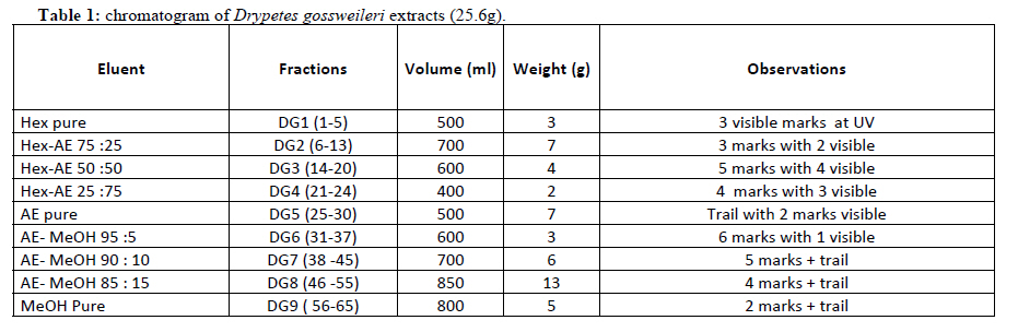
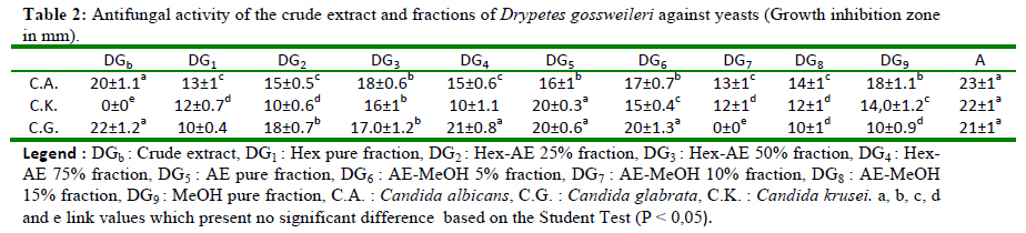
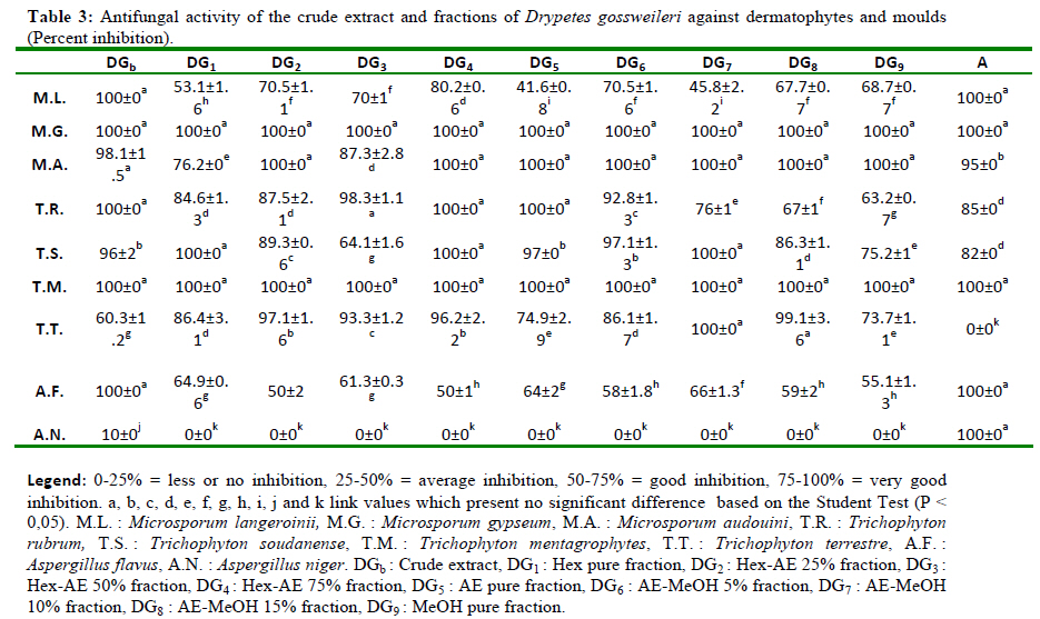
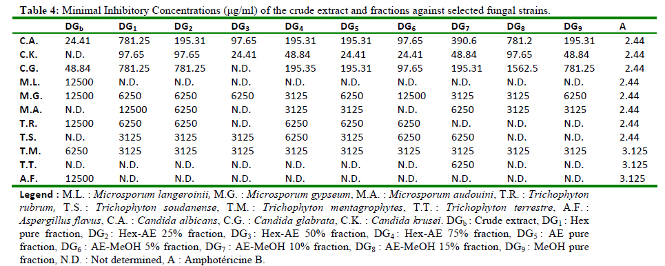
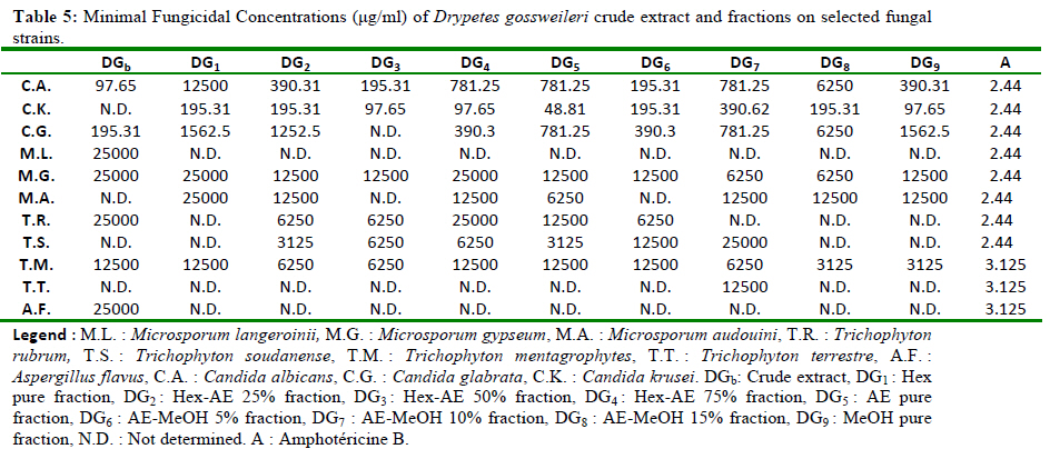
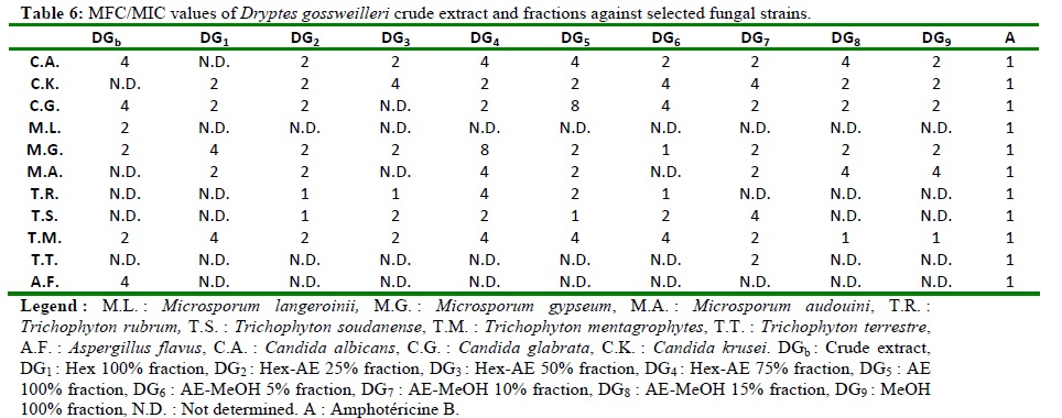
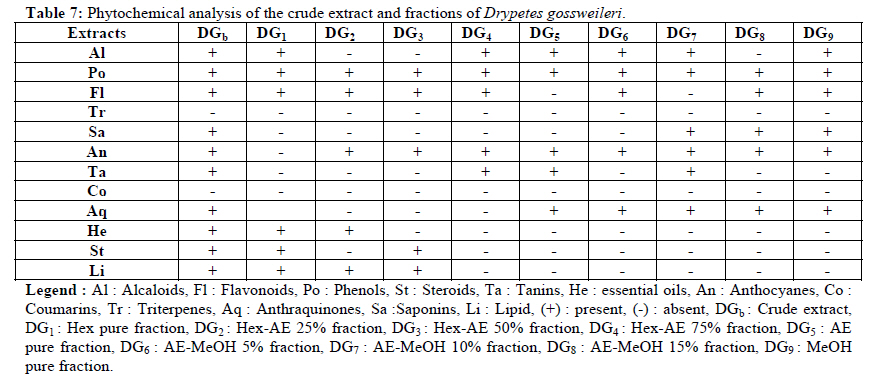
Yaoundé I, P.O. Box 812, Yaoundé, Cameroon, Code Number: tc11042 Abstract Drypetes gossweilleri S. Moore is a plant used in traditional medicine in Cameroon. The antifungal properties of its stem-bark crude extract and fractions DG1, DG2, DG3, DG4, DG5, DG6, DG7, DG8 and DG9 were assayed by agar and broth dilution methods on solid and liquid media against C. Krusei, C. albicans, C. glabrata, T. mentagerophytes, M. langeroinii, M. gypeum, M. audouini, T. rubrum, T. soudanense, T. terrestre, A. flavus and A. niger. The results revealed a substantial antifungal effect with minimal inhibitory concentrations ranging respectively from 24.11µg/ml to 1562µg/ml for yeasts and from 3125µg/ml to 12500µg/ml for filamentous fungi. Among the fractions, fraction DG4 exerted the highest antifungal activity. Moreover, no toxic effect was noticed in male and female albinos Wistar rats treated per os with the crude stem bark’s extract of Drypetes gossweileri at a dose up to 12g/kg of body weight. The phytochemical screening of the crude extract and fractions showed the presence of alkaloids, phenols, flavonoids, saponins, anthocyanines, anthraquinones, sterols, lipids and essential oils. Therefore, Drypetes gossweileri may be safe as phytomedecine for the treatment of fungal infections. Key words: Antifungal activity, Drypetes gossweileri, acute toxicity. Introduction Numerous studies have recently shown increases in fungal infections and many others established the antifungal potentials of plants (Amvam et al., 1998; Ngono et al., 2000). The advent of synthetic drugs in the health care system coupled with industrialization, in developed countries has made the use of herbal productions as medicine to decline gradually right from the beginning of the 20th century up to the 1970s. In the course of last three decades, there has been renewal and growing interest in the use of plant-derived biologically active compounds as drugs or leads in the pharmaceutical industries (Houghton and Raman, 1998). Today highly effective pharmaceutical drugs are of plant origin. Drypetes gossweileri is a common Euphorbiaceae in Central Africa, where it is widely used to treat helminthic diseases and rheumatism (Walker and Sillians, 1961). In the Congo, the root of the plant is employed in the treatment of wounds and toothache (Troupin, 1983). Many researches described the antibacterial (Ijah and Oyebanji, 2003), antioxidant and antiradical (Agnaniet et al, 2003) effects of Drypetes gossweileri. The previous phytochemical analysis of the extracts indicated the presence of steroids, triterpenoids, alkaloids, saponins with antimicrobicidal properties (Dupont et al, 1997). The present study aims to evaluate the antifungal properties of the stem bark extracts of Drypetes gossweileri and the acute toxicity of its crude extract. Material and Methods Plant material Drypetes gossweileri stem bark were collected at the Eloundem Mountain, and authenticated by Mr Nana victor at the Cameroon National Herbarium, Yaoundé where a vouched specimen was conserved under the identification number 5746/SRF/Cam. Extraction and fractionation The plant material was dried at room temperature and ground to powder. The powder (2516 g) was macerated in 8l CH2Cl2 /MeOH (1:1) for 48h. The filtrate was concentrated to dryness under vacuum to obtain the dark- purple residue. The percent extraction yield was 4.53%. The crude extract (113.97 g) was fractionated by flash chromatography on silica gel (70230 mesh, 120g) column using an increasing polarity solvent system: Hexane (Hex), Hexane-Ethyl Acetate (Hex-AE), Ethyl Acetate (AE), Ethyl Acetate-Methanol (AE-MeOH), and Methanol (MeOH) gradients (table 1). The afforded fractions were pooled according to their TLC (Thin Layer Chromatography) profile. These extract and fractions were kept at 4°C prior to testing. Before use, they were dissolved in 10% DMSO to give away different concentrations to be used in the test. Phytochemical screening The methods described by Harbone (1976) and Odebeyi and Sofowora (1978) were used to assess the main group of chemical substances (alkaloids, anthraquinones, anthocyanes, flavonoids, phenols, steroids, tannins, triterpenes, saponins, sterols, lipids, reducing sugars, essential oils, coumarins) present in the stem bark crude extract and fractions of Drypetes gossweileri and its frations. Antifungal assays Microorganisms Eleven pathogenic strains of fungi were used in the study. Three yeasts (Candida albicans, Candida krusei and Candida glabrata), seven dermatophytes (Microsporum gypseum, Microsporum langeroinii, Microsporum audouini, Trychophyton rubrum, Trychophyton soudanense, Trychophyton mentagrophytes, Trychophyton terrestre) and two moulds (Aspergillus flavus and Aspergillus niger). These strains were kindly provided by the “Centre Pasteur du Cameroun”. They were maintained in culture on Sabouraud-Glucose (4%) Agar (SGA) medium. Antifungal screening test The preliminary antifungal screening on filamentous fungi was done by the food poisoning method (Ngono et al., 2000). The strains were cultured on SGA medium in 55 mm Petri dishes. For each extract or fraction, 100μl were aseptically mixed with 1,9 ml of SGA to final concentrations of 50mg/ml for the crude extract and 25mg/ml for the fractions. 10% DMSO and amphotericine B were used as negative and positive control respectively. After solidification, an explant of 6 mm diameter of a particular dermatophyte or mould was inoculated at the center of the Petri dish and incubated at 25°C for 7 days and 5 days for dermatophytes and moulds respectively. Growth diameters were there after measured and used to calculate the percentage of inhibition. For yeasts, SGA was poured on 90 cm Petri dishes. After solidification, an inoculum of yeasts strains standardized at 2.5 x 10⁵ CFU/ml on Malassiez cell was spread on the surface of the solid medium. Following the pre-incubation time of 15 mins, wells were hollowed and 100μl of the crude extract (50 mg/ml), fractions (25 mg/ml) and the positive control amphotericine B from Sigma (100μg/ml) were introduced in individual wells respectively. This was done in triplicate. Inhibition zone diameters were measured after 24 hrs of incubation at 37°C. Minimum inhibitory concentration (MIC) The MIC which is the concentration of an extract that inhibits any visible growth of the microorganism was determined by the broth microdilution method on yeasts (Ngono et al., 2000). SGB (Sabouraud Glucose 2% Broth) and each extract or fraction was mixed and a serial two-fold dilution was done ranging from 24.41 to 25000µg/ml and 50 µl of a suspension of spores (2.104 CFU/ml) was introduced into each well of a 96 wells microtiter plate. The negative control wells received 10% DMSO. Amphotericin B (Sigma) was used as positive control, at concentration ranging from 0.012 to 12.5µg/ml. The microplates were incubated for 48 hrs at 37°C and the MIC determined as the lowest concentration which did not discolored the phenol red used as indicator of yeast growth. For dermatophytes, the food poisoning technique was used as previously described. Each extract concentration ranged from 3125 to 25000µg/ml and the MIC determined as the lowest concentration of the extract that exhibited 100% inhibition. Minimum fungicidal concentration (MFC) Incubated microtiter plates (for yeasts) and Petri dishes (for dermatophytes and moulds) were subcultured on free- extract culture media and incubated at 37 and 25°C respectively and the MFC determined as the lowest concentration at which no growth was observed. Acute toxicity The plant crude extract was tested for acute toxicity on male and female albino Wistar rats according to the WHO experimental procedure (1992). Extract doses of 0, 4, 8, and 12g/Kg body weight were administered per os to 4 groups of animals of both sex. The rats were observed first 48hrs for death and for 7 days for toxic effects. Results and Discussion The preliminary antifungal activities of the extracts are presented in Tables 2 and 3. The fraction DG5 was found to be the most active fraction against yeasts with an average inhibition zone diameter of 18.66 mm (Table 2). C. albicans was the most sensitive among the yeast. In Table 3, we noticed a significant activity of the crude extract and fractions on the dermatophytes and moulds tested. The fraction DG4 present the highest antidermatophytic activity (80.71%) as shown in Table 3. In addition, M. gypseum and T. mentagrophytes were the most sensitive dermatophytes whereas Aspergillus Niger the less sensitive. The above results prompted the analysis of antifungal effect shown in Table 4. The crude extract and the fractions were active against all tested strains. MIC ranged from 24.41 to 1562.5 µg/ml for the yeast and 3125 to 12500 µg/ml for dermatophytes and the moulds. C. krusei was the most sensitive yeast with MIC ranging from 24.41µg/ml to 97.65µg/ml. The most interesting activity was found with fraction DG4. From the results presented in Tables 5 and 6, overall extracts of Drypetes gossweileri showed MFC values ranging from 48.81 to 6250µg/ml for yeasts and 3125 to 25000µg/ml for dermatophytes and moulds, with the majority of fungicidal indices ≤ 4. The results presented in Table 7 revealed that the most common constituents found in the extract and fractions are alkaloids, phenols, flavonoids, saponines, anthraquinones, tannins, anthocyanins, sterols, lipids and essential oils. The alkaloids, flavonoids, saponines, tannins, anthraquinones, coumarine, essential oil are known to possess antifungal activities (Bouchet et al., 1986, Cowan, 1999; Bruneton, 1999; Sautour et al., 2004). Anthocyanins, flavonoids, and tannins, have been reported by Barnabas and Nagarajan (1988), and Burapedjo and Bunchoo (1995), to inhibit cell wall formation in fungi leading to the death of the microorganism. This supports the fungicidal activities exerted by the extract and fraction of Drypetes gossweileri. The differential distribution of the bioactivity among fractions might be explained by proportions of different bioactive components from each fraction acting either in a synergetic or potentiating ways. The study of the acute toxicity of the crude extract of Drypetes gossweileri stem bark showed no acute toxic effect in rats at doses ≤ 12g/Kg through oral route. Conclusion Results achieved in this study, in addition to the lack of toxicity observed in rats support further investigation of extracts of Drypetes gossweileri stem bark as potential antifungal agents. Acknowledgements The authors acknowledge the assistance of Mr. Nana Victor from Cameroon National Herbarium, Yaoundé for identifying and collecting plant sample, and the ‘’Centre Pasteur du Cameroun’’ for providing fungal strains. References
Copyright 2011 - African Journal of Traditional, Complementary and Alternative Medicines The following images related to this document are available:Photo images[tc11042t5.jpg] [tc11042t2.jpg] [tc11042t1.jpg] [tc11042t6.jpg] [tc11042t7.jpg] [tc11042t3.jpg] [tc11042t4.jpg] |
| |||||||||
