 |
| About Bioline | All Journals | Testimonials | Membership | News |
|
||||||
|
||||||
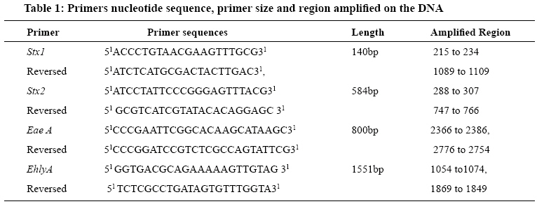
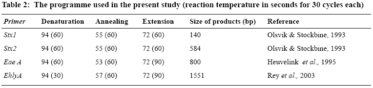
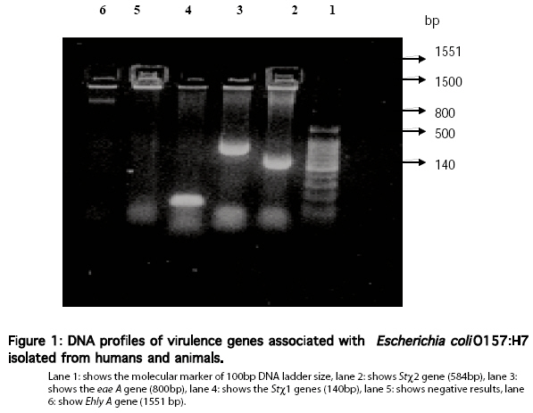
Tanzania Journal of Health Research, Vol. 10, No. 3, July, 2008, pp. 151-158 Prevalence and characterization of verotocytoxin producing Escherichia coli O157 from diarrhoea patients in Morogoro, Tanzania M.A. Raji1, 2*, U.M. Minga2 and R.S. Machang’u2 1Department of Veterinary Pathology and Microbiology, Ahmadu Bello University Zaria, Kaduna State, Nigeria Received 7 January 2008; Revised 7 June 2008 Accepted 12 June 2008 Abstract Escherichia coli O157:H7 is an important agent of haemorrhagic colitis and haemolytic uraemic syndrome in children less than five years old and elderly people. The objective of this study was to investigate the prevalence of verotocytoxin producing E. coli 0157 (VTEC O157) among human patients with diarrhoea in Morogoro, Tanzania. Faecal samples originating from 275 human patients with diarrhoea were screened for presence of E. coli O157:H7. A total of 96 E.coli isolate were identified. Of these, 10 isolates were grouped into sorbitol non-fermenting and glucuronide negative and 49 isolates were sorbitol positive and glucuronide positive. The remaining 37 were sorbitol negative and glucuronide positive. Using the polymerase chain reaction techniques, a total of ten verotocytocin producing E. coli isolated in this study were used. The overall two (15%) and one (7%) of the isolated of E. coli possessed both attaching and effacing (eae A) and enterohemolysin (ehly) A genes respectively. Other enterobacterial agents including Pseudomonas spp, Proteus spp and coliforms were also isolated. The VTEC O157 isolates were 100% resistant to oxytetracycline, chloramphenicol, streptomycin, and amoxyclav. In conclusion, the isolation of diarrhoeaogenic E. coli O157:H7 in this region suggests that the pathogen is an important aetiology of acute gastroenteritis in Tanzania. There is therefore, need to improve sewage and refuse disposal system, the provision of safe potable water, sanitation, personal hygiene and health education in order to reduce infection with this and other enteric pathogens. Keywords: verotoxigenic, Escherichia coli O57, diarrhoea, children, antibiotics, resistance, Tanzania Introduction Verotoxigenic producing Escherichia coli (VTEC) or Shiga toxin-producing strains of Escherichia coli (STEC) are recognized as an important human pathogen of public health concern (Bettelheim & Beutin, 2003). Whereas STEC isolates belong to many different serotypes, E. coli O157:H7 and an occasional non-motile variant O157:H are the most common serotypes associated with human illness. Isolates of this pathogen are a major cause of haemorrhagic colitis (HC) and mild diarrhoeal illness and are the major aetiological agent of haemolytic uraemic syndrome (HUS) (Pulz et al., 2003). HUS is characterized by a prodrome of gastroenteritis, frequently including bloody diarrhoea, followed by acute haemolytic anaemia, thrombocytopenia and renal failure. Infections are usually linked to the consumption of VTEC-contaminated and improperly cooked beef, faeces-contaminated vegetables, apple cider, water and direct transmission of VTEC from animals to man (Griffin & Tauxe, 1991). The VTEC are each capable of producing one or two potent toxins called Shiga toxins (st1 and st2) because of their cytotoxic effects on African Green Monkey kidney (Vero) cells in cultures (Konowalchuck et al., 1977). In addition to toxin production, another virulence-associated factor expressed by VTEC is a protein called intimin, which is responsible for intimate attachment of VTEC to the intestinal epithelial cells, causing attaching and effacing (AE) lesions in the intestinal mucosa (Paton& Paton, 1998). Intimin is encoded by the chromosomal gene eae A, which is part of a pathogenicity island termed the locus for enterocyte effacement (LEE). A factor that may also affect virulence of VTEC is the enterohemolysin (enterohaemorrhagic E. coli haemolysin, EHEC-HlyA) (O´Brien et al., 1982; Schmidt et al., 1995). Microbial resistance to antibiotics is a worldwide problem. The single main factor contributing for the increase in the antibiotics resistance is irrational use of antibiotics. Animals, which are asymptomatic carriers of E. coli O157, including EHEC, when exposed to antimicrobial agents, may serve as a reservoir for antimicrobial-resistant bacteria (Schroeder et al., 2002). The objective of this study was to investigate the prevalence of VTEC in faeces of humans with diarrhoea in Morogoro, Tanzania and to characterize the isolates for the presence of virulence genes (stx1, stx2, eaeA and EHEC-hlyA). Materials and Methods Study area and subjects This study was conducted in peri-urban areas of Morogoro Municipality (6O 49’S; 37O40’E) in eastern Tanzania. The annual rainfall and air temperature range between 800-1000 mm and 18 OC -32 OC respectively. The area experiences two rainy seasons; long rains in March to May and short rains in October-December. A total of 275 faecal swab samples were collected randomly from patients with diarrhoea in Morogoro. Samples were acquired from the Morogoro Regional Hospital, Sokoine University ofAgriculture Dispensary and Upendo Medical Diagnostic Laboratory. The age of the patients in this study group ranged between 0 to 72years. The sex of the patients was also considered. There was no information available on the recent history of antibiotic use and the occupational status by any of the patients sampled. Ten to twenty humans stool swab samples were collected per day (depending on the number of diarrhoea patients). Each swab was then put into a bijou bottle containing 10 ml of Modified Trypticase Soya broth (mTSB)(Oxoid Ltd, UK) sealed and then put into a cool box with ice (+4OC) and transported to the laboratory, where the microbiological examination was started within 20 hours as described by Ritchie et al. (1992). Isolation of O157:H7 VTEC and serotyping of E. coli In the laboratory the samples were first screened by inoculating them onto the surface of Cefixime-tellurite Sorbitol-MacConkey agar (CT-SMAC) with 5-bromo4-chloro-3 iodo-‘β-D-glucuronide (BCIG) (Oxiod, UK). After an incubation period of 18hours suspected colonies of E. coli O157 were then picked from CT-SMAC-BCIG and inoculated onto mTSB broth for Immunomagnetic separation (IMS) (Thran et al., 2001). After 18 hours of incubation of suspected colonies from preliminary screening of the samples on CT-SMAC-BCIG agar were inoculated onto mTSB and about 5ml of each broth culture was used for immunomagnetic separation (IMS). IMS with magnetic beads coated with antibody to O157 (Dynal) was performed according to the instructions of the manufacturer. The concentrates were inoculated onto CT- SMAC and the plates were incubated at 37OC for 18 to 20 hours. The β–glucuronidase activity was accessed using CT-SMAC containing BCIG (Oxiod, UK) as described previously by Aleksic et al. (1992). Presumptive O157:H7 VTEC isolates (those with a typical E. coli metallic sheen on L-EMB; and isolates that gave agglutination with E. coli latex test kit (Oxoid) and BCIG negative was confirmed to be E. coli by using an biochemical tests for the ability to ferment lactose and sucrose in Triple Sugar Iron agar (TSI Difco) slants, indole production, methyl red and Voges Proskauer reactions and citrate utilization (IMVIC tests) as described by Ritchie et al. (1992). O and H grouping was carried out by bacterial agglutination (Orskov & Orskov, 1977) with antiserum against E. coli groups O157 and H7 (Siitonen, 1992). The isolates that gave clumping with 4% saline were defined as rough. The O157 and H7 antigens were tested with the E. coli O157 antigen detection kit (Oxoid, UK). The ability of the isolate to produce VT1 and /or VT2 was determined by a reverse latex agglutination test (Vertox F; Deka Seiken, Tokyo, Japan) according to the manufacturer’s instructions (Ratnam et al., 1988). Antimicrobial sensitivity Antimicrobial susceptibility testing was done using the Bauer et al. (1996) method in which selected antimicrobial agents for the treatment of diarrhoea in humans were used. An 8 to 12 hours broth culture was prepared for ten isolate obtained from human faeces. Using a sterile cotton swab, an entire surface of dried Muller Hinton agar plates with 4mm of agar depth, was streaked uniformly with the swab previously dipped in the test E. coli O157 culture suspension after squeezing off extra fluid on the wall of the tube. The inoculated plate was allowed to dry for 5min and appropriate antibiotic disks from commercial sources were then applied using sterile forceps and incubated at 37OC overnight. The antibiotic disks used were: amoxyclav, cephtriaxone, norfloxacin, ofloxacin, nitrofurantoin, nalidixic acid, gentamicin, and sulphamethoxazole supplied by Hi-media Laboratory Ltd, India. Streptomycin, chloramphenicol, cephalexin, oxytetracycline, and neomycin were from Oxoid UK. The inhibition zones were interpreted by measuring the diameter of zone of inhibition. For analytical purposes, isolates that were moderately sensitive were taken as fully sensitive. Polymerase chain reaction (PCR) For PCR analysis, the primer sequences selected for the amplification of the stx1, stx2, eae A and ehlyA genes completely matched the sequences of the corresponding genes encoding stx toxin, eae A gene of EPEC and haemolysin ehly A in the GenBank/EMBL database libraries (Table 1). The oligonucleotides used as primers were purchased from Synthengen®, USA. The bacterial isolates were cultured on Sorbitol MacConkey agar at 37OC for 24 hours. A loopful of bacterial culture from the agar plate was suspended in 200 μl of sterile distilled water in Eppendorf micro centrifuge tube, and boiled in a water bath at 80OC for 20min, and then centrifuged at 12,000 rpm. The supernatant served as DNA source as previously described by Madico et al. (1995). All reactions were performed using USA Technologies Rapid Cycler® MJ Research brand thermal Cycler (DNA amplifier). The PCR mixture consisted of 1µl of 10X PCR buffer, 0.2μl of Taq polymerase, 5.98μl of double deionized DNA free water, 0.8μl of 2.5mM MgCl2, 0.02μl of 0.2mM(dATP, dCTP, dGTP and dTTP), 0.5μl of 100pmole (each) of the stx-specific primer pair, molecular weight marker (Promega®, USA) was run with each gel. Positive samples were identified based on the presence of bands of appropriate sizes for stx 1, stx 2, eae A and Ehly A. Data analysis Association of the isolated VTEC pathogens with respect to age and sex was analysed using Chi-square and 1µl of DNA in a final volume of 10µl. In the PCR assays, DNA was amplified by stx 1, ehly A, stx2 and eae A primers separately (Table 2). The PCR was started with 0.2 μl of 5U of Taq polymerase (PromegaR, USA). The PCR mixture was overlaid with mineral oil and run in the thermal Cycler (Gannon et al., 1993; Rey et al., 2003). All reactions were performed using USA Technologies and 1μl of DNA in a final volume of 10μl. In the PCR Rapid Cycler® MJ Research brand thermal Cycler (DNA assays, DNA was amplified by stx 1, ehly A, stx2 and amplifier). The PCR mixture consisted of 1μl of 10X eae A primers separately (Table 2). The PCR was started PCR buffer, 0.2µl of Taq polymerase, 5.98µl of double with 0.2 µl of 5U of Taq polymerase (PromegaR, USA). deionized DNA free water, 0.8µl of 2.5mM MgCl2, The PCR mixture was overlaid with mineral oil and 0.02µl of 0.2mM(dATP, dCTP, dGTP and dTTP), 0.5 run in the thermal Cycler (Gannon et al., 1993; Rey et µl of 100pmole (each) of the stx-specific primer pair, al., 2003). Electrophoresis of amplified products Aliquots of 10μl of amplified products of PCR were analysed by electrophoresis in a 1.5% agarose gels (SeaKem; FMC Bio-products, Rockland, Maine USA). The gels were stained with ethidium bromide (0.5µg/ ml). 1XTris-borate/EDTA electrophoresis buffers were used and the electrophoresis was run at 100V for 30min. The 6X loading dye from PromegaR, USA was used for loading the PCR amplified products. The comb size of 2mm was used for making the wells. The gels were then photographed under UV transillumination. A 100bp test. The prevalence of the verocytotoxin E. coli was calculated as percentage. The data was analysed using SPSS statistical package and a p-value of <0.05 was considered significant. Results Isolation rate of Escherichia coli O157 Faecal samples were collected from 275 human patients with diarrhoea. A total of A total of 96 (34.9%) E. coli isolates were identified from faecal samples. The isolates exhibited three distinct colonial morphologies on CT-SMAC-BCIG). The non-sorbitol fermenting and glucuronidase negative isolates accounted for 3.6%, while the sorbitol positives and glucuronidase positives, which appeared as purple colonies on CT-SMACBCIG, accounted for 17.8%. The sorbitol positives and glucuronidase negative isolates which appeared as pink colonies accounted for 13.46%. Coliforms were also isolated from 25.46% of human samples. This was also recognized on the basis of the mucoid appearances on eosin methylene blue agar for Klebsiella spp while other coliforms, such as Citrobacter spp and Enterobacter spp, formed brownish colonies on eosin methylene blue agar and pinkish (ferment lactose) on MacConkey agar. The identification of VTEC O157 isolates was based on colonial morphology, sorbitol negative and glucuronidase negative properties of Verotoxigenic E. coli, which appeared as colourless on Sorbitol MacConkey agar with BCIG. This was further confirmed on the basis of a greenish metallic sheen produced on EMB and pink mauve on CHROMR agar TM O157. The serotyping of E. coli O157 was demonstrated using Oxoid latex agglutination kit. All the isolates from humans agglutinated with E. coli O157 latex. All of the presumptive verotoxigenic E. coli isolated from humans were therefore E. coli O157 on the basis of the agglutination with Oxoid E. coli O157 latex test kit. The presence of verotoxin was demonstrated with reverse passive latex verotoxin agglutination kit (Denka Seiken, Japan). The results indicated that six males and four females were infected with verotoxigenic E. coli out of the total 10 samples that were positive for VTEC. There was, no statistically significant difference between sex (χ2=3.750, P=0.153)) or age (χ2=2.000, P=0.368) in the distribution of infection in this study. Of the 13 antimicrobial agents tested against ten verotoxigenic E. coli isolates, resistance was recorded against sulphonamide, streptomycin, and chloramphenicol (either alone or in combinations).Amoxyclav (either alone or combined with other antibiotics), which was the most common antibiotics against which VTEC showed resistance (100%) against humans isolates. Resistance to sulphamethoxazole was noted in 60% of human isolates, and was the second most frequently found resistance pattern. Humans VTEC strains showed 100% resistance against chloramphenicol, oxytetracycline, and streptomycin. Many of these isolates demonstrated multiple resistances to antibiotic. In general, isolates from the humans were resistant more frequently to the different tested antimicrobial agents, with the exception of ofloxacin, norfloxacin and gentamicin (Table 3). Molecular characterization Ten isolates that were sorbitol negative and glucouronidase negative were used for PCR investigation. PCR was positive for some of the isolates DNA templates except the negative control (no DNA template). One (10%) of the human VTEC strains was positive only for stx1. The stx2 gene alone was detected in one (10%) human isolate. The stx1 and stx2 gene in combination was not present in any human strain. Eae A was detected in 2 (20%) human strains. The stx 1, eae A and Ehly A were present in only one of human strain (Figure 1). Discussion Since there is no data available in Tanzania regarding the prevalence of VTEC in humans, this study aimed at determining the prevalence of VTEC in humans with diarrhoea. The isolation of 3.6% from patients with diarrhoea was lower than those reported by Olorunshola et al. (2000) and Akinyemi et al. (1998) in Nigeria and Galane & Roux (2001) in South Africa. A much lower prevalence has been reported in China (Li et al., 2002). On the other hand, Kaddu-Mulindw et al. (2001) could not isolate VTEC O157 in urban children in Uganda. The predisposing factors such as age and sex were also investigated in human VTEC infection in our study. Males and females, both young and adults, were equally infected with VTEC o157. Although reported outbreaks of E. coli O157:H7 in Africa have been few to date, available information indicates that the pathogen has a wide geographical distribution (Raji et al., 2003; 2006). Since the 1992 outbreak, in South Africa, culture-proven E. coli O157:H7 diarrhoea illness has been reported in several countries, including Kenya, Nigeria, Ivory Coast, and Central African Republic (Sang et al., 1996; Germani et al., 1998). Since E. coli O157:H7 is not detected by the usual methods used to isolate and identify typical enteric bacterial pathogens, microbiological laboratories in many countries in Africa do not routinely test for this pathogen (E.coli O157:H7), and infections may go unrecognized (WHO, 1995; Wittenberg, 1999). Reports on African dysentery outbreaks attributed to Shigella dysenteriae sometimes indicate that specimens were not tested for VTEC until several months into the outbreak, or did not use laboratory methods that are suitable for detection of E.coli O157:H7 (Aragon et al., 1995; Malakooti et al., 1997). The majority of outbreaks caused by VTEC are missed due to this laxity. This is unfortunate because the spectrum of clinical illness resulting from Shigella infection overlaps considerably with that of E.coli O157:H7 and mixed outbreaks have been reported (Cunin et al., 1999). The differences in the results of Stx assay using VTEC-Screen SEIKEN and PCR may be attributed to the fact that VTEC-Screen SEIKEN has the ability to cross-react with the variant of Stx 1 and Stx 2 whereas PCR primer Stx 1 and Stx 2 are specific for these genes (Beutin et al., 2002). The losses of Stx1 and Stx2 prior to testing by PCR have been documented (Karch et al., 1998; Feng et al., 2001; Blanco et al., 2003). This may be plausible explanation for the difference between the result of VTEC-RPLA and PCR obtained in this study. Susceptibility to fourteen antimicrobial agents used in human and veterinary medicine were determined. Of the 10 human E. coli isolates characterized in this study, half displayed resistance to one or more antimicrobials including sulphamethoxazole, cephalexin, oxytetracycline and streptomycin and chloramphenicol. These data are in agreement with reports from previous studies, which suggested that the use of these drugs had been a key factor in the emergence of antimicrobial-resistant E. coli (Meng et al., 1998; Teshager et al., 2000; Zhao et al., 2001; Schroeder et al., 2002). In conclusion, the isolation of diarrhoeaogenic bacterial pathogens, particularly E. coli, O157:H7 in Morogoro, Tanzania, suggests that this pathogen is likely to be an important aetiology of acute gastroenteritis in the country. The prevalence of VTEC O157: H7 would have possibly been higher if this study was to target patient with Haemolytic uraemic syndrome and haemolytic colitis. There is a need to improve sewage and refuse disposal system, the provision of safe potable water, sanitation, personal hygiene, health education and disease control strategies, in Tanzania, in order to reduce infection with this and other enteric pathogens. Acknowledgements The authors thank DAAD-ANSTI for award of PhD fellowship to Sokoine University of Agriculture, Morogoro, Tanzania. Our sincere thanks also go to the technical staff at the Microbiology laboratory at Sokoine University ofAgriculture, Morogoro, Tanzania. We also thank Professor P.S. Gwakisa, the Head of Department of Parasitology and Microbiology for his support and assistance rendered during the period of study. References
© Copyright 2008 - Health User's Trust Fund (HRUTF) The following images related to this document are available:Photo images[th08026f1.jpg] [th08026t2.jpg] [th08026t1.jpg] [th08026t3.jpg] |
| |||||||||