 |
| About Bioline | All Journals | Testimonials | Membership | News |
|
||||||
|
||||||
Streptomycetes from Jordan soils active against Agrobacterium tumefaciens
I. SAADOUN and F. AL-MOMANI
Department of Biological Sciences, Jordan Univ. of Science & Technology, Irbid 22110, Jordan
Code Number: AC97005
Sizes of Files:
    Text: 20.2K
    Graphics: Photograph (jpg) - 40.7K
ABSTRACT. During an investigation on soil actinomycetes from two plant nurseries in Jordan, sixty streptomycetes were recovered. Of these 73% showed antibiotic activity against bacteria and eleven also against Agrobacterium tumefaciens. In order to exploit their potential for biological control of crown gall, the latter were investigated in detail and identified to the species level.
Streptomycetes are known antibiotic producers and have been also investigated for their in vitro activity against soil-borne pathogens (Turhan, 1981; Rothrock & Gottlieb, 1981). Tulemisova & Nikitina (1989) reported that 44.1% of the Streptomyces spp., isolated from the rhizosphere of sugar beet, were antagonistic to phytopathogenic fungi, mainly against Fusarium solani. The issue is of particular interest since chemical control of soil-borne pathogens is difficult, economically demanding and often ineffective because of the appearance of resistant strains (Papavizas, 1973). The biological control of crown gall, a world-wide and economically important plant disease caused by Agrobacterium tumefaciens, has been approached by Kerr and associates (New & Kerr, 1972; Htay & Kerr, 1974; Kerr & Htay, 1974). using non-pathogenic agrobacteria. In the present study a number of streptomycetes, isolated from two nursery soils in Jordan, have been characterised and investigated for their activity against the phytopathogen.
MATERIALS and METHODS
Sampling. Samples were collected from six fields of two government nurseries in Jordan, established to supply farmers with plant seedlings. The seedlings grown in each field were as follows:
- Al-Waleh Nursery. WO: olives; WA: almonds; WC: untilled control.
Samples were collected monthly, starting in the summer (June-August) with an auger. These were taken up to 10cm depth, after removing approximately 3cm of the soil surface, from six different sites within each field. Two auger holdings were collected from each site. Samples were placed in polyethylene bags, closed tightly and stored in a refrigerator.
Isolation. Samples were mixed thoroughly and sieved (2mm mesh). Subsamples of 1g were suspended in 100ml distilled water on a reciprocal shaker (190 rpm, 30 min), serially diluted up to 10^-6 and spread (0.1ml) over the surface of agar plates with sterile L-shaped glass rods. Triplicate plates of Standard Plate Count agar (Merck) and glycerol nitrate casein agar (Kuster & Williams, 1964) were used for total bacteria and streptomycete counts respectively. Plates were incubated at 27 C and the number of colonies was determined after 48hrs (bacteria) and 10 days (streptomycetes). Dilutions that gave 20-200 colonies were chosen for the isolation. Selected colonies were purified by repeated streaking.
Characterisation of the isolates. Streptomyces colonies were characterised morphologically and physiologically following the directions given for the International Streptomyces Project (ISP) (Shirling & Gottlieb, 1966). General morphology was determined on oatmeal agar plates, incubated in the dark at 27 C for 21dd, by direct light microscopy examination of the surface of crosshatched cultures.
Spore surface was examined by scanning electron microscopy (SEM). Surface growth from 14-21dd cultures was deposited onto aluminium stubs which were then placed in a sputter coater (E 5000, Polaron Equipment Ltd.) for 2-3min. (1-4 kV, 20 mA, 0.1 torr; approximately 150A layer of gold). Specimens were examined under a scanning electron microscope (Lutz 100 A) operated at 20 kV.
Colours were determined according to Prauser (1964) and isolates were grouped as proposed by Nonomura (1974).
Antimicrobial activity. This was tested by the plate diffusion method against Staphylococcus aureus, Bacillus subtilis, Escherichia coli and Agrobacterium tumefaciens, strain Ab 136. Isolates were grown on oatmeal agar for 14 days; discs (9mm in diameter) were cut and placed on nutrient agar seeded with the test organisms and incubated at 27 C. Inhibition zones were checked after 24 hrs.
RESULTS and DISCUSSION
The average microbial count ranged between 10^6 to 10^9 and 10^4 to 10^6 CFU/gm dry soil for bacteria and streptomycetes respectively.
A total of 60 streptomycetes were recovered from the^ samples. The isolates were distributed into seven series according to the colour of their mature sporulated aerial mycelium (Table 1). Eighteen were isolated from Al-Faysal, and 42 from Al-Waleh nurseries.
Table 1. Distribution of the Streptomyces isolates in different fields. Numbers in parentheses represent the colour series percentage (* see Text).
---------------------------------------------------------------------------
                              Colour Series
        -------------------------------------------------------------------
Site*    Grey    Red    Green    Blue    White   Yellow   Violet    Total
---------------------------------------------------------------------------
FA        4       1       1        1       1        0       0        8
FP        3       0       2        0       0        0       0        5
FC        3       0       2        0       0        0       0        5
WA        2       2       1        0       0        0       0        5
WO       10       5       2        2       1        3       1       24
WC        6       1       0        3       3        0       0       13

Total(%) 28(47)  9(15)   8(13)    6(10)   5(8)     3(5)    1(2)    60(100)
---------------------------------------------------------------------------
Data show that the grey series includes most (47%) of the isolates. This is in agreement with data obtained by Hamdi et al. (1980) and Abussaud & Saadoun (1988) who found that 50% and 51% of their Streptomyces isolates were grey. Of the 60 isolates, 18 produce melanin, 30 show distinctive reverse side pigment and 12 produce soluble pigments (Table 2).
Table 2. Pigmentation and sporophore morphology of the 60 Streptomyces isolates.
---------------------------------------------------------------------------
              Character              No. of Isolates       %
---------------------------------------------------------------------------
Pigment         Melanin                  18               30
production      Reverse side             30               50
                Soluble                  12               20

Sporophore      Spiral                   40               67
morphology      Straight                 13               22
                Flexuous                  5                8
                Retinaculum Apertum       2                3
--------------------------------------------------------------------------
With reference to the morphology of spore bearing hyphae most isolates show spiral sporophores (Table 2). Hamdi et al. (1980) in Iraq and Coelho & Drozdowicz (1979) in Brazil found that most of their isolates had straight and spiral sporophore, respectively. In a previous study, Abussaud & Saadoun (1988) reported that most of Streptomyces isolates recovered from Jordan Valley soils had spiral sporophore. Climate, soil type and vegetation may be responsible for species composition in different environments.
Antibacterial activity of the isolates against the tested bacteria is shown in Table 3.
Table 3. Antibacterial activity of the Streptomyces isolates.
---------------------------------------------------------------------------
                                Colour Series
                -----------------------------------------------------------
Antibiosis      Grey   Red   Green   Blue   White   Yellow  Violet   Total
---------------------------------------------------------------------------
Isolates         28      9      8      6       5        3      1       60
Active Isolates  20      7      8      2       4        2      1       44
%                71     78    100     33      80       67    100       73

S.aureus         20      7      8      2       4        2      1       44
%               100    100    100    100     100      100    100      100

E.coli            6      2      4      1       2        1      1       17
%                30     29     50     50      50       50    100       39

B.subtilis        7      2      4      1       2        1      1       18
%                35     29     50     50      50       50    100       41

A.tumefaciens     5      4      2      0       0        0      0       11
%                25     57     25      0       0        0      0       25
---------------------------------------------------------------------------
Seventy-three per cent of the isolates were active against one or more of the test organisms, however the percentage of active isolates varies within each colour series, tending to be higher in the green and violet series and lower in the blue one. All of the isolates were active against S. aureus. However, 41, 39 and 25% of them were active against B. subtilis, E. coli, and A. tumefaciens respectively. Hussein et al. (1980) reported that most of the grey series isolates recovered from Egyptian soils were inactive against Gram negative bacteria. Whereas 38% of Streptomyces series isolated from Iraqi soils were active against Gram-negative bacteria.
Data indicated that the different colour series of Streptomyces have the same antibiotic activity against the crown gall pathogen (A. tumefaciens) in agreement with previous results of Abussaaud & Saadoun (1988) for soils in the Jordan Valley.
With reference to the results obtained, more detailed investigations were carried out on the strains active against Agrobacterium tumefaciens in order to determine their taxonomic status at the species level.
Morphological characteristics and pigment production of 10 active strains are summarised in Table 4.
Table 4. Morphological and cultural characteristics of the Streptomyces isolates inhibiting Agrobacterium tumefaciens Ab 136.
--------------------------------------------------------------------------
                                 No of isolates/ Colour series
                                --------------------------------
Characteristic                   Grey     Red    Green    Total
----------------------------------------------------------------
Number of strains                 5        4       1       10

Pigment Production:
Melanin                           2        1       0        3
Reverse Side                      4        3       0        7
Soluble                           1        1       0        2

Sporophore Morphology:
Spiral                            3        3       0        6
Rectus                            1        1       0        2
Flexuous                          1        0       1        2

Spore Surface:
Smooth                            3        4       1        8
Spiny                             2        0       0        2
Hairy                             0        0       0        0
--------------------------------------------------------------------------
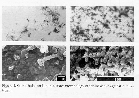
The isolates belong to grey, red and green colour series and the majority of them shows spiral spore chains and smooth spore surface (Fig. 1). No isolates of the white, yellow, blue and violet colour series are active against A.tumefaciens.
Figure 1. Spore chains and spore surface morphology of strains active against A.tumefaciens.
These results are in agreement with those presented by Abussuad & Saadoun (1988) with differences imputable to soil habitat and vegetation. Table 5 shows the complete identification of the Streptomyces isolates based on morphological and biochemical characteristics. All isolates are able to utilise arabinose, fructose, rhamnose and sucrose as carbon sources.
However, half of the isolates do not utilise raffinose.
Table 5. Characteristics of the Streptomyces strains inhibiting A.tumefaciens Ab 136
--------------------------------------------------------------------------
       Cultural      Morphology    Sugar  Utilisation
       Characters
No   ---------------  --------  ------------------------     Species
    AM    ME  RP  SP   SC  SS   A  X  I  M  F  R  S  Ra
---------------------------------------------------------------------------
1  Grey   -   -   -    Sp  SP   +  +  +  +  +  +  +  +  S.antimycoticus
2  Grey   -   +   +    Re  SM   +  -  -  +  +  +  +  -  S.cyanocolor
3  Red    -   -   -    Sp  SM   +  +  +  +  +  +  +  -  S.vinaceus-drappus
4  Grey   +   +   -    Sp  SM   +  +  +  -  +  +  +  +  S.resistomycificus
5  Grey   +   +   -    Fl  SM   +  +  +  -  +  +  +  +  S.aurantiogriseus
6  Red    -   +   -    Sp  SM   +  -  -  +  +  +  +  -  S.aurantiacus
7  Green  -   -   -    Fl  SM   +  +  -  +  +  +  +  -  S.psammoticus
8  Red    -   +   +    Sp  SM   +  +  -  +  +  +  +  -  S.murinus
9  Grey   -   +   -    Sp  SP   +  +  +  +  +  +  +  +  S.griseoruber
10  Red   +   +   -    Re  SM   +  +  +  +  +  +  +  +  S.racemochromogenus
---------------------------------------------------------------------------
[Cultural characters: AM = aerial mycelium colour; ME = melanin pigments; RP = reverse colour; SP = soluble pigments; Morphology: SC = spore chains (Sp = Spirales, Re = Recti; Fl = Flexibiles); SS = spore surface (SP = spiny; SM = smooth); Sugar utilisation: A = arabinose; X = xylose; I = inositol; M = mannitol; R = rhamnose; S = sucrose; Ra = raffinose]
Streptomycetes by virtue of their wide distribution, filamentous growth and antibiotic production are active components the microbial equilibrium in soil and may represent a factor affecting the incidence of certain soil-borne plant pathogens (Waksman, 1967).
One possible approach to biological control of crown gall disease would be inoculating soil with selected antagonists. This however requires a preliminary investigation on the conditions which favour the survival of the antagonists, since soil is a very complex substrate in which numerous factors influence the number of microorganisms as well as the qualitative composition of its microflora. In this study, we have attempted to isolate and examine the characteristics of the Streptomyces microflora in two plant nurseries. Results may represent a preliminary stage of the possibility of using streptomycete strains, isolated from different habitats, as a source of antibiotics or as a means of biological control of some plant diseases.
ACKNOWLEDGEMENTS. This research was financed by a grant No. 24/96 from the Deanship of Scientific Research at Jordan University of Science and Technology. The technical assistance of Maher Obiedat are greatly appreciated.
REFERENCES
Abussaud, M.J. & I.M.Saadoun (1988). Isolation, characterization and taxonomy of Streptomyces sp. isolated from Jordanian soils and antagonistic to Agrobacterium tumefaciens. Egypt. J. Microbiol., 23: 597-609
Coelho, R. R. & A.Drozdowicz (1979). The occurrence of actinomycetes in cerrado soil in Brazil. Rev. Ecol. Biol. Sci., 15: 459-474
Hamdi, Y.A., D.Ahmed & A.M.Al-Tai (1980) Genera and species of actinomycetes isolated from Iraqi soils. Egypt. J. Microbiol., 15: 7-22
Htay, K. & A.Kerr (1974). Biological control of crown gall: seed and root inoculation. J. Appl. Bacteriol., 37: 525-530
Hussein, A.M., A.M.Rajab, A.A.Elgammal, F.A.Mansour, E.Sami, M.Helmy & N.E.Shehata (1980). Taxonomy of gray pigmented Streptomyces spp. isolated from Egyptian soil. Egypt. J. Bot., 23: 9-16
Kerr, A. & K.Htay (1974). Biological control of crown gall through bacteriocin production. Physiol. Plant Pathol., 4: 37-44
Kuster, E. & S.T.Williams (1964). Selection of media for isolation of streptomycetes, Nature, 202: 928-929
New, P.B. & A.Kerr (1972). Biological control of crown gall: field measurements and glasshouse experiments. J. Appl. Bacteriol., 35: 279-287
Nonomura, H. (1974). Key for classification and identification of 485 species of the streptomycetes included in the ISP. J. Ferm. Tech., 52: 78-92
Papavizas, G.C. (1973). Status of applied biological control of soil-borne plant pathogens. Soil Biol .Biochem., 5: 709-720 Prauser, H. (1964). Aptness and application of colour for exact description of colours of Streptomyces. Zeitsch. Allgem. Mikrobiol., 4: 95-98 Rothrock, C.S. & D.Gottlieb (1981). Importance of antibiotic production in antagonism of selected Streptomyces species to two soil-borne plant pathogens. J. Antibiot., 88: 830-835
Shirling, E.B. & D.Gottlieb (1966). Methods for characterization of Streptomyces species. Int. J. Syst. Bacteriol., 16: 313-340
Tulemisova, E. & T.Nikitina (1989). Search for actinomycetes antagonists of fungi causing sugar beet root rot. Acta Biotechn., 9: 389-391
Turhan, G. (1981). A new race of Streptomyces ochraceiscleroticus in the biological control of soil-borne pathogens: I. Effects of the isolate C/2-9 on some of the most important soil-borne fungi in vitro. J. Plant Diseases and Protection, 88: 373-381
Waksman, S.A. (1967). The Actinomycetes: A Summary of Current Knowledge. Ronald Press Co., New York. Copyright 1997 C.E.T.A., The International Centre for Theoretical and Applied Ecology, Gorizia The following images related to this document are available:Photo images[ac97005a.jpg] |
| |||||||||