 |
| About Bioline | All Journals | Testimonials | Membership | News |
|
||||||
|
||||||
The Journal of Food Technology in Africa, Vol. 6, No. 1, Jan-Mar, 2001, 5-7 Sensitivity of the improved Dutch tube diffusion test for detection of antimicrobial residues in Kenyan milk.
Shitandi. A. Dept. of Dairy & Food Science, Egerton University, P.O. Box 536 Njoro, Kenya Code Number: ft01002
Summary
The sensitivity of the improved two-tube test for detection of antimicrobial residues in Kenyan milk was investigated by comparison with the commercial Delvo test SP. Suspect positive milk samples (n =244) from five milk collection centers, were analyzed with the improved two-tube and the commercial Delvo SP test as per manufacturer, weekly over a ten-week period. The 't' test statistic was utilised based on the hypothesis: H0: Ud = 0 (U2 U1 = Ud) and Ha: Ud > 0 (one-sided test) to analyse the results. The t calculated was compared to the tabulated t value at p = 0.05 for ten degrees of freedom. The results suggest that the improved two-tube test has some improved effect on the sensitivity for antimicrobials residues in comparison to the commercial Delvo test. Utilizing the improved two-tube test could lead to an improvement in sensitivity for antimicrobial residues.
Key words: milk, antimicrobial residues, Bacillus stearothermophilus var. calidolactis. Maximum Residual Limits (MRLs), Improved two-tube test. Introduction
Microbiological assays are widely used to detect inhibitory substances in many countries (Booth & Harding, 1986; Carlsson & Björck 1989; International Dairy Federation, 1991; Honkanen - Buzalski & Reybroeck, 1995;). The improved Dutch tube diffusion test is one such method, developed to detect a broad spectrum of antibiotics at EU MRL levels (Nouws et al, 1995; Rikilt-dlo., 1998; Nouws et al, 1999;). It involves the inhibition of the test organism, Bacillus stearothermophilus var. calidolactis if an antibiotic is present in milk. The test organism is cultured in two tubes in the presence of nutrients and bromecresol purple as the indicator dye. The pH is modified to 7.00 and 8.00 to cater for different antibiotics. Under normal conditions as the culture grows the dye color is changed from purple to yellow. If an antibiotic is present the culture is killed and the dye remains purple.
In Kenya there is no national control program to ensure milk is free from potentially harmful drugs, which may be used, for both therapeutic and prophylactic purposes. This study evaluated whether the improved Dutch tube diffusion test had any improved effect on the sensitivity for antimicrobials in suspect milk samples at the European union (EU) maximum residual levels. Materials and Methods Sampling Five milk collection centers were randomly selected within the Nakuru district in the Rift Valley of Kenya. Each center was visited three times weekly for samples over ten weeks between May and August 2000. Suspect positive samples from field screening tests in an on going project were tested. Sampling was done as per IDF standards 50 B, 1995. 244 samples, which were suspect positive, were tested using the improved two-tube diffusion test and the commercial Delvo SP test. The analysis was at Egerton University, department of dairy technology, microbiology section. The raw milk was transported chilled within 2 hrs to the laboratory. Liquid samples were tested within 8-10 hours after sampling, maintaining them at not more than 6C between sampling and analysis. The samples were stored frozen at 20C for further analysis, which was done within one week. Samples were heated at 80C for 10 minutes in order to eliminate natural inhibitory substances.
Blank control milk, free of antimicrobial substances was collected from the Egerton University farm Kenya. The milk was stored frozen at -20C and used within one month. Positive control solutions of the tube diffusion test were prepared as described by Rikilt.dlo., 1998. Apparatus and Reagents
The apparatus were pH meter capable of calibration at 50C and 63C, balance sensitive to 0.1 mg and 0.01 mg, water baths at 50 ± 1C, 63 ± 1C, 70 ± 1C, and 80 ± 1C. An autoclave operating at 121C /atm, incubators at 30 ± 1 C, 37 ± 1 C, 55 ± 1C, micropipettes for volumes 100 - 200ml, test tubes 16 mm internal diameter, 80 mm length, sterilized plates 140 mm internal diameter and a microscope.
The solutions used in the two-tube diffusion test were 1M sodium hydroxide solution, 0.01M. 0.1M and 1M hydrochloric acid solutions, 0.1M and 1M bromocresol purple solution, chloramphenicol solution (200,000 mg/Kg), trimethoprim solution (50,000mg/kg), phenylbutazone working solution (20mg/ml), physiological saline (0.85 %) and 0.1M Phosphate buffer pH 8.0. The preparations were as per Nouws et.al.(1995).
Five mg of pure antimicrobial drugs were weighed and dissolved in 5 ml of the following reagents: Distilled water (benzylpenicillin), methanol (sulphamethazine); methanol (dapsone), 0.1 M HCl (oxytetracycline), 0.1 M phosphate buffer pH 8.0 (spiramycin), 0.1 M phosphate buffer pH 8.0 (dihydrostreptomycin). They were then made up to 100 ml with distilled water. The stock solutions were stored at -20 C and used within two weeks as per IDF - Group E 503, (1997). The reagents were purchased from Sigma Chemical Co. (St. Louis, Missouri - USA) and Merck Darmastadt, Germany to ensure conformity to tested methods. The plate count agar, Nutrient broth and agar (Difco, Difco Laboratories, Detroit, M1, USA) were prepared as per the manufacturer and autoclaved at 121 C/15 min. The test microorganisms Bacillus stearothermophilus var. calidolactis C 953 spore suspension (107 spores/ml) was supplied by Riklt-dlo laboratory, the Netherlands and propagated as per Nouws (1995). Test Procedures
Plate count agar medium was melted and kept at 63C in a water bath. Bromecresol purple solution (2ml) and B. calidolactis spore suspension (2 ml of 107 spores/ml) was pipetted into the medium (100-ml). The medium was thoroughly mixed with a final spore concentration estimated at 2 x 10⁵ spores/ml medium. Chloramphenicol solution 1.5 ml, was added to 100 ml inoculated agar to prepare medium A and 0.3 ml trimethoprim solution added to 100 ml inoculated agar to prepare medium B. The mediums A and B were adjusted to pH 7.0 ± 0.02 and 8.0 ± 0.02, respectively, by 1 M NaOH solution at 63C. The media was distributed in 1ml portions in test tubes, placed uprightly and agar let to solidify at room temperature. The prepared test tubes were used the same day or kept for a maximum of 24 h at 15C.
The suspect milk samples (10 ml of each sample) were pre-heated at 80C for 10 min to inactivate natural inhibitory substances and kill contaminating bacteria. The samples were then let to cool to room temperature. Phenylbutazone working solutions (200 ml) was added to each milk sample (10 ml). The final phenylbutazone concentration was about 400-mg/ml milk. Positive control solutions were prepared as described by Rikilt-dlo., (1998). The suspect samples (0.33 ml) were pipetted to tubes A and B, and left at 25 oC for one hour to allow the milk to diffuse into the medium. After decanting the remaining milk, the tubes with the positive and negative controls, were all covered with aluminium foil to protect against dehydration and heat treated in a water bath at 70oC for 10 min to activate the growth of the spores. The tubes were then incubated in a water bath at 63oC until the tubes with negative positive control milk turned yellow and positive control remained purple. This was after 5-6 hrs.
A color index with different levels of resulting colors from yellow to purple was used to interpret the results. A sample was considered positive if the color of the tube medium was in agreement with colors 4, 5, 6 of the color index. A negative result was given if the color of the tube medium was in agreement with color 1, 2, 3 of the index. Statistical analysis
The 't' test statistic was utilised based on the hypothesis: H0: Ud = 0 (U2 U1 = Ud) and Ha: Ud > 0 (one-sided test). The t calculated was compared to the tabulated t value at p = 0.05 for ten degrees of freedom and inferences drawn. Results and Discussion
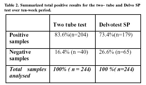
Based on the qualitative colour index the results for the weekly screened suspect milk from the collection centres using the two-tube test and the commercial Delvo test were interpreted as in table 1.
From table 1; tcal > 1.833 at p = 0.05, the Ho was rejected and it was concluded that the available data provided sufficient evidence that the improved two-tube test leads to an improvement in sensitivity of antimicrobial residues. The interpretation of the two-tube test is based on a color change of the medium, which if small can be interpreted either way. 16.4% (n=40) of the samples were thus classified as inconclusive in the two tube test, since they had passed the field screening test but failed the two tube on analysis in the laboratory. 26.6 %(n=65) were also inconclusive for the Delvotest. This could be attributed to the samples having low concentrations of antimicrobials, which on storage decomposed or false positive results in the screening at the field. The samples could also contain other inhibitors, which were not sufficient to cause inhibition. It would be of interest to repeat the study with a more objective test such as the ELISA. Conclusions
From the foregoing data the results suggest that the two-tube test has some improved effect on the sensitivity for antimicrobials residues in comparison to the commercial Delvo test. The two-tube test could be a suitable method for a developing dairy industry such as Kenya where previous studies indicate an urgent need for control of antimicrobial residues at the milk collection centers, (Shitandi., 2000). The method as developed claims detection limits for beta-lactams, tetracyclines, sulphonamides, trimethoprim, macrolides and aminoglycosides below or near the respective MRLs, (Nouws et al, 1995; Rikilt-dlo., 1998; Nouws et al, 1999;). It offers the advantages of easy performance, good response to compositional changes, low cost per sample and broad-spectrum detection.
It is crucial that the hygienic and compositional quality of milk be safeguarded in the long-term development of a dairy industry. To ensure technological and toxicological safety, an integrated detection system for antimicrobials must be developed. Acknowledgements
The studies were carried out at the Department of Food Science, Egerton University Njoro- Kenya. I am very grateful to the Swedish Institute (SVENSKA), the scholarship donor and the Swedish Dairy Training program who kindly provided financial support during the research period. I am also grateful to Associate Professor Åse Sternesjö for her devoted guidance throughout the research, Mrs. Lotta Wall, for excellent technical input. Associate Professor Symon Mahungu, for permission to use the Egerton University, dairy department facilities for the local research. References
Copyright 2001 The Journal of Food Technology in Africa, Nairobi The following images related to this document are available:Photo images[ft01002t1.jpg] [ft01002t2.jpg] |
| |||||||||