 |
| About Bioline | All Journals | Testimonials | Membership | News |
|
||||||
|
||||||
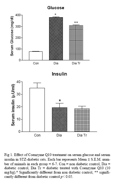
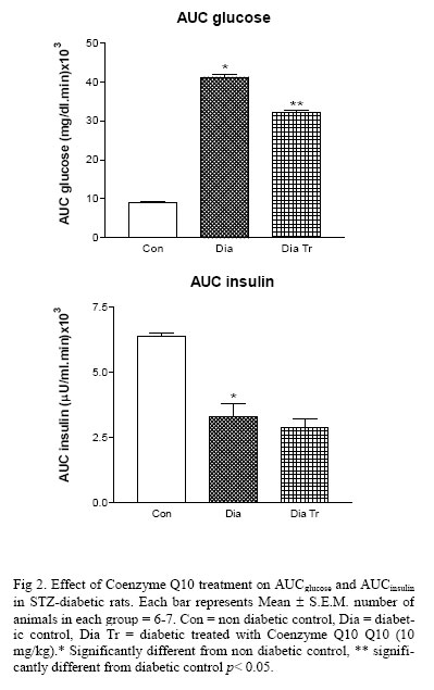
Iranian Journal of Pharmacology & Therapeutics, Vol. 5, No. 1, 2006, pp. 61-65 Research Article Beneficial Effects of Coenzyme Q10 in Streptozotocin-Induced Type I Diabetic RatsKETAN P. MODI, SANTOSH L. VISHWAKARMA, RAMESH K. GOYAL and PARLOOP A. BHATT Current Author Addresses Ketan P. Modi, Department of Pharmacolgoy, Shri B. M. Shah College of Pharmaceutical Education & Research, Modasa, India; Department of Pharmacology, L. M. College of Pharmacy, Ahmedabad, India. Email: ketan_modi11@rediffmail.com. Received January 27, 2006; Revised May 31, 2006; Accepted June 1, 2006 Code Number: pt06010 ABSTRACT The present investigation was undertaken to study the benefical effects of Coenzyme Q10 in streptozotocin (STZ)-induced type I diabetic rats. STZ-diabetes produced a significant increase in fasting glucose levels that was associated with decrease in serum insulin levels. STZ also produced hypercholesterolemia, hypertriglyceredemia, increase in lipid peroxidation and decrease in high density lipoprotein (HDL) levels. Treatment with Coenzyme Q10 produced a significant decrease in fasting glucose levels without affecting insulin levels. Coenzyme Q10 was also found to decrease significantly AUCglucose and no significant change in AUCinsulin values in STZ-diabetic rats. Treatment with Coenzyme Q10 also caused decrease in serum cholesterol, serum triglyceride levels and an increase in HDL levels. Coenzyme Q10 treatment also reduced lipid peroxidation in diabetic rats. The elevated blood pressure in diabetic rats was also lowered. Our data suggest that Coenzyme Q10 has beneficial effects in diabetes induced complications. Keywords: Coenzyme Q10, Streptozotocin, Diabetes It is widely accepted that there is oxidative stress in diabetes mellitus [1]. Hyperglycemia in diabetes mellitus generates free radicals by mechanisms that are thought to involve metal-catalyzed oxidation of glucose, oxidative degeneration and protein glycation [2, 3]. Enzymes that normally detoxify free radicals may also be partially incapacitated by non-enzymatic glycation in diabetic individuals. The presence of these free radicals may account for many of the complication of diabetes [4, 5]. Even in diabetic individuals using insulin, oxidative stress may be due to the recurrence of transiently high blood glucose concentration as a result of inexact exogenous control of circulating insulin levels [6]. Degeneration of vital tissues leading to diabetic complications may be due to increased oxidative stress that is a reason to hope that chronic antioxidant therapies may be useful in decreasing the risk of diabetic complications. Coenzyme Q10 is an endogenous antioxidant that scavenges free radicals directly, inhibits biomolecule oxidation and affects antioxidants in vivo [7-9]. Although its structural characteristic (delocalized p-electrons, adjacent electron-donating heteroatoms and a long isoprenoid chain) allow Coenzyme Q10 to diffuse into the membrane phospholipid bilayer, where it serves as an electron transfer intermediate in the mitochondrial respiratory chain, its reduced form is a powerful antioxidant [10]. Coenzyme Q10 regulates oxidative phosphorylation and prevents lipid peroxidation [11]. Coenzyme Q10 has been reported to have a beneficial effect on different symptoms in mitochondrial myopathy, encephalopathy, lactic acidosis and stroke-like episodes (MELAS) and Kearns-Sayare syndrome [12, 13]. Rauscher et al. reported various effects of coenzyme Q10 treatment on antioxidant pathways in normal and streptozotocin-induced diabetic rats [14]. Coenzyme Q10 reduces blood pressure and insulin resistance in hypertensive patients with coronary artery disease [15]. In this light the objective of the current investigation was to study beneficial effects of coenzyme Q10 in streptozotocin-induced diabetic rats. Materials and Methods Animals Male Sprague Dawley rats (weighing between 200-260 g each) were used for the study. They were maintained under standard environmental conditions and were fed a standard pellet diet with water ad libitum. Change in body weight, food intake and water intake were recorded at interval of 4 weeks. Induction of DiabetesDiabetes was induced by single tail vein injection of STZ (45 mg/kg) [Sigma, St. Luis, MO, USA] to male Sprague Dawley rats (200-260 g). Animals showing glucosuria more than 2% (Diastix, Bayer Diagnostics, India) or blood glucose level (>140 mg/dl) 48 h after STZ injection were selected for the experiment. Animals were divided into three groups: non diabetic control, diabetic control and diabetic treated(n = 6 -7 in each group). Treatment groups received Coenzyme Q10 at the dose of 10 mg/kg i.p. [14] (Rauscher et al., 2001) daily for four weeks.Control group received the vehicle i.e. dimethyl sulfoxide. During the study standard food and water were provided ad libitum. Changes in body weight, food intake and water intake were recorded. Blood Sampling and Biochemical Analysis At the end of four-week treatment, blood samples were collected from the tail vein into centrifuge tubes and allowed to clot for 30 minutes at room temperature. Blood samples were centrifuged at 3000 rpm for 20 minutes. Serum was separated and stored at -20°C until analysis was done. Serum samples were analyzed spectrophotometrically for glucose, cholesterol, triglycerides and HDL (Bayer Diagnostics Kit, India). Serum insulin levels were estimated by radioimmunoassay method using the kit from Bhabha Atomic Research center, Mumbai, India. VLDL and LDL were calculated as per Friedevald’s equation. VLDL = Total serum triglycerides/ 5, while LDL = total serum cholesterol- total serum triglycerides/5 – HDL. Oral Glucose Tolerance Test Rats were subjected to an oral glucose tolerance test (OGTT). Glucose (1.5 g /kg) was administered to 12 hours fasted rats. Blood samples were collected at 0, 30, 60, 120 minutes. Serum was separated immediately and analyzed for glucose and insulin. The results of OGTT were expressed as integrated areas under the curves for glucose (AUCglucose) and insulin (AUCinsulin) over a period of 0-120 minutes. Measurement of Blood PressureBlood pressure was recorded by the tail-cuff method using the Harvard blood pressure monitor (Kent, UK). Rat was placed into a restrainer and its tail was introduced into the cuff. The initial gain set was established by means of a pulse sensor to get monitor deflection. The pressure was first raised to 200 mm Hg and then slowly released by means of a screw attachment. During this decline of pressure, the point at which there is an increase in magnitude of deflection of the pulse analyzer was considered as the systolic blood pressure of the rat. At this point the heart rate was measured by increasing chart speed and recording the number of beats per min. Blood pressure recording were repeated three times to obtain consistent results. Lipid PeroxidationTwenty-four hours after the last antioxidant or vehicle dose, animals were anesthetized with sodium pentobarbitone (40 mg/kg i.p.). Livers was excised and immediately frozen in dry ice and stored at -20 OC. Frozen tissue from each rat was homogenized in ice cold 0.1 M Tris-HCl buffer (pH 7.4) and assayed for degree of lipid peroxidation by measuring thiobarbituric acid reactive substances (TBARS) according to Ohkawa et al.[16]. To 0.5 ml tissue homogenate, 0.5 ml saline and 1.0 ml 10% TCA were added, mixed well and centrifuged at 3000 rpm for 20 minutes. To 1.0 ml of the protein-free supernatant, 0.25 ml of thiobarbituric acid (TBA) reagent was added; the contents were mixed and heated for 1 hour at 95ºC. The tubes were cooled to room temperature under running water and absorbance measured at 532 nm. The levels of lipid peroxides were ex-pressed as micro moles of thiobarbituric acid reactive substances (TBARS)/mg protein. Statistical AnalysisThe results were analyzed statistically using one way ANOVA followed by Tukey's multiple tests to determine level of significance. Value of p < 0.05 was considered significant. Results General Features of Diabetic Animals Animals, which received STZ, showed a significant reduction in weight gain, increase in water intake and food intake as compared to control animals (p < 0.05) (Table 1). Treatment with Coenzyme Q10 did not alter the body weight, water intake and food intake. Serum Glucose, Insulin and Lipid Levels of Diabetic AnimalsSTZ-rats exhibited a significant hyperglycemia and hypoinsulinemia as compared to non diabetic control animals. Treatment with Coenzyme Q10 significantly prevented STZ-induced hyperglycemia. STZ-induced decrease in insulin levels was further decreased but it was not significant (Fig 1). STZ-injection caused a significant increase in AUCglucose associated with a significant decrease in AUCinsulin values in diabetic control animals as compared to control. Treatment with Coenzyme Q10 significantly prevented STZ-induced increase in AUCglucose but did not show any effect on AUCinsulin (Fig 2). STZ-diabetic rats produced significant increase in cholesterol, triglyceride, VLDL and LDL while decrease in HDL, as compared to non diabetic control animals. Treatment with Coenzyme Q10 showed decrease in cholesterol, triglyceride, VLDL and LDL levels while increased HDL levels (Table 1).
Blood Pressure The diabetic animals showed higher blood pressure and bradycardia as compared to the non diabetic control groups (Table 1). Treatment with Coenzyme Q10 produced a significant decrease in blood pressure but no significant effect was observed on heart rate in diabetic animals (Table 1). Lipid PeroxidationThe diabetic animals showed higher lipid peroxidation when compared to non diabetic control groups. Treatment with Coenzyme Q10 produced a significant decrease in lipid peroxidation (Table 1). DiscussionIntravenous administration of STZ produced cardinal symptoms such as hyperglycemia, hypoinsulinemia, loss of body weight, polyphagia, polyurea and polydypsia. These findings are consistent with earlier findings [17, 18]. Treatment with Coenzyme Q10 did not produce any change in body weight, hyperphagia and polydypsia in diabetic rats. STZ diabetic rats showed significant increase in glucose levels and decrease in insulin levels. Coenzyme Q10 is reported to have significant decrease in fasting and postprandial glucose and insulin levels in trans fatty acid rich diet [19]. In our study Coenzyme Q10 significantly decreased serum glucose and AUCglucose levels, insulin and AUCinsulin levels were decreased but the changes were insignificant in diabetic treated rats. Glucose autoxidation and glycation of protein leads to generation of oxygen free radicals, which can enhance lipid peroxidation and oxidation of LDL [2]. STZ diabetic rats showed significant increase in serum cholesterol, triglycerides, VLDL, LDL and decrease in HDL levels. Treatment with Coenzyme Q10 significantly decreased serum cholesterol, triglycerides, VLDL, LDL and increased HDL levels in diabetic treated rats. Singh et al. reported that Coenzyme Q10 decreased the aortic cholesterol, triglycerides, sudanophilia and aortic and coronary artery plaque sizes, coronary atherosclerosis index, aortic and coronary atherosclerosis scores in trans-fatty rich diet [20]. The reduction in lipid levels and increase in HDL levels may be due to inhibition of LDL oxidation and reduce oxidative stress. STZ induced diabetic rats showed increase in blood pressure and bradycardia. Coenzyme Q10 reported to decreases the left ventricular pressure in STZ induced diabetic rats [21]. Singh et al. reported that Coenzyme Q10 decreased blood pressure in coronary artery disease [15]. In our study, treatment with Coenzyme Q10 significantly lowered the blood pressure. This effect may be due to decrease in oxidative stress and lower blood viscosity. In STZ induced diabetes due to oxidative stress there is generation of free radical which promotes lipid peroxidation. Coenzyme Q10 supplementation was associated with significant reduction in thiobarbituric acid reactive substances, malondealdehyde and diene conjugates in coronary artery disease patients [22]. In present study Coenzyme Q10 significantly decreased the lipid peroxidation which may be due to decreased oxidative stress. In conclusion, Coenzyme Q10 has beneficial effect in complications associated with diabetes due to decrease in oxidative stress in diabetes. AcknowledgementThe authors are thankful to Troikaa Pharmaceuticals Ltd. India for providing us gift sample of Coenzyme Q10. References
Copyright © 2005 by Razi Institute for Drug Research (RIDR) The following images related to this document are available:Photo images[pt06010f1.jpg] [pt06010f2.jpg] | ||||||||||||||||||||||||||||||||||||||||||||||||||||||||||||||||||||||||||
| |||||||||